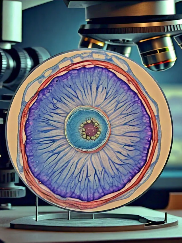
Microscopes have been around for the ages. Roman philosophers had mentioned “burning glass” within their works. However, the first microscope of this type was not invented until the 1300’s. Two lenses were set on opposite sides of the tube. This tube of magnifying power was the basis for our modern-day microscope.
Contents
First Microscope
Grinding glass used to make magnifying glasses and spectacles was commonplace throughout the early 13th century. In the 16th century, several Dutch lens makers created instruments that magnified objects however, around 1609 Galileo Galilei perfected the first microscope.
Dutch manufacturer of optical spectacles Zaccharias Janssen, and Hans Lipperhey are noted as the first people to come up with the idea of the compound microscope.
By putting various types and sizes of lenses at opposite tubes, they found that tiny objects were expanded.
Lens Improvement
In the 16th century Anton van Leeuwenhoek began polishing and grinding lenses after the discovery was made that certain types of lenses could increase the size of an image.
The lenses made of glass that were invented by him could magnify the size of an object by many times. The lens’s quality enabled him, for the first time in time, to observe the various microscopic animals and bacteria as well as the fine detail of everyday objects.
Leeuwenhoek is regarded as the pioneer of the field of microscopy. He important role played in the creation of the cell theory.
Achromatic Lens
The microscope was used for more than 100 years before the next major advancement was made. Utilizing early microscopes was challenging. Light reflected when passing through the lenses and changed the way the image appeared to be.
The achromatic lens was invented for spectacles, through Chester Moore Hall in 1729 The quality of the microscopes improved. With these lenses, many would keep improving the visual quality by using the microscope.
Mechanical Improvements
In between the 19th century and 18th, a lot of changes were made in the design of the housing as well as what microscope technology was used to create.
Microscopes were smaller and more stable. Lens improvements helped solve several optical problems that were prevalent when using earlier version.
The history of the microscope broadens and extends from this point by bringing together people from all over the globe working on similar improvements and lens technologies in the same time.
August Kohler is credited with the idea of providing uniform illumination for microscopes that enabled subjects to be photographed. Ernst Leitz devised a way to permit various magnifications with one microscope by placing several lenses on a moveable rotating turret on the other end of the tube.
Are you looking for a way to permit more light-spectrum colors to be seen, Ernst Abbe designed a microscope that, in just a few years would supply Zeiss with the equipment needed to build an ultraviolet-focused microscope.
Modern Technology Improving Microscopy
The development of the microscope has allowed researchers and scientists to study the microscopic creatures that inhabit the world that surround them.
In learning about the development of the microscope, it is essential to know that before the discovery of these microscopic creatures the reasons for illnesses and diseases were thought to be but were not fully understood.
The microscope enabled humans to escape the realm of things that were not visible and into an environment where the diseases causing agents were identified, identified and, in time, eliminated.
Charles Spencer demonstrated that light changed the way images were perceived. It took more than 100 years to create the first microscope working without light. First electron microscopes were created around 1930 in the 1930’s by Max Knoll and Ernst Ruska.
Electron microscopes are able to provide images of the smallest of particles, however they aren’t able to study living creatures. The resolution and magnification are incomparable to that of a light microscope. To study live specimens, you will require an ordinary microscope.
Scanning probe microscopy permits the examination of specimens at the atomic level . This was first introduced with the invention of the scanning tunneling microscope around the year 1981 by Gerd Bennig and Heinrich Rohrer.
Later , Bennig and his coworkers were able to go further to develop an atomic force microscope which brought an time of research on nanoparticles. Microscopes have been around for many centuries and Leeuwenhoek’s initial design has not changed from the 1600’s.
Microscope Timeline From 13th Century to Today!
A microscope is an optical instrument employed to look at tiny objects. Thus, the goal of microscopes is to magnify an object and make the image big enough to allow an even better perspective. The timeline of the history is traced way back to the first and third century , when the antiquated Romans as well as Egyptians were studying and developing glass.
Although they did not create microscopes like we see them , they studied the ways in which different types of glass created objects to appear larger and the ability to bend light. For example at the turn of the century Claudius Ptolemy published a paper describing how sticks appeared to be bent when placed in a lake of water. This led him to determine the angle. This is where it becomes clear that curiosity about the various properties of glass started in the beginning. In this article, we will take a look at the past or time line of the microscope.
13th Century
In the 13th century (1284) Salvino D’Armato degli Armati from Florence (Italy) created the wearable eyeglass which magnifies objects, which allowed the user to view more clearly. Through some study, Salvino discovered that convex pieces of glass were able to magnify the appearance of objects were magnified. This enabled him to design the eyeglasses.
16th Century
The development of what is considered to be”the first compound microscope” attributable to two Dutch spectacle makers, Zacharias Jansen, and Hans Jansen, his father. Hans. After conducting experiments with lenses Jensen was able to create the first microscope made up out of 3 draw tubes as well as lens (Bi-convex Eyepiece as well as Plano-convex objective) placed at the two ends.
Through adjusting (through moving) through the tube the out Jansen could narrow the focus of the microscope. Even though the images were unclear, it was able to allow to magnify the images by 9x when extended, and 3x when it was closed.
17th Century
Galileo Galilei (1609)
In 1609, when attempting to build a telescope Galileo Galilei used lenses that had a shorter focal length to transform his telescope into an microscope which was able to increase the size of small objects. The microscope was equipped with two lenses. These were an objective with a bi-convex as well being a bi-concave lens.
While Galileo also invented a microscope in 1624, which utilized three bi-convex lens, it was not able to provide greater magnification than the earlier model he invented. The Galileo microscope had the equivalent of 30x magnification.
The term “microscope” to refer to Galileo’s microscope compound was invented by Giovanni Faber in 1625
Robert Hooke (1665)
It was in 1665 that Robert Hooke, an English natural philosopher and physicist, published his “Micrographia” In it, he jotted down the results of his observations made using the microscope. Utilizing a microscope that was primitive, Hooke was able to see a variety of things, including corks and fleas.
In this instance, he was able to see small scales of hair on the flea and cork’s pores (which Hooke described as cell). But what Hooke did not know at the moment was that he had just discovered plants cells. Hooke’s microscope was one lens that was lit by candles. Have a look at the cell theories he proposed.
Anton Van Leeuwenhoek (1674)
In the late 1670s Leeuwenhoek was an Dutch tradesman and scientist, utilized his expertise as a lens grinding machine to create a microscope capable of greater magnification. The microscope he designed was tiny (about two inches in length and 1 inch wide). The microscope was made up of two metal plates that were riveted together, with tiny bi-convex lenses in between. This microscope was capable of offering magnifications ranging from 70x and 270x.
Leeuwenhoek’s lens was of top quality in comparison to other lenses of the same time period, with the thickness of just one millimeter, and a curvature radius of approximately 0.75 millimeter. With this microscope could be used to look at bacteria.
19th Century
Joseph Jackson Lister (1826)
The year was 1826. Joseph Jackson Lister, an English wine merchant and scientist, was successful in developing an achromatic lens, which eliminated the effect of chromatic (spherical aberration). In this case, Lister used several weak lenses in conjunction at certain distances that resulted in high magnification but and blurred images. This was an important advancement in microscopy, and has made microscopes an essential tool for medical research.
“In 1874 Ernst Abbe came up with the theoretical resolution of the light microscope. This is because Abbe created an equation that correlated the power of resolving with the light wavelength, making it possible to determine the theoretical resolution of the microscope.
20th Century
The 20th Century saw more developments within the area of microscopy. making various microscopy techniques which are becoming increasingly important in the present. This includes:
- The Transmission Electron Microscope (1931) – It was created and constructed by Ernst Ruska and Max Knoll following the designs that came from Leo Szilard. The microscope was powered by electrons rather than light.
- Phase Contrast Microscope (1932) – A phase-contrast microscope designed in the laboratory of Frits Zernike, in the year 1932 in order to imaging transparent specimens. The imaging of specimens using this microscope without staining is accomplished through interference rather than absorbance of light.
- Scanning Electron Microscope (1942) – It was created by Ernst Ruska and worked by transmitting electrons over the surface of the specimen.
- Confocal imaging principle (1957) – This method was invented by Marvin Minsky. It utilizes lasers to offer the same resolution but with a slight improvement to microscopes that utilize light. With this method it is simpler to view virtual slices in the thick specimen.
- First CAT scanner (1972) – The technology was invented in 1972 by Godfrey Hounsfield and Allan Cormack and involves the fusion of several pictures of X-rays (with the assistance of computers) to assist in producing cross-sectional views and 3D images.
- Electron backscatter patterns observed (1973) – Developed by John Venables and CJ Harland in 1973, this technique can be used to give precise microstructural data about materials as minerals, metals and ceramics for instance.
- Confocal laser scanning microscope (1978) – Developed by Thomas and Christoph Cremer, the confocal laser scanning microscope assists in scanning objects with a laser beam.
- Scanning Tunneling Microscope (1981) – In the year 1981 Gerd Binnig along with Heinrich Rohrer created this method that is used to measure interactions between atoms.
- Green Fluorescent Protein (1992) – It was discovered in 1992. Green fluorescent protein first first discovered in the year 1962, through Osamu Shimomura Frank Johnson and Yo Saiga It was then replicated in 1992 and its derivatives used for fluorescence microscopy.
Related Posts
- Trinocular Microscope – Definition, Principle, Parts, Protocol, Uses
- Coarse Adjustment and Fine Adjustment Knob of Microscope
- Working Distance – Definition, Measurement, Types, Importance
- Pocket Microscope – Definition, Parts, Principle, Uses, Types
- Polarizing Microscopes – Principle, Definition, Parts, Applications


