Contents
What is Polarizing Microscope?
A polarizing microscope is a type of microscope that uses polarized light to view specimens. It is commonly used to observe minerals, crystals, and other transparent or semi-transparent materials, as well as to analyze the structure and properties of these materials.
Polarized light is light that vibrates in a specific plane, rather than vibrating randomly in all directions like normal light. When light passes through certain types of materials, it can become polarized. A polarizing microscope uses this property of light to reveal the internal structure and properties of a specimen.
A polarizing microscope typically consists of a microscope body with an objective lens and eyepiece, a light source, and two polarizing filters called polarizers. The polarizers are placed at the light source and at the eyepiece, and they are oriented so that the planes of polarization are perpendicular to each other. When a specimen is placed between the polarizers, it absorbs or reflects the polarized light differently depending on its properties, allowing the internal structure and properties of the specimen to be observed.
Polarizing microscopes are commonly used in scientific research, mineralogy, and other fields where the analysis of transparent or semi-transparent materials is important. They can be used to identify minerals, analyze the structure and composition of crystals, and study the properties of other transparent materials.
Non-Polarized to Polarized Light Convertion
- Polarized light microscopes transform unpolarized light to polarised light in order to function. This can be accomplished by absorbing the vibrational movement of light in a certain direction.
- Certain natural minerals, such as tourmaline, or manufactured films that perform the same function can do this.
- Polaroid filters are composed of microscopic crystallites of iodoquinine sulphate that are aligned in the same direction and incorporated into a polymeric filter.
- This embedding is performed to prevent crystal migration and orientation alteration. A polarizer is a device that chooses plane-polarized light from natural or unpolarized light.
Principles of Polarized Light Microscopes
In a polarised light microscope, the light source and sample are separated by a polarizer. Before reaching the sample, the polarised light source is transformed to plane-polarized light. This polarised light strikes a material with double refraction, which forms two wave components at right angles to one another. These two waves are known as common and exceptional light beams.
The specimen is traversed by the waves in several phases. An analyzer then combines them using constructive and destructive interference. This results in the final creation of an image with strong contrast.
Parts of a Polarized Light Microscope
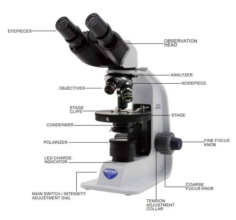
1. Polarizers
- Polarizing filters are the most necessary component of a polarised light microscope. Typically, polarising filters consist of two components: the polarizer and the analyzer.
- The polarizer is positioned beneath the specimen stage and may be turned 360 degrees. It aids in polarising the light falling on the specimen.
- The analyzer is positioned above the objective and is sometimes rotatable. It mixes the many rays emitted by the specimen to get the final image.
2. Specialized Stage
- This is the specimen stage, which can rotate 360 degrees to ensure the specimen is properly aligned with the objective plane.
- In several stages, a Vernier scale is also included to offer 0.1° of accuracy in the stage’s rotational angle.
3. Strain-Free Objectives
- Any strain placed on the objective during installation can alter the optical properties of the lens, resulting in a reduction in performance.
- Additionally, if the lens is attached too tightly on the frame, strain can be introduced. In addition, anti-reflection coatings and refractive characteristics must be precisely evaluated to guarantee polarisation and enhanced contrast.
4. Revolving Nosepiece
- As the stage and objectives of many polarising microscopes can rotate, a rotatable nosepiece is frequently included so that the specimen can be observed in the centre of the field of vision even when the stage is rotated.
5. Compensator and Retardation Plates
- Multiple polarisation microscopes are equipped with compensators and/or retardation plates. This is put between the crossed polarizers in order to increase the optical path difference within the specimen. This would raise the contrast of the image even further.
- Consequently, polarising microscopes are employed to enhance image contrast in order to visualise several anisotropic subcellular features.
6. Light intensity adjustment
- Operate on the light intensity adjustment dial to turn ON / OFF the microscope and to increase / decrease the illumination voltage.
7. Coarse focus tension adjustment
- Adjust the tension using the provided tool. The tension of the coarse focusing knob is factory preset.
- To modify the tension according to personal’s needs, rotate the ring. Clockwise rotation increases the tension. If the tension is too loose, the stage could go lower by itself or the focus easily lost after fine adjustment. In this case, rotate the knob in order to increase the tension.
8. Adjust the interpupillary distance
- Hold the right and left parts of the observation head using both hands and adjust the interpupillary distance by turning the two parts until one circle of light can be seen.
- The graduation on the interpupillary distance indicator, pointed by the spot “.” on the eyepiece holder, shows the distance between the operator’s eyes.
- The range of the interpupillary distance is 48- 75 mm.
9. Diopter adjustment
- Look into the right eyepiece with your right eye only, and focus on the specimen.
- Look into the left eyepiece with your left eye only. If the image is not sharp, use the dioptric adjustment ring to compensate.
- The adjustment range is ±5 diopter. The number indicated on the adjustment ring graduation should correspond to the operator’s dioptric correction.
10. Aperture diaphragm
- The Numerical Aperture (N.A.) value of the aperture diaphragm affects the image contrast. Incre- asing or reducing this value one can vary resolution, contrast and depth of focus of the image.
- Move the diaphragm lever toward left or right to decrease or increase the N.A. value. With low contrast specimens set the numerical aperture to about 70%-80% of the objective’s N.A. If necessary, remove on eyepiece and, looking into empty sleeve, adjust the condenser’s diaphragm in order to obtain an image.
11. Rechargeable batteries
- When the microscope is plugged with the power supply, the LED indicator for the battery recharge is lit.
- LED red: battery under charge
- LED green: battery fully charged. When the microscope is unplugged, the LED is off.
- During the normal use with batteries, LED is always OFF.
12. Strain Free Condenser
- Several characteristics are shared by condensers built for polarised light microscopy, including the use of strain-free lenses.
- Some condensers include a socket for the polarizer, while others have the polarising element installed directly into the condenser, beneath the aperture diaphragm.
- Numerous polarised light condensers contain a top lens that may be withdrawn from the light path (a swing-lens condenser) to produce almost parallel illumination wavefronts for low magnification and birefringence investigations.
13. Eyepieces
- The eyepieces of a polarised light microscope have a cross-wire reticle (or graticule) to mark the centre of the field of view.
- Frequently, the cross wire reticle is replaced by a photomicrography reticle that aids in focusing the specimen and composing photographs with a set of frames enclosing the viewfield to be shot digitally or on film.
- A point pin that slips into the observation tube sleeve ensures the correct orientation of the eyepiece relative to the polarizer and analyzer.
14. Bertrand Lens
- A specialised lens fitted within an intermediate tube or the observation tubes, a Bertrand lens brings an interference pattern created at the objective back focal plane into sharp focus at the microscope picture plane.
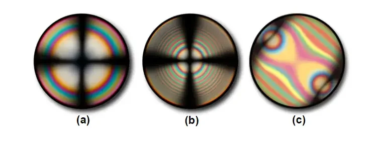
- The lens is designed to permit simple observation of the objective rear focal plane, precise adjustment of the illuminating aperture diaphragm, and visualisation of interference patterns comparable to those shown in Figure 2.
- Note that the interference patterns in Figures 2(a) and 2(b) represent those found with a uniaxial crystal in polarised light, but the pattern in Figure 2(c) is representative of a uniaxial crystal with a first order retardation plate added into the optical channel.
Basic Properties of Polarized Light
Polarized light is light that vibrates in a specific plane, rather than vibrating randomly in all directions like normal light. Some basic properties of polarized light include:
- Polarization plane: The plane of vibration of the light waves is called the polarization plane. Polarized light can be horizontally polarized, vertically polarized, or polarized at any angle in between.
- Polarization direction: The direction of the electric field in the light waves is called the polarization direction. Polarized light can be linearly polarized, meaning that the electric field is in a single direction, or circularly polarized, meaning that the electric field rotates around the direction of propagation.
- Extinction: When the plane of polarization of the light is perpendicular to the plane of polarization of the analyzer (a polarizing filter), the light is extinguished, meaning that it is completely absorbed by the analyzer. This property is used in polarizing microscopes to analyze the birefringence of materials.
- Brewster’s angle: The angle at which light is completely polarized when it is incident on a transparent medium at a particular angle is called Brewster’s angle. This angle is dependent on the refractive index of the medium and the wavelength of the light.
- Double refraction: Some materials, such as crystals, have the property of double refraction, meaning that they can split light into two different beams that vibrate in different planes. This property is known as birefringence, and it can be studied using a polarizing microscope.
Operating Procedure of Polarized Light Microscope
Use in brightfield
- Move the analyzer slider to the right to remove analyzer filter from the light path.
- In this way it is possible to work in brightfield.
Use in polarized light
- Move the analyzer slider to the left to insert analyzer filter into the light path.
- Remove one eyepiece from the observation head.
- Insert 10X objective into the light path.
- Remove the specimen from the stage.
- Rotate the polarizer filter until a complete dark field of view can be achieved. Now the
“crossed-Nicol” position is obtained and it is possible work in polarized light. - Put a specimen on the stage.
- Insert the desired objective.
- Focus the specimen.
- Begin the observation.
- Polarizer can be removed from the light path simply rotating the polarizer unit toward right.
How does a Polarized Light Microscope Works?
Compared to approaches such as darkfield and brightfield illumination, differential interference contrast, phase contrast, Hoffman modulation contrast, and fluorescence, polarised light is a contrast-enhancing technique that improves the quality of the image acquired using birefringent materials. Polarized light microscopes have a high level of sensitivity and can be used for quantitative and qualitative research on a wide variety of anisotropic objects. Numerous tomes are devoted to the topic of qualitative polarising microscopy, which is widely used in practise. In contrast, the quantitative features of polarised light microscopy, which is largely used in crystallography, are a far more complex topic that is typically reserved to geologists, mineralogists, and chemists. Biologists are now able to investigate the birefringence of numerous anisotropic sub-cellular assemblies due to the continuous progress gained over the previous decade.
The polarised light microscope is designed to study and photograph objects that are predominantly visible due to their anisotropic optical properties. In order to achieve this task, the microscope must be supplied with a polarizer positioned in the light path before the specimen and an analyzer (a second polarizer; see Figure 1) positioned between the objective back aperture and the observation tubes or camera port. Image contrast results from the interaction of plane-polarized light with a birefringent (or doubly-refracting) specimen, which generates two wave components polarised in mutually perpendicular planes. The velocities of these components, known as the ordinary and extraordinary wavefronts (Figure 1), change with the direction of propagation through the specimen and are distinct. After leaving the specimen, the light components fall out of phase; however, they are recombined by constructive and destructive interference as they pass through the analyzer. Figure 1 illustrates these notions for the wavefront field formed by a hypothetical birefringent material. In addition, the picture depicts the essential optical and mechanical components of a contemporary polarised light microscope.
Similar to brightfield illumination, polarised light microscopy may provide information on absorption colour and optical path boundaries between minerals with different refractive indices, but it can also distinguish between isotropic and anisotropic material. In addition, the contrast-enhancement technique takes advantage of the optical features unique to anisotropy and reveals vital identification and diagnostic information regarding the structure and composition of materials.
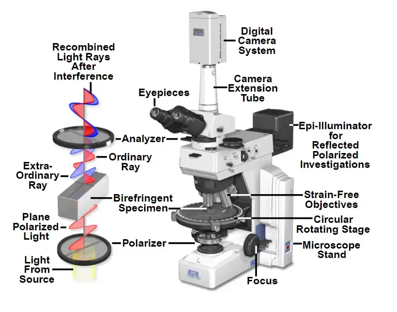
Probed in all directions, isotropic materials, which include a range of gases, liquids, unstressed glasses, and cubic crystals, exhibit the same optical properties. These materials have a single refractive index and do not restrict the direction of light’s vibration when it passes through them. In contrast, the optical characteristics of anisotropic materials, which account for 90 percent of all solid substances, vary with the orientation of incident light along the crystallographic axes. They exhibit a range of refractive indices dependent on the direction of light propagation through the substance and the vibrational plane coordinates. Moreover, anisotropic materials function as beamsplitters and separate light rays into two orthogonal components (as illustrated in Figure 1). The technique of polarising microscopy extracts information about anisotropic materials by utilising the interference of light rays recombined along the same optical path.
Polarized light microscopy is likely most well-known for its applications in the geological sciences, which largely examine minerals in thin rock sections. Polarized light can be used to study a wide range of different materials, including natural and artificial minerals, cement composites, ceramics, mineral fibres, polymers, starch, wood, urea, and several biological macromolecules and structural assemblies. The approach is an excellent tool for the materials sciences, geology, chemistry, biology, metallurgy, and even medicine, as it can be applied qualitatively and quantitatively with equal success.
Understanding the analytical techniques of polarised microscopy may be more difficult than other forms of microscopy, but it is well worth the effort due to the additional information that can be collected in comparison to brightfield imaging. Understanding the fundamental concepts underpinning polarised light microscopy is also required for the accurate interpretation of differential interference contrast (DIC).
Applications of Polarized Light Microscope
Polarized light microscopes are used in a variety of applications where the analysis of transparent or semi-transparent materials is important. Some common applications of these microscopes include:
- Mineralogy: Polarizing microscopes are commonly used in mineralogy to identify and analyze minerals. They can be used to study the crystal structure, refractive index, and other properties of minerals, which can help to identify them and understand their properties.
- Material science: Polarizing microscopes are also used in material science to study the structure and properties of transparent or semi-transparent materials. They can be used to analyze the microstructure of materials, such as fibers, films, and coatings, and to study the effects of different processing techniques on the material.
- Industrial inspection: Polarizing microscopes are often used in industrial settings to inspect and analyze materials for quality control purposes. They can be used to detect defects, such as cracks, voids, and inclusions, in transparent or semi-transparent materials, such as plastics, ceramics, and glass.
- Biology: Polarizing microscopes are also used in biology to study the structure and properties of biological specimens, such as cells, tissues, and organelles. They can be used to analyze the organization and orientation of molecules within cells and to study the effects of different treatments on biological samples.
- Geology: Polarizing microscopes are used in geology to study the structure and properties of rocks and minerals. They can be used to identify minerals, analyze the composition of rocks, and study the effects of different geological processes on the materials.
Advantages of Polarized Light Microscope
- High contrast: Polarized light microscopes provide high contrast images of transparent or semi-transparent materials, making it easier to see small details and structures within the specimen.
- Analysis of birefringence: These microscopes can be used to analyze the birefringence of materials, which is the ability of a material to split light into two different beams that vibrate in different planes. This can provide important information about the structure and properties of the material.
- Non-destructive analysis: Polarizing microscopes do not require any preparation or sample preparation, making them suitable for non-destructive analysis of materials.
- Versatility: These microscopes can be used to study a wide range of materials, including minerals, crystals, fibers, films, and biological specimens.
- High-precision rotating stage: The stage is spacious, pre-adjusted, and has 45° click stops. The stage’s smooth motion enables stable and simple rotation, resulting in great operability and high-quality polarised images. Because the stage is supported from the bottom near the optical axis and is equipped with steel cross roller guides, it is exceptionally stable and durable. The focus stroke has been increased to 30mm, making it easier to observe tall samples. Clamp-type upper limit focusing mechanism makes sample exchange simple and secure.
Limitations of Polarized Light Microscope
- Limited to transparent or semi-transparent materials: Polarizing microscopes are not effective at analyzing opaque materials, as they rely on the transmission of light through the specimen.
- Complex to use: Polarizing microscopes can be more complex to use compared to other types of microscopes, as they require the use of polarizing filters and the correct orientation of these filters.
- High cost: Polarizing microscopes can be more expensive compared to other types of microscopes, due to the specialized components and features they require.
- Limited to transmitted light: These microscopes can only be used to study specimens in transmitted light, meaning that they cannot be used to observe surface features or structures.
Polarized Light Microscope Images


References
- https://microscopeinternational.com/optika-b-150p-brpl-binocular-led-polarizing-microscope-rechargeable/
- https://www.microscope.healthcare.nikon.com/products/polarizing-microscopes
- https://www.microscopyu.com/techniques/polarized-light/polarized-light-microscopy
- https://www.olympus-lifescience.com/en/microscope-resource/primer/techniques/polarized/configuration/
- https://www.microscopeworld.com/t-polarizing_microscopes.aspx
- https://www.microscope.com/specialty-microscopes/polarizing-microscopes
- https://www.azolifesciences.com/article/What-are-Polarized-Light-Microscopes-and-How-Do-They-Work.aspx
- https://www.sciencedirect.com/topics/chemistry/polarizing-microscopy
- https://meijitechno.com/meiji_old/polarizing_applications.htm
- https://www.news-medical.net/Life-Science-and-Laboratory/Polarizing-Microscopes
- https://zeiss-campus.magnet.fsu.edu/referencelibrary/polarizedlight.html
- https://www2.humboldt.edu/scimus/HSC.36-53/Descriptions/AOPolScp.htm
- https://www.optikamicroscopes.com/optikamicroscopes/product/pol-series/
- https://www.motic.com/As_Polarized_microscope/
- https://amscope.com/collections/polarizing-microscopes
Related Posts
- Trinocular Microscope – Definition, Principle, Parts, Protocol, Uses
- Coarse Adjustment and Fine Adjustment Knob of Microscope
- Working Distance – Definition, Measurement, Types, Importance
- Pocket Microscope – Definition, Parts, Principle, Uses, Types
- How are samples prepared for a transmission electron microscope?


