| Genus and species | Entamoeba histolytica |
| Domain | Eukaryota |
| Phylum | Amoebozoa |
| Family | Entamoebidae |
| Etiologic agent of: | Amoebiasis; amoebic dysentery; extraintestinal amoebiasis, usually amoebic liver abscess; “anchovy sauce”); amoeba cutis; amoebic lung abscess (“liver-colored sputum”) |
| Infective stage | Tetranucleated cyst (having 2-4 nuclei) |
| Definitive host | Human |
| Portal of entry | Mouth |
| Mode of transmission | Ingestion of mature cyst through contaminated food or water |
| Habitat | Colon and cecum |
| Pathogenic stage | Trophozoite |
| Locomotive apparatus | Pseudopodia (“false foot””) |
| Motility | Active, progressive and directional |
| Nucleus | ‘Ring and dot’ appearance: peripheral chromatin and central karyosome |
| Mode of reproduction | Binary fission |
| Pathogenesis | Lytic necrosis (it looks like “flask-shaped” holes in Gastrointestinal tract sections (GIT) |
| Type of encystment | Protective and Reproductive |
| Trophozoite stage | |
| Pathognomonic/diagnostic feature | Ingested RBC; distinctive nucleus |
| Cyst Stage | |
| Chromatoidal body | ‘Cigar’ shaped bodies (made up of crystalline ribosomes) |
| Number of nuclei | 1 in early stages, 4 when mature |
| Pathognomonic/diagnostic feature | ‘Ring and dot’ nucleus and chromatoid bodies |
Contents
Entamoeba Histolytica Introduction
- Entamoeba histolytica can be described as an anaerobic parasite amoebozoan that is part of the Genus Entamoeba. It is a major cause of infection for humans and other primates , causing amoebiasis. E. histolytica is estimated to affect between 35 and 50 million people across the globe.
- E. histolytica is thought to kill over 55,000 people a year.
- In the past, it was believed to be that 10% of globe’s population was infected. However, these numbers predate the discovery that at minimum 90% of the illnesses were due to an additional type of organism, E. disper.
- Animals like cats and dogs can be affected in a brief period, but they do not seem to play a major role in transmission.
Entamoeba Histolytica Definition
- Amoebae is an Greek word which means to change.
- It comes in two forms, like Trophozoite form, which is the Latin word which means to feed. It is the motile, living, feeding stage of humans. It also has a Cyst type, which is the resting, non-motile or infective form. Cysts are able to survive outdoors (around 3-months) and are invulnerable to the elements.
- E. histolytica is a unicellular organism that has a single protozoan nucleus. It affects between 10% and 12percent of the global population. The majority of cases occur located in the tropical regions. It is common among male homosexuals ranging from 20 to 32%.
- Entamoeba Histolytica causes amoebiasis. This is due to the histo (tissue) and histo (tissue) and (destruction).

Distribution of Entamoeba Histolytica
Entamoeba histolytica is distributed globally (cosmopolitan). Nonetheless, it is more prevalent in tropical and subtropical climates than in temperate regions. The most epidemic cases of this parasite have been reported in Mexico, China, India, the Philippines, and Thailand.
Its frequency is disproportionately higher in rural and heavily populated metropolitan regions, particularly in areas with poor sanitation. In India, its influence is more pronounced in humid climes than in dry and cold ones. Children and adults are infected more frequently, but interestingly, males are infected more frequently than females.
Morphology of Entamoeba Histolytica
Entamoeba histolytica exists in 3 morphological forms Trophozoite, Pre-cyst, Cyst – uninucleate, binucleate, and quadrinucleate.
1. Trophozoite
- It is the developing and feeding stage of the parasite Sape, which is not fixed since its position is continually shifting.
- Size: varying from 18-40 µm; average being 20-30 µm
- Cytoplasm: The cytoplasm is comprised of two distinct components, ectoplasm and endoplasm. Endoplasm contains ingested RBCs, tissue granules, and food particles as well.
- Nucleus: It has a spherical shape and a diameter ranging from 4-6µ. The nucleus comprises a core karyosome and fine chromatin on its periphery.
- With the aid of pseudopodia, trophozoites are actively mobile.
- Trophozoites are anaerobic parasite ( present in large intestine)
2. Pre cyst
- It is the transitional phase between trophozoite and cyst.
- It is 10-20µ less in size.
- It is spherical or slightly oval with a blunt pseudopodium extending from the margin.
- There are no red blood cells or food particles on its endoplasm.
3. Cyst
- It is the sort of parasite that is infectious.
- It has a circular, round, or oval shape with a diameter of 12 to 15 µm.
- It is enclosed by a membrane known as the cyst wall that is very retractile. The cyst wall is resistant to digestion by the human stomach’s gastric juice.
- A developed cyst’s nucleus is quadri-nucleated.
- Cytoplasm: There are chromatid bars and glycogen masses in the cytoplasm, but no RBCs or food particles.
- A mature cyst was expelled in the faeces of an infected patient and stayed dormant in the soil for a few days.
Transmission of Entamoeba Histolytica
The active (trophozoite) stage can only be found in the host , and also in fresh feces. Cysts remain outside the host, in water, in soils and on food items, especially in moist conditions in the last. The infection may occur when someone puts something in their mouth that has been in contact with the feces of someone affected by E. histolytica, swallows something, like food or water, which is contaminated by E. histolytica, or takes in E. histolytica cysts (eggs) collected from surfaces that are contaminated or on fingers.
The cysts are easily destroyed by heat or frigid temperatures. They last for just some months away from the host. If the cysts are swallowed they can cause infection by excysting (releasing the stage of trophozoites) within the digestive tract. There are many symptoms that can be present, including bleeding diarrhea, fulminating dysentery as well as weight loss as well as abdominal pain, fatigue and amoeboma. Amoeba’s can bore in the intestinal walls, which can cause lesions as well as intestinal discomfort and can also enter into the bloodstream. It can then be able to reach the vital organs of your body. This is primarily the liver, but also the brain, lungs and the spleen. The most common result of the invading of tissues is an abscess, which could be fatal if left untreated. Red blood cells that are digested are often found in the amoeba cells cytoplasm.
- Geographical distribution: All over the world and more frequent in subtropics and tropics particularly in areas of inadequate sanitation (developing and under-developed nations).
- Habitat: Trophozoites from E. histolytica reside in the submucosal and mucosal layer of the intestine of a man. The life-cycle of Entamoeba histolytica is divided into two stages that includes a motile trophozoite as well as a non-mot cyst. Trophozoites are present inside intestinal lesions lesions that extend into the intestine as well as diarrheal stools. Likewise, cysts are predominant in stools with no diarrhea.
- Infective form: A mature quadrinucleate cyst. it has a spherical form with a refractile wall.
Life Cycle of Entamoeba Histolytica in brief
Many protozoan species within the genus Entamoeba are found in humans, however not all are linked to disease. Entamoeba histolytica is known for its role as an ameba that causes disease that is associated with extraintestinal and intestinal infections. Other species are also crucial because they could be misinterpreted as E. histolytica during the course of diagnostic tests.
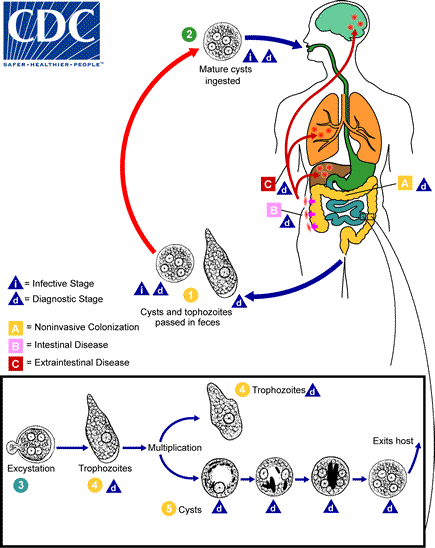
- Cysts and trophozoites can be found through feces. Cysts are usually found in stool that is formed while trophozoites are usually found in diarrheal stools.
- Infection caused by Entamoeba histolytica is caused by the ingestion of mature cysts from water, food or on hands.
- Excystation is a common occurrence inside the small intestine.
- The trophozoites then are released and then are then transported to the large intestine.
- The trophozoites multiply through binary fission. They create cysts (5) The cysts and both stages are absorbed into the feces. Due to the protection provided by their walls cysts can last from days to weeks in the environment and can be responsible for transmitting.
- The toxins in stool are destroyed quickly once they leave the body. Moreover, if taken in, would not survive exposure to the gastric environment.
- In many cases, trophozoites are confined to within the lumen of the colon (A: non-invasive infection) of people who suffer from asymptomatic carriers. passing cysts through their stool.
- In some cases, trophozoites can invade the intestinal mucosa (B IBD) or, via the bloodstream, sites outside of the intestinal tract such as the liver the lungs, and brain (C ex: extra-intestinal illness) and cause pathological manifestations.
- It is now known that the noninvasive and invasive forms are two distinct species, E. histolytica as well as E. disper. These two species are morphologically indistinguishable unless E. histolytica is observed with ingested red blood cells (erythrophagocystosis).
- Transmission could also happen through the contact with feces during sexual contact (in this case, not just cysts however, but also trophozoites can be infective).
Trophozoite is responsible for disease conditions
- The trophozoites infiltrate and invade the colonic epithelium, and release the necrosis-causing enzymes. There is a small amount of inflammation on the location.
- As the lesion progresses to the muscle layer and the muscularis layer, it is a common “flask-shaped” ulcer forms, which could cause damage to vast portions of intestinal epithelium.
- The progression into submucosa results in the invasion in the portal circulation with trophozoites.
Genome of Entamoeba Histolytica
- The genome of 20 million basepairs assembly includes 8,160 predicted genes The transposable elements have been identified and identified.
- The largest group of transposable components in E. histolytica are non-LTR retrotransposons. These are classified into three families, namely EhLINEs as well as EhSINEs (EhLINE1,2,3 and EhSINE1,2,3).
- EhLINE1 encodes one of the enzymes known as endonuclease (EN) protein (in addition to reverse transcriptase and nucleotide binding ORF1) that share a similarities with the restriction endonuclease of bacterial species. The similarity to bacterial proteins suggests that transposable elements were obtained from prokaryotes through horizontal gene transfer within these protozoan parasites.
- A genome from E. histolytica has been identified to have snoRNAs containing characteristics reminiscent of opisthokonts.
- The E. histolytica U3 snoRNA (Eh_U3 snoRNA) has shown the same structural and sequence characteristics the Homo sapiens U3 snoRNA.
Oro-fecal mode of spread of Entamoeba Histolytica/ Different Stages of Entamoeba Histolytica
Cystic stage
- The cysts are then ingested, and are absorbed into the lower part in the small intestine.
- The cysts are able to survive in the air, for almost 3 months.
- Cysts can withstand the environment.
- Cyst, under the influence of enzymes breaks down and transforms into an ameba quadrinucleate.
- This ameba can be able to escape the walls and create 8 trophozoites.
- A cyst is immune to dehydration, and can even withstand certain chemicals, such as fluorides and chlorinated compounds.
- The cyst can live in water for about a month, while cysts in feces found on land could endure for up to 12 days ( another source says that it can last for 3 months).
- The cyst is able to tolerate temperatures as high as 50 degrees Celsius.
- The cyst is able to tolerate the stomach’s acidity , and eventually reach in the small intestinal tract, the ileum which is where excystation takes place. Intestinal alkaline media is ideal for excyctation.
- Following excyctation the tetranuclear bacterium undergoes mitosis and gives birth to 8 trophozoites that are metacystic.
- They’ll move on to the large intestine where they eat as they grow and reproduce.
- They will be deposited in the cecal region.
Trophozoites stage
- Trophozoites migrate to the large intestine , and create an infection in the colon. (The most common location is the cecal colon).
- The trophozoites in the digestive tract create the flask-like ulcer.
- In these ulcers, trophozoites could enter bloodstreams and cause abscesses in the extraintestinal tract.
- Trophozoites are found inside the intestinal lumen.
- They infiltrate the crypts they feed upon RBCs.
- In the end, the ulcer.
- Trophozoites get into the bloodstream and then spread to other areas such as the liver.
- Trophozoite changes into a precyst that has two nuclei as well as chromatoid bodies.
- Precyst changes into a cyst which contains approximately 4 nuclei.
- Trophozoites change into cysts and then pass through the stool.
- Trophozoites are not able to be found outside, so they aren’t as infectious as cysts are.
- Trophozoite can be found in the intestine.
- They are able to eat bacteria.
- Protozoa can be eaten.
- They can also eat intestinal cell.
- They may ingest RBC into their bodies.
- E. Histolytic secrets battery of enzymes. One of which is known as histolysin. This enzyme allows trophozoites to infiltrate the submucosal tissues.
- In an infected patient who suffers from dysentery, the mucosal ulceration grows and causes an extensive the intestine that is destroyed. The epithelium overlying it will slough away and expose the necrotic areas.
- The destructive process is generally followed by a process of regenerative growth, that leads to the growth of the wall as a result of the fibrosis.
- Trophozoites are found in the portal circulation and can cause liver abscess.
- This can lead to hepatic amoebiasis, and possibly amoebic Hepatitis.
- The abscess hepatic could be multiple or single.
- From there , the diaphragm, leading to a lung abscess.
- Other organs that are affected are the brain, heart and spleen. They also have gonads, spleen and the skin, which can cause secondary amoebiasis.
- Cockroaches and fleas serve as mechanical vectors of spread.
Epidemiology
- E. histolytica was first reported in 1875 by an untrained Russian peasant from the harbor of Arkhangelsk. It’s a cosmopolitan illness.
- Distribution is mostly due to insufficient environmental sanitation and inadequate personal hygiene. It’s not related to the climate.
- It is thought that Entamoeba histolytica is infecting 10 percent of the world’s population. The actual incidence is perhaps one percent of the world population.
- The disease is more prevalent in subtropical and tropical nations in areas with poor sanitation.
- It is found in numerous regions of subtropical and tropical Africa, Asia, Mexico, China, and South America.
- Method of transmission The feco-oral route that is associated with the infective cyst can be ingested by drinking food, water or hands that have been contaminated.
- Amoeba can be described as the third leading cause of death due to parasites in the world. Around 500 million people suffer from the disease and around 100,000 die every year.
- It was first discovered in 1873 by a clinical associate, D.F. Losch, in St.Petersburg, Russia. He discovered the abundance of amoeba within the stool of the peasant.
- The finding was verified after forty years.
- Asymptomatic carriers are those whose cysts and the trophozoites have not been ingested by RBCs.
- A disease that is active when Toxizites are found to have consumed RBCs.
Laboratory Diagnosis of Entamoeba Histolytica
The following procedures are utilized to determine the presence of Entamoeba histolytica.
- The most frequent is direct Fecal Smear (DFS) as well as staining (but cannot allow identification to the level of species).
- Enzyme immunoassay (EIA)
- Indirect Hemagglutination (IHA)
- Antigen detection – monoclonal antibody
- A PCR is used to identify species
- Sometimes, only the use of an antibiotic (formalin) can be effective in diagnosing cysts.
- Culture: From faecal samples – Robinson’s medium, Jones’ medium.
Prevention/Prophylaxis of Entamoeba histolytica
- Beware of fecal contamination in the environment by using the nearby latrines.
- Make sure to avoid contamination of water sources by the Fecal matter.
- Always wash your hands at minimum three times using soap following defecation.
- Cleanse your hands prior to starting eating.
- Inhibit the spread of disease through flies that have cysts.
- Keep food safe from Cockroaches.
- Boil water until at 55°C, E. histolytica cysts are destroyed.
- Avoid raw or green salads that could have cysts.
- Cleansing thoroughly all fruits and vegetables thoroughly.
- Health education, specifically for school food handlers and health centers for community members.
- Beware of fertilizers made from human feces.
- A good sanitation system for disposing of waste.
- The food processing industry must screen the transporter.
Treatment
There are numerous effective drugs. A variety of antibiotics are readily available for treating Entamoeba histolytica. Infected patients is treated with just one antibiotic, if there is no evidence that the E. histolytica infection has not caused the person to become sick and they will likely receive two prescriptions in the event that the patient has felt sick. In the event that they are not sick, there are alternative alternatives for treatment.
- Intestinal infection: Usually, nitroimidazole variants (such like metronidazole) are employed, as they are extremely efficient against the trophozoite type of amoeba. Because they are not effective on amoeba cysts the procedure is followed by an antibiotic (such as diloxanide or paromomycin furoate) which acts on the organism inside the lumen.
- Liver abscess: In addition treating the presence of organisms in the tissues, most commonly using drugs such as metronidazole and chloroquine. Treatment of liver abscess should include agents that work in the lumen of intestinal tract (as in the previous paragraph) to stop an re-invasion. Surgery is rarely required, unless rupture is in the near future.
- People who do not have symptoms: For those who do not have symptoms (otherwise called carriers who are not symptomatic) Non-endemic areas must be treated with paromomycin. alternative treatments include diloxanide furoate and Iodoquinol. There have been problems with the use of iodoquinol and iodochlorhydroxyquin, so their use is not recommended. Diloxanide furoate may also be utilized by patients with mild symptoms who have just passed cysts.
Diseases
- Non-invasive infections: In many instances, the trophozoites are in the intestinal lumens of patients who are non-symptomatic carriers as well as cysts passers.
- Intestinal disease: In a few patients, trophozoites infiltrate the mucosa of the intestinal tract,
- Extra-intestinal illness: via the bloodstream, trophozoites enter extraintestinal organs like the brain, liver and lungs, causing symptoms that are pathological.
Pathogenesis
1. Mode of infection
- Entamoeba histolytica is a parasitic protozoan responsible for amoebiasis. Entamoeba histolytica is transmitted through the eating of cysts, which are the parasite’s dormant and infectious form. Infected individuals shed cysts in their faeces, which can contaminate food, water, and surfaces that come into touch with excrement. Once swallowed, the cysts travel through the stomach and into the small intestine, where they change into active trophozoites capable of invading the intestinal wall and causing tissue damage. The trophozoites can also form cysts, which are then expelled in the faeces to perpetuate the infection cycle. Inadequate sanitation, a lack of clean water, and overcrowding can all contribute to the spread of Entamoeba histolytica infection.
2. Virulence factors
- Cyst wall: the cyst wall is resistant to low pH and stomach acid.
- Lectin: The surface of trophozoites contains a lectin specific to lingards (N-acetyl-galactosamine and galactose sugar) that are found on the surface of intestinal epithelium.
- Ionophore-like protein: It induces ion leakage from target cells, including Na+, K+, and Ca++.
- Hydrolytic enzymes: Phosphatase, proteinease, glycosidase, and RNase are hydrolytic enzymes that induce tissue degradation and necrosis.
- Toxin and haemolysin
3. Pathogenesis
- The parasites express an abundance of virulence factors, including as lectin, lytic peptide, cysteine, proteinases, and phospholipases.
- The intestinal cyst releases four trophozoites, which eventually colonise the large intestine. In the pathophysiology, the binding of trophozoites to the colonic epithelium is a dynamic process. Following adhesion, the ionophore-like protein of the trophozoite lyses the target cell by causing ion leakage from the cytoplasm. Intestinal amoebiasis is characterised by the loss of tissue caused by proteolytic enzymes released by amoebae, resulting in a flask-shaped amoebic ulcer.
- Trophozoites invade the columnar epithelium of the mucosa, producing lysis, and travel deep within until they reach the submucosal layer, at which point they reproduce quickly. Amoebas ultimately kill a substantial portion of the submucosa, resulting in the creation of an abscess that deteriorates into an ulcer. The ulcer has the shape of a flask with a narrow neck and a broad base. The ulcer may be confined in the ileocaecal region or widespread throughout the large intestine.
- Blood circulation may transport parasites from the colon to other critical organs such as the liver, heart, and brain. Frequent are pulmonary and hepatic amoebic abscesses, while cerebral, cutaneous, and splenic amoebic abscesses are uncommon.
Amoebic liver abscess
- Around 2-10% of people who are infected by E. histolytica have liver complications.
- In 50 percent of cases, ameobic dysentery is not always evident.
- Toxizoites in E.histolytica are transported in emboli via the portal vein’s radicles at the base of an amoebic ulcers inside the large intestinal.
- The liver’s capillary system is a filter that works effectively and helps trophozoites grow within liver cells. It then is responsible for cytolytic action.
- This can cause obstruction of the circulation and causes the portal venules to thrombosis (sinusoids) which results in necrosis in the liver cells.
- Degradation of liver cells’ concentric layers is the norm.
- Large-sized abscess develops through the coalescence of the abscess of the miliary.
Amoebic liver abscess
- The liver abscess amebic is distinguished by pain in the right upper quadrant along with weight loss, swelling, and fever. It can also be accompanied by a tender liver that is enlarged.
- Right-lobe abscesses may penetrate the diaphragm, causing the lung condition (pulmonary amoebiasis)
- Other metastatic lesions
- Cerebral amoebiasis
- Amoebic pericarditis.
- Cutaneous amoebiasis
- Splenic abscess etc.
- The majority of cases of amebic liver abscess happen in patients who haven’t experienced any intestinal amebiasis.
- The aspiration of the liver abscess produces brownish-yellow pus that has the consistency similar to that of anchovy paste.
Clinical Findings
- Acute intestinal amebiasis
- dysentery (i.e. bloody, mucus containing diarrhea)
- lower abdominal discomfort,
- flatulence.
- Chronic amebiasis: symptoms of low-grade like occasional weight loss, diarrhea and fatigue can also occur.
- About 90% of affected patients are not aware of the infection, however they could be carriers.
- Ameboma, a lesion that is granulomatous, may develop in the rectosigmoid or cecal parts of the colon, in certain patients. These lesions look like an adenocarcinoma in the colon and need to be distinguished from.
FAQ
How to prevent Entamoeba histolytica?
Prophylaxis of Entamoeba histolytica involves taking measures to prevent the transmission of the parasite. This includes practicing good hygiene, ensuring safe water and food, sanitizing surfaces, practicing safe sex, seeking medical care, and taking precautions when traveling to areas with poor sanitation.
How common is Entamoeba histolytica?
Entamoeba histolytica infection is estimated to affect up to 50 million people worldwide, with the highest prevalence in developing countries with poor sanitation and hygiene practices. It is more common in areas with inadequate access to clean water and proper sanitation facilities. The infection can cause a range of symptoms, from mild diarrhea to severe dysentery, and can be life-threatening if left untreated.
Where is Entamoeba histolytica found?
Entamoeba histolytica is found in areas with poor sanitation, where contaminated food or water can spread the parasite. It is commonly found in developing countries with inadequate sanitation and hygiene practices.
What does Entamoeba histolytica eat?
Entamoeba histolytica is a parasitic protozoan that feeds on bacteria and other microorganisms found in the intestine of its host, which can include humans and other animals.
What is Entamoeba histolytica?
Entamoeba histolytica is a parasitic protozoan that can cause amoebiasis, a disease that affects the intestines and can spread to other parts of the body.
How is Entamoeba histolytica transmitted?
Entamoeba histolytica is transmitted through ingestion of cysts, which are the dormant and infective form of the parasite. Cysts can contaminate food, water, or surfaces that come into contact with fecal matter.
What are the symptoms of Entamoeba histolytica infection?
Symptoms of Entamoeba histolytica infection can range from mild diarrhea to severe dysentery, and may include abdominal pain, bloody stools, and fever.
How is Entamoeba histolytica infection diagnosed?
Entamoeba histolytica infection can be diagnosed through stool samples, blood tests, and imaging studies such as ultrasound and CT scans.
What is the treatment for Entamoeba histolytica infection?
The treatment for Entamoeba histolytica infection typically involves a course of antibiotics and supportive care to manage symptoms.
Can Entamoeba histolytica infection be prevented?
Entamoeba histolytica infection can be prevented by practicing good hygiene, ensuring safe water and food, sanitizing surfaces, practicing safe sex, seeking medical care, and taking precautions when traveling to areas with poor sanitation.
Who is at risk for Entamoeba histolytica infection?
People who live in or travel to areas with poor sanitation and hygiene practices are at higher risk for Entamoeba histolytica infection.
Can Entamoeba histolytica infection be fatal?
In severe cases, Entamoeba histolytica infection can be life-threatening if left untreated or if it spreads to other parts of the body.
Is there a vaccine for Entamoeba histolytica infection?
There is currently no vaccine for Entamoeba histolytica infection.
How common is Entamoeba histolytica infection?
Entamoeba histolytica infection is estimated to affect up to 50 million people worldwide, with the highest prevalence in developing countries with poor sanitation and hygiene practices.
References
- https://www.onlinebiologynotes.com/entamoeba-histolytica-morphology-life-cycle-pathogenesis-clinical-manifestation-lab-diagnosis-treatment/
- https://universe84a.com/collection/entamoeba-histolytica/
- https://www.vedantu.com/biology/entamoeba-histolytica-life-cycle
- https://microbeonline.com/entamoeba-histolytica-life-cycle-diseases-laboratory-diagnosis/
- https://labpedia.net/amoebiasis-entamoeba-histolytica-life-cycle-diagnosis-and-intestinal-amoebas/
- https://www.sciencedirect.com/topics/agricultural-and-biological-sciences/entamoeba
- https://cpha.tu.edu.iq/images/E.histolytica.pdf.pdf
- https://www.biologydiscussion.com/parasites/the-structure-and-life-cycle-of-entamoeba-with-diagram/2735
- http://epgp.inflibnet.ac.in/epgpdata/uploads/epgp_content/S000035ZO/P000888/M027352/ET/1519016883M18Morphology,Lifecycle,PathogenecityEntamoebaPart1Quad1.pdf
- http://lamarck.unl.edu/Parasitology-UNL/Lecture/notes/Protista.pdf
Related Posts
- Ascaris lumbricoides – Structure, Reproduction, Life Cycle, Characteristics
- Ancylostoma duodenale – Structure, Life Cycle, Habitat, Characteristics
- Cysticercosis – Definition, Symptoms, Treatment, Diagnosis
- Host-Parasite Interactions – Definition, Types, Mechanism
- Fasciola hepatica – Definition, Structure, Reproducation, Life Cycle


