

Plasma membrane models are scientific representations developed to explain the organization of plasma membrane, as its fine structure could not initially be resolved directly. Different models were then proposed with better experimental evidences, and the lipid bilayer became the basic structural foundation of membrane structure. Later biochemical studies, microscopy and experiments on movement of membrane components developed a more detailed and dynamic view of the membrane. The fluid mosaic model, proposed by S. J. Singer and G. L. Nicolson in 1972, remains the foundational general model of biological membranes. Modern studies, however, have refined its simplified textbook view, showing that membrane lipids and proteins are not always freely and uniformly distributed throughout the membrane.
What Are Plasma Membrane Models?
Plasma membrane models are scientific representations used to describe the arrangement and behaviour of different components of the plasma membrane. These models explain how lipids are organized into a bilayer and how membrane proteins are associated with this lipid framework. The lipid bilayer forms the basic structural foundation of biological membranes.
The models are also used to understand membrane permeability and fluidity, including movement of lipids and many proteins within the plane of membrane. Another feature is membrane asymmetry, where the two sides of the bilayer are not identical in their lipid and protein organization. Thus, different membrane models provide a way to represent these basic structural and dynamic properties without describing every function of the plasma membrane.
Why Scientists Developed Different Membrane Models?
Different plasma membrane models were developed because the earlier models could not explain all experimental observations of the membrane. Initial studies gave information mainly about lipids and membrane permeability. Later experiments supported the lipid bilayer and provided new evidence about membrane proteins, membrane thickness and molecular arrangement. Electron microscopy also changed the earlier structural view of membrane.
Further biochemical and membrane-dynamics experiments showed that lipids and many proteins have lateral mobility, and membrane is not simply a fixed layered structure. This evidence resulted in the development of the fluid mosaic model and its later modifications.
Thus, each major model preserved useful elements of earlier ideas while correcting important limitations. The general development can be represented as: model → new evidence → revised model.
Historical Development of Plasma Membrane Models
The concept of plasma membrane structure developed with different experiments carried out at different times. Early studies mainly gave information about the lipid nature of cell boundary. Later, experiments on lipids, proteins, electron microscopy and membrane movement changed the earlier views. Different membrane models were proposed from these findings.
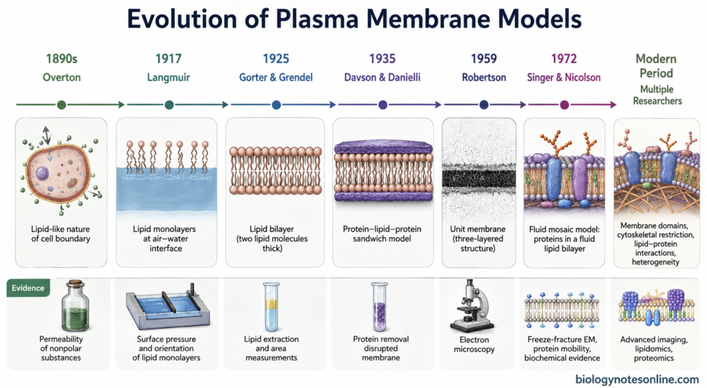
| Period | Scientist(s) | Main contribution |
|---|---|---|
| 1890s | Ernest Overton | From permeability studies, he suggested that the cell boundary has lipid-like properties. |
| 1917 | Irving Langmuir | Studied the behavior and orientation of lipid monolayers at the air-water surface. |
| 1925 | Evert Gorter and François Grendel | Proposed that the cell boundary is made of a lipid bilayer, two lipid molecules thick. |
| 1935 | Hugh Davson and James Danielli | Proposed the protein-lipid-protein sandwich model. |
| 1959 | J. David Robertson | Proposed the unit membrane model based mainly on electron microscopic observations. |
| 1972 | S. J. Singer and G. L. Nicolson | Proposed the fluid mosaic model, where proteins are associated with a fluid lipid bilayer. |
| Modern period | Multiple researchers | Studies showed membrane domains, cytoskeletal restriction, lipid-protein interactions and membrane heterogeneity. |
1. Overton’s Lipid Hypothesis
Observations Made by Overton
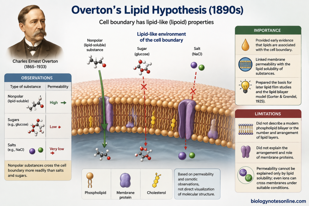
- During the 1890s, Charles Ernest Overton studied the permeability of living plant and animal cells. Nearly 500 different compounds including salts, sugars and nonpolar substances were examined during his studies.
- He observed that many nonpolar or lipid-soluble substances could pass across the cell boundary more readily. Salts and many sugars showed much lower permeability.
- From these observations, Overton suggested that the cell boundary must contain a lipid-like or lipoid environment. He also considered substances such as cholesterol and phospholipids as possible components.
- His work was mainly based on permeability and osmotic observations. It was not a direct observation of membrane molecular structure.
Importance of Overton’s Work
- Overton’s studies provided important early evidence that lipids are associated with the cell boundary. This became one of the basic ideas for later studies of membrane structure.
- His findings also connected membrane permeability with the lipid solubility of different substances. The lipid nature of membrane was therefore supported before its detailed structural arrangement was known.
- These observations prepared the basis for later experiments on lipid films and finally the lipid bilayer model proposed by Gorter and Grendel in 1925.
Limitations of the Lipid Hypothesis
- Overton did not describe a modern phospholipid bilayer. His work mainly indicated that the cell boundary had lipid-like properties. The number of lipid layers and their molecular arrangement were not established.
- The hypothesis also did not explain the arrangement and role of membrane proteins. Later studies showed that proteins are major membrane components and are required for many forms of selective ion movement.
- Permeability could not be explained only by simple lipid solubility. Later experiments showed that even inorganic ions can cross biological membranes under suitable conditions, making the membrane more complex than a lipid-like barrier alone.
2. Gorter and Grendel Lipid Bilayer Model
Gorter and Grendel Experiment
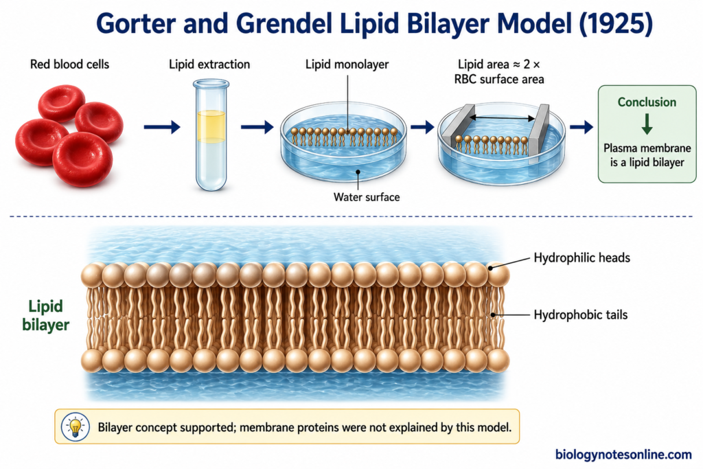
- In 1925, Evert Gorter and François Grendel studied the membrane lipids of red blood cells from humans and several other mammals. Red blood cells were suitable because these cells provided a simple system for studying the cell boundary.
- The cells were separated from plasma and washed. Their lipids were then extracted, mainly using acetone in the original experiment.
- The extracted lipids were spread over a water surface to form a monomolecular layer or monolayer. The area occupied by this lipid film was measured.
- The experimental sequence can be written as: red blood cells → extraction of membrane lipids → spreading as monolayer → measurement of lipid area.
- The area of the lipid monolayer was about twice the calculated total surface area of the red blood cells. From this result, Gorter and Grendel proposed that the cell boundary contained a layer of fatty substances two molecules thick.
Proposed Structure of the Lipid Bilayer
- The model proposed two layers of lipid molecules, rather than a single lipid layer. This became the basic lipid bilayer concept.
- Membrane lipids are mainly amphipathic molecules, having a polar region and a nonpolar hydrocarbon region.
- In a bilayer, the hydrophilic or polar regions are directed towards the aqueous environment on the two sides of membrane.
- The hydrophobic regions remain towards the inner part, where they are kept away from water. Two lipid layers are arranged tail-to-tail in this manner.
Evidence Supporting the Model
- The major evidence came from comparison of the surface area of erythrocytes with the area occupied by their extracted lipids when arranged as a monolayer. The approximate 2:1 relationship supported a membrane two lipid molecules thick.
- Their results from different mammalian red blood cells were considered compatible with the same bimolecular lipid arrangement.
- Later studies confirmed that a continuous lipid bilayer forms the basic structural framework of biological membranes.
- The erythrocyte experiment remains one of the important early observations in the development of membrane biology.
Limitations of the Gorter-Grendel Model
- The experiment mainly established the bilayer nature of membrane lipids. It did not give a complete molecular structure of the plasma membrane.
- Membrane proteins were not properly included in this model. It is now known that proteins are associated with the lipid bilayer in several different ways and many extend through the bilayer.
- A lipid bilayer alone could not explain many specific membrane functions. Most receptors, enzymes and transport processes depend mainly on membrane proteins.
- The original experiment also contained limitations in lipid extraction and estimation of cell surface area. These did not remove the importance of the bilayer concept, but the experimental measurements were not as exact as the simple textbook description suggests.
3. Davson–Danielli or Sandwich Model
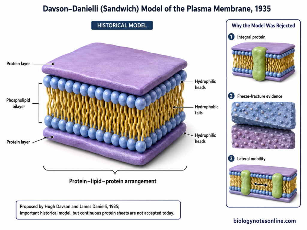
The Davson–Danielli model is an early structural model of biological membrane in which a lipid bilayer was associated with protein layers on its two surfaces. It is also referred to as the sandwich model or paucimolecular model. The lipid bilayer idea was retained, while proteins were added to explain some properties of natural membranes.
Who Proposed the Davson-Danielli Model?
- The model was proposed by James Frederic Danielli and Hugh Davson in 1935. Their work was published as A Contribution to the Theory of Permeability of Thin Films.
- They used the earlier lipid bilayer concept but considered that lipid alone was not sufficient to describe the cell boundary.
- Proteins were therefore placed in association with the two surfaces of the lipid bilayer. The model was mainly developed from physicochemical and permeability observations available at that time.
Structure of the Sandwich Model
- A phospholipid bilayer forms the central part of the membrane.
- The hydrophobic portions of lipids remain directed towards the interior of the bilayer. Polar regions remain towards the aqueous sides.
- A layer of protein was proposed over each surface of the lipid bilayer. This gave a protein-lipid-protein arrangement.
Protein layer
↓
Phospholipid bilayer
↓
Protein layer
- In this arrangement, proteins were mainly considered as surface coatings. They were not distributed through the bilayer in the way known for many membrane proteins today.
Main Features of the Davson-Danielli Model
- It is based on a lipid bilayer covered by protein on both sides.
- The membrane was considered to have a rather ordered and layered molecular arrangement.
- Protein layers were added to account for properties that could not be explained properly by lipid alone.
- The model connected membrane structure with permeability and other physicochemical properties of the cell boundary.
- It became an important membrane model before more direct information about membrane proteins was available.
Evidence Supporting the Model
- The already known Gorter and Grendel lipid bilayer provided the lipid foundation on which this model was developed.
- Studies of membrane and lipid-film properties suggested that proteins could be associated with membrane lipids. This helped in proposing protein material at the lipid-water interfaces.
- Later electron microscopic observations showed membranes with a dark-light-dark appearance. At that time, this trilaminar appearance seemed compatible with protein layers present on both sides of a lipid region.
- The electron microscopic pattern was not direct proof of two continuous protein sheets. Its interpretation was later changed as more information about membrane organization became available.
Limitations of the Davson-Danielli Model
- The model showed proteins mainly as uniform continuous sheets. Biological membrane proteins do not have this simple arrangement.
- Different biological membranes contain different amounts and types of proteins. For example, protein-to-lipid proportion differs considerably between plasma membrane and some organelle membranes.
- Many proteins are integral membrane proteins. Their hydrophobic regions remain inside the lipid bilayer, and some proteins extend completely across it. This could not be represented by simple outer protein coatings.
- Freeze-fracture studies showed particles present on the internal fracture faces of membranes. These findings supported macromolecules located within the membrane rather than proteins being restricted only to its outer surfaces.
- The model was also relatively static. Experiments later showed that many membrane proteins can move laterally in the plane of membrane. The mouse-human cell fusion experiments of Frye and Edidin in 1970 provided important evidence for such movement.
Why Was the Davson-Danielli Model Rejected?
- The proposed protein-lipid-protein sandwich could not fit the growing structural evidence for proteins embedded within the lipid bilayer.
- Freeze-fracture observations did not show a simple membrane covered everywhere by continuous protein sheets. Instead, intramembrane particles were observed within fracture faces.
- Biochemical studies showed integral and transmembrane proteins having hydrophobic regions that interact directly with the hydrophobic interior of the bilayer.
- Membrane proteins also showed lateral mobility, which did not fit well with a fixed continuous protein coating.
- These findings required another arrangement of lipids and proteins. In 1972, Singer and Nicolson proposed the fluid mosaic model, where proteins are associated with the fluid lipid bilayer in different ways rather than forming two uniform outer sheets.
4. Robertson’s Unit Membrane Model
What Is the Unit Membrane Model?
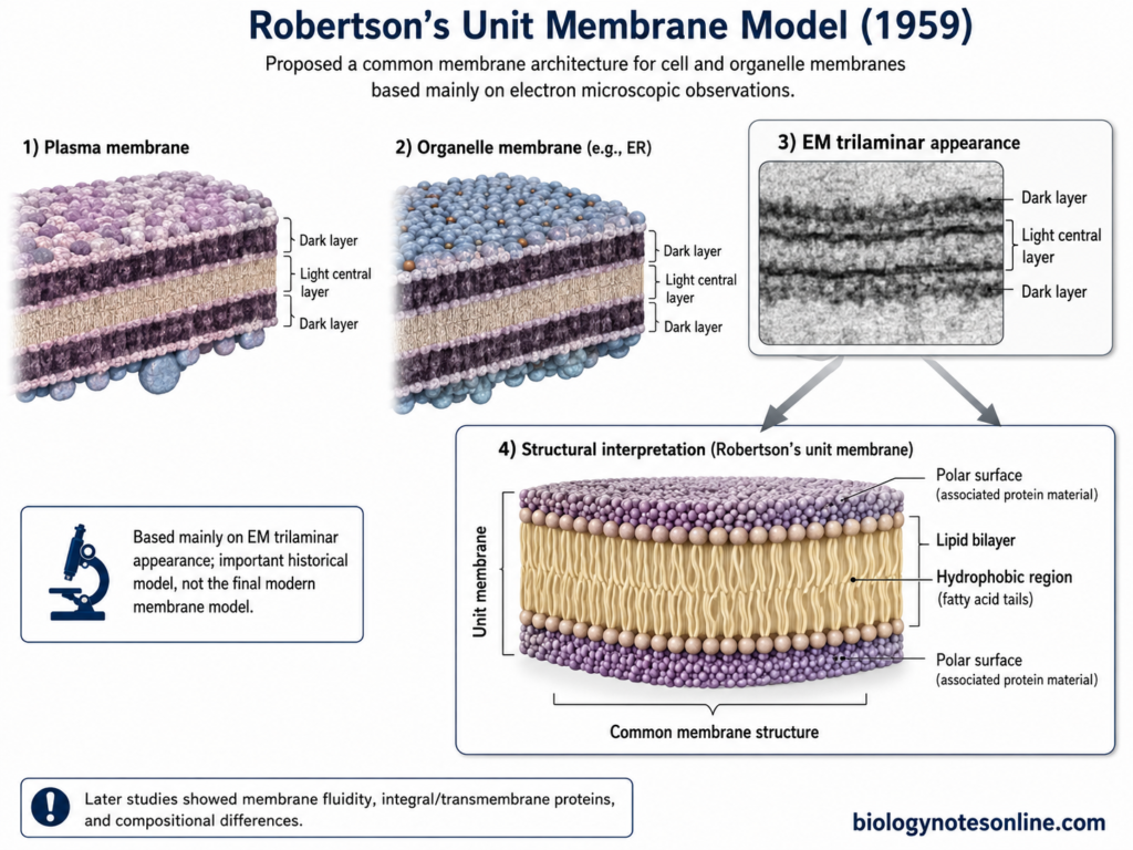
- The unit membrane model was proposed by J. David Robertson from electron microscopic studies of biological membranes. His major description of the model was published in 1959.
- Robertson observed a similar thin membrane pattern around the cell and different cell organelles. From this, he proposed that biological membranes have a common basic molecular organization.
- The model was strongly based on electron microscopy, unlike the earlier models which were developed mainly from permeability and physicochemical experiments.
Electron Microscopic Evidence
- Robertson studied membranes after suitable fixation and heavy-metal staining. Potassium permanganate (KMnO₄) was also used to improve membrane contrast in his studies.
- Under the electron microscope, the membrane showed a characteristic dark–light–dark appearance. This is referred to as the trilaminar appearance.
- Two electron-dense lines were present on the two sides with a lighter region between them.
- Similar trilaminar appearance was observed at the plasma membrane and membranes surrounding different cellular compartments. This observation was important for the development of the unit membrane concept.
Dark layer
↓
Light central layer
↓
Dark layer
Structure Proposed by Robertson
- Robertson considered a lipid bilayer as the basic central structure of biological membrane.
- The central lighter region was interpreted as the hydrophobic lipid region. The two electron-dense regions were interpreted in relation to the polar lipid surfaces and associated protein material.
- His interpretation was related to some features of the earlier Davson–Danielli model, but the unit membrane model was mainly developed from electron microscopic observations.
- The important part of Robertson’s proposal was that this basic membrane architecture was not restricted only to plasma membrane. It was considered common to many cellular and organelle membranes.
Why Is It Called the Unit Membrane Model?
- The term unit membrane was used because Robertson considered the trilaminar membrane as a common structural unit of biological membranes.
- Plasma membrane and intracellular membranes appeared to follow a similar general pattern under electron microscope.
- A common lipid bilayer-based organization was therefore proposed for these membranes. The bilayer is also accepted as the basic structural foundation of modern biological membranes.
Importance of Robertson’s Model
- The model provided important electron microscopic evidence for a common membrane structure.
- It strengthened the concept that the lipid bilayer is widely present in biological membranes.
- Membranes of intracellular organelles were included together with the plasma membrane under a general structural concept. This was an important feature of Robertson’s work.
- The characteristic trilaminar or railroad-track appearance became an important observation in membrane ultrastructure studies.
Limitations of the Unit Membrane Model
- Biological membranes do not have exactly the same molecular composition. The amount and types of lipids and proteins vary considerably among different membranes.
- The interpretation of proteins mainly at membrane surfaces was too simple. Many proteins are integral proteins, and several of them pass completely through the lipid bilayer.
- Freeze-fracture electron microscopy later showed particles within the internal faces of membranes. These are associated with transmembrane proteins and could not be explained by simple continuous outer protein layers.
- The trilaminar electron microscopic appearance alone could not give the complete molecular arrangement of lipids and proteins.
- Robertson also suggested at that time that cellular membranes formed a continuous interconnected barrier. This part of the proposal was not supported by later studies.
- The model did not properly represent the fluid and dynamic behavior of membrane lipids and proteins. Modern membrane studies show lateral movement of both components within the bilayer.
5. Fluid Mosaic Model of Plasma Membrane
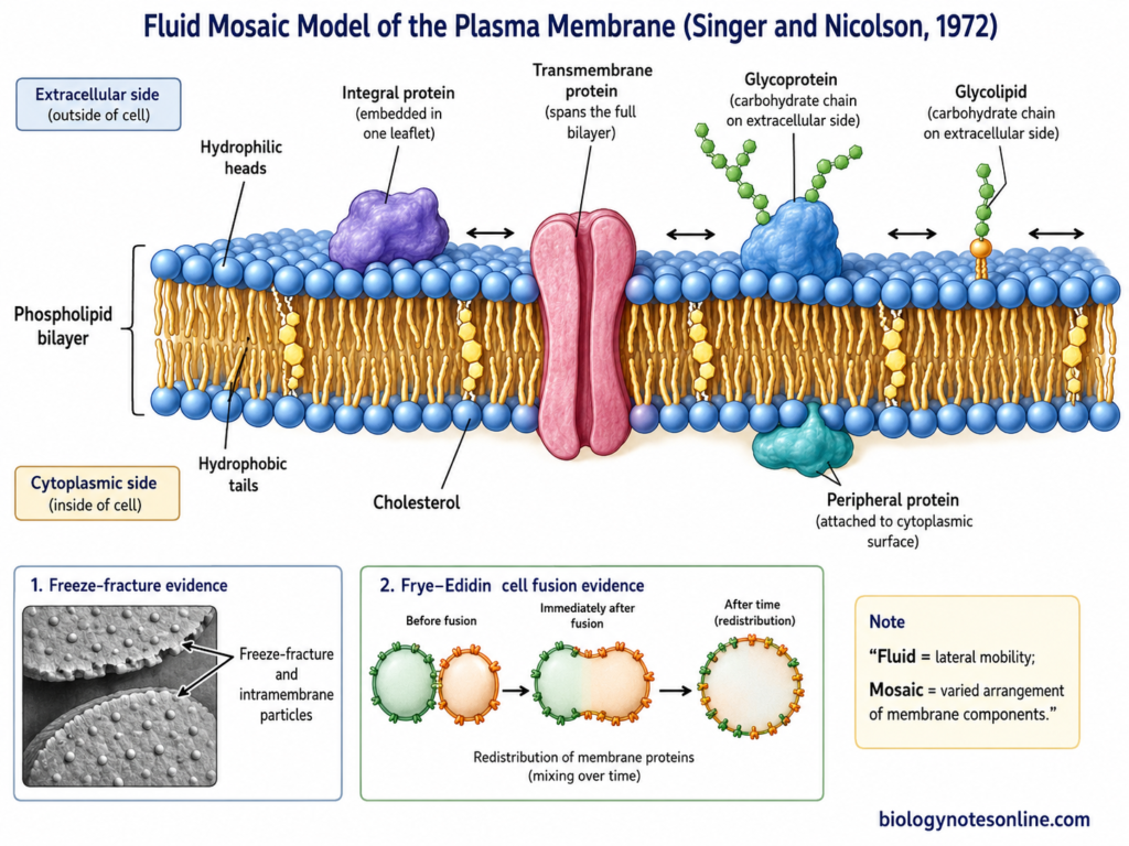
The fluid mosaic model describes the plasma membrane as a fluid lipid bilayer containing proteins in different arrangements. Lipids form the basic continuous part, while proteins are embedded in or associated with the membrane. This model remains the basic general model used for biological membranes.
Who Proposed the Fluid Mosaic Model?
- The fluid mosaic model was proposed by S. J. Singer and Garth L. Nicolson in 1972.
- Their model was published in Science under the title The Fluid Mosaic Model of the Structure of Cell Membranes.
- It replaced the earlier idea of proteins being present mainly as continuous sheets on the two sides of membrane.
- In this model, membrane proteins have different positions. Some are embedded deeply in the lipid bilayer, while others are associated more loosely with the membrane surface.
Why Is It Called the Fluid Mosaic Model?
- The term fluid refers to the dynamic nature of the lipid bilayer. Individual lipid molecules can move laterally within their own layer, and many membrane proteins also show lateral movement.
- The membrane therefore is not a rigid fixed sheet. However, movement of every membrane component is not equally free.
- The term mosaic refers to the different proteins and other components present in different positions in the lipid bilayer. Proteins do not form a uniform layer over the membrane.
- Thus, fluid represents mobility, while mosaic represents the varied arrangement of membrane components.
Structure of the Fluid Mosaic Model
- The main structural foundation is a phospholipid bilayer. Phospholipids are amphipathic, with hydrophilic regions towards water and hydrophobic regions directed inside the bilayer.
- Integral membrane proteins are embedded firmly within the lipid bilayer. Their hydrophobic portions interact with the hydrophobic region of membrane lipids.
- Transmembrane proteins are integral proteins that extend across the complete lipid bilayer. They have regions exposed on both sides of membrane.
- Peripheral membrane proteins do not enter deeply into the hydrophobic core. These proteins are associated with membrane surfaces or attached indirectly to integral proteins.
- Carbohydrates are present mainly on the non-cytosolic or extracellular surface as parts of glycoproteins and glycolipids. This contributes to membrane asymmetry.
- Sterols are also present in many biological membranes. In animal plasma membrane, cholesterol is an important lipid component present between other membrane lipids.
- The two leaflets of plasma membrane are not identical. Lipids, proteins and carbohydrates can have an asymmetric distribution across the membrane.
Main Features of the Fluid Mosaic Model
- The membrane has a continuous lipid bilayer as its main structural framework.
- Proteins occur in different arrangements and are not present as two continuous outer protein coatings.
- Both lipids and many proteins can move laterally in the plane of membrane. This gives the membrane its dynamic nature.
- Integral and peripheral proteins have different associations with the bilayer.
- The membrane is asymmetric. The molecular composition of the two surfaces is different.
- Different biological membranes also contain different proportions and types of proteins, depending upon their functions.
- The basic membrane therefore is not considered as a fixed protein-lipid-protein sandwich.
Experimental Evidence Supporting the Fluid Mosaic Model
Freeze-Fracture Electron Microscopy
- Freeze-fracture electron microscopy provided important structural evidence about the internal organization of membrane.
- During freeze-fracture, the membrane tends to split through the weakly bonded interior of the lipid bilayer. This exposes internal fracture faces of membrane.
- Electron microscopy of these fracture faces showed numerous intramembrane particles. The observations were consistent with proteins being present within or extending through the lipid bilayer, rather than forming only continuous surface layers.
- Later freeze-fracture studies also associated many of these particles with integral membrane proteins.
- These findings supported the mosaic arrangement of proteins within a lipid bilayer.
Frye and Edidin Cell-Fusion Experiment
- In 1970, Larry Frye and Michael Edidin carried out an important experiment using mouse and human cells.
- The experiment can be represented as: mouse cell + human cell → cell fusion → differently labelled surface antigens → redistribution with time.
- Mouse and human cells were fused to form heterokaryons. Surface antigens from the two cells were identified using differently labelled fluorescent antibodies.
- Immediately after fusion, the mouse and human surface markers remained mainly on their original sides.
- With time, these markers mixed over the surface of the fused cell. Extensive mixing was observed within about 40 minutes at 37°C in most heterokaryons.
- This experiment provided strong evidence that membrane proteins can show lateral mobility in the plane of the membrane.
Biochemical Evidence for Integral and Peripheral Proteins
- Biochemical studies showed that membrane proteins do not all have the same association with membrane lipids.
- Peripheral proteins can generally be removed without disrupting the lipid bilayer, because they are attached mainly through noncovalent interactions with membrane surfaces or other proteins.
- Integral proteins are much more strongly associated with membrane. Their removal generally requires detergents or other treatments that disrupt lipid-protein interactions.
- Many integral proteins contain hydrophobic amino acid regions located inside the lipid bilayer. Transmembrane proteins extend across the bilayer.
- These observations fitted the Singer-Nicolson idea of proteins having different positions within and around the lipid bilayer.
Advantages of the Fluid Mosaic Model
- The model retained the well-established lipid bilayer and gave a more suitable arrangement for membrane proteins.
- It explained why proteins can be present both at the membrane surface and inside the lipid bilayer.
- Lateral mobility of membrane lipids and many proteins could be included in this model.
- It also allowed biological membranes to have different protein compositions rather than having one uniform protein coating.
- Membrane asymmetry could be represented because the two membrane surfaces do not need to contain identical components.
- The general lipid-protein arrangement proposed in this model still forms the basic foundation for present membrane structure.
Limitations of the Original Fluid Mosaic Model
- The original model gave a useful general arrangement, but actual plasma membranes are more heterogeneous than a simple fluid lipid sea with independently distributed proteins.
- Later studies showed that membrane lipids and proteins can form domains and molecular complexes. Their distribution is not always random.
- Movement of membrane proteins can also be restricted by interactions with the cytoskeleton, extracellular matrix, other proteins and neighboring cells.
- Some membrane components remain confined within particular regions instead of undergoing unrestricted lateral diffusion.
- Modern studies also give greater importance to membrane crowding, lipid-protein interactions, membrane domains and local heterogeneity. These features were not represented fully in the original simple model diagram.
- The fluid mosaic model is therefore still the basic membrane model, but its simple 1972 representation has been modified with these additional levels of membrane organization.
What the Fluid Mosaic Model Explains?
- The fluid mosaic model explains the basic organization of biological membranes as a fluid lipid bilayer with proteins present in different arrangements. Integral proteins are partly or completely embedded in the bilayer.
- It explains membrane fluidity. Lipids and many membrane proteins can move laterally in the plane of membrane. The membrane is therefore not considered as a rigid fixed layer.
- The model explains the mosaic arrangement of proteins. Different proteins have different sizes, structures and positions instead of forming a continuous protein coating on both membrane surfaces.
- It provides a structural basis for selective permeability. The hydrophobic lipid region forms a barrier to many water-soluble substances, while particular membrane proteins can provide specific routes across the membrane.
- Membrane asymmetry can also be represented by this model. Membrane proteins have specific orientations in the bilayer, and the two sides of a membrane are not structurally equivalent.
- The model explains why membrane components can undergo redistribution. In the Frye and Edidin experiment, surface antigens of fused mouse and human cells intermixed with time, showing lateral movement of membrane components.
- It also provides a framework for different membrane-mediated processes, where movement and interaction of membrane proteins are involved. Singer and Nicolson specifically used the model for considering changes and interactions occurring at cell surfaces.
Which Plasma Membrane Model Is Accepted Today?
The fluid mosaic model proposed by S. J. Singer and Garth L. Nicolson in 1972 remains the foundational model for biological membrane organization, although modern research has substantially refined its original representation.
According to this model, a lipid bilayer forms the basic membrane structure and different membrane proteins are embedded in or associated with it. Many lipids and proteins also show lateral movement within the membrane.
The original model is not considered a complete description of all membrane organization. Modern studies have shown membrane domains, protein complexes, lipid-protein interactions and cytoskeletal restrictions on movement of membrane components.
Plasma membrane is therefore not a completely uniform two-dimensional fluid. Some membrane components move freely, while others remain clustered or restricted within particular regions.
These findings are generally considered refinements of the fluid mosaic model, rather than one completely new model replacing it. The lipid bilayer and mosaic arrangement of membrane proteins still remain its basic structural concepts.
Plasma Membrane Models at a Glance
- Overton → Lipid nature
Cell boundary was suggested to have lipid-like properties from permeability observations. - Gorter and Grendel → Lipid bilayer
Proposed that membrane lipids are arranged in two molecular layers. - Davson and Danielli → Sandwich model
Proposed a protein–lipid–protein arrangement around the lipid bilayer. - Robertson → Unit membrane model
Described a common trilaminar membrane structure from electron microscopic observations. - Singer and Nicolson → Fluid mosaic model
Proposed a fluid lipid bilayer with proteins present in different positions and many components showing lateral mobility. - Modern view → Dynamic, heterogeneous and organized membrane
Membrane contains domains, protein complexes and restricted molecular movement, along with the basic fluid bilayer organization.
Comparison of Different Plasma Membrane Models
Different plasma membrane models were proposed as new experimental findings became available. The major differences are mainly in the arrangement of lipids, position of proteins, membrane mobility and interpretation of membrane structure.
| Plasma membrane model | Main proposal | Lipid arrangement | Protein arrangement | Main evidence or basis | Major limitation / present status |
|---|---|---|---|---|---|
| Overton’s Lipid Hypothesis (1890s) | Cell boundary has lipid-like properties. | No definite molecular arrangement was proposed. | Not explained. | Lipid-soluble substances generally entered cells more readily than many water-soluble substances. | It was a precursor, not a complete structural membrane model. It did not describe a phospholipid bilayer or membrane proteins. |
| Gorter and Grendel Model (1925) | Cell membrane contains a lipid bilayer, two lipid molecules thick. | Polar regions directed towards aqueous sides and hydrophobic regions towards the interior. | Not adequately included. | Extracted erythrocyte lipids spread as a monolayer occupied approximately twice the calculated cell surface area. | Established the important bilayer concept, but could not explain membrane proteins and complete membrane organization. |
| Davson–Danielli Sandwich Model (1935) | Lipid bilayer is present between proposed protein layers. | Lipid bilayer forms the central part. | Proteins placed mainly as coatings on the two membrane surfaces. | Based on lipid-film properties, permeability and physicochemical observations available at that time. | Uniform protein sheets could not explain integral and transmembrane proteins, membrane heterogeneity or protein mobility. |
| Robertson’s Unit Membrane Model (1959) | Biological membranes have a common basic unit membrane organization. | Lipid bilayer forms the central structural basis. | Surface-associated protein material was included in the molecular interpretation. | Electron microscopy showed a characteristic dark–light–dark trilaminar appearance in many cellular membranes. | Did not properly explain different protein arrangements, membrane composition and dynamic behavior. |
| Fluid Mosaic Model (1972) | Membrane is a fluid lipid bilayer containing proteins in different arrangements. | Fluid phospholipid bilayer with lateral lipid mobility. | Integral, transmembrane and peripheral proteins occur in different positions. | Biochemical evidence, membrane asymmetry, protein mobility and structural studies supported this type of organization. | Remains the basic general model, but its original simple representation does not show all membrane domains and restricted molecular movement. |
| Modern Refinements | Plasma membrane is fluid but also highly heterogeneous and organized. | Lipids can occur in local domains and different compositions. | Proteins may form complexes, clusters and show restricted movement. | Advanced microscopy and membrane-dynamics studies show domains, cytoskeletal confinement and molecular heterogeneity. | These are mainly refinements of the fluid mosaic framework rather than one single replacement model. |
Difference Between Davson-Danielli and Fluid Mosaic Models
| Feature | Davson-Danielli Model | Fluid Mosaic Model |
|---|---|---|
| Proposed by | Hugh Davson and James Danielli, 1935 | S. J. Singer and Garth L. Nicolson, 1972 |
| Basic structure | Lipid bilayer present between two protein layers. | Fluid lipid bilayer with proteins present in different positions. |
| Protein arrangement | Proteins form continuous layers on both membrane surfaces. | Proteins may be integral, transmembrane or peripheral. |
| Nature of membrane | Membrane was considered relatively fixed and uniform. | Membrane is fluid and dynamic. |
| Protein mobility | Lateral movement of proteins was not included. | Many membrane proteins can move laterally within the membrane. |
| Membrane heterogeneity | Could not properly explain differences and irregular protein distribution. | Explains the mosaic arrangement and different membrane components. |
| Present status | Rejected as an adequate membrane model. | Remains the basic general model, with modern modifications. |
Difference Between Unit Membrane and Fluid Mosaic Models
| Feature | Unit Membrane Model | Fluid Mosaic Model |
|---|---|---|
| Proposed by | J. David Robertson, 1959 | S. J. Singer and Garth L. Nicolson, 1972 |
| Basic structure | Membrane has a common trilaminar unit structure based around a lipid bilayer. | Membrane is a fluid lipid bilayer containing proteins in different arrangements. |
| Protein arrangement | Proteins were mainly interpreted in association with membrane surfaces. | Proteins may be integral, transmembrane or peripheral. |
| Main evidence | Based strongly on electron microscopic dark–light–dark appearance. | Supported by biochemical studies, freeze-fracture and membrane-mobility experiments. |
| Nature of membrane | Membrane was represented more as a fixed common structural unit. | Membrane is dynamic and fluid. |
| Protein mobility | Lateral movement of proteins was not properly included. | Many proteins can move laterally in the plane of membrane. |
| Present status | Important historical model, but insufficient for complete molecular organization. | Remains the basic general model with later refinements. |
Importance of Plasma Membrane Models
- Plasma membrane models are important for understanding how lipids and proteins are organized in biological membranes. The lipid bilayer forms the basic structural framework, with different proteins associated with it.
- These models helped in developing the present concept of membrane structure from early lipid observations to a more detailed lipid-protein organization. Different experimental findings corrected the earlier structural ideas.
- They provide a structural basis for selective permeability. The hydrophobic interior of the lipid bilayer acts as a barrier to many polar molecules and ions, while membrane proteins provide specific pathways for their movement.
- Membrane models also explain fluidity and lateral movement of lipids and many proteins in the plane of membrane. These are important properties of a biological membrane.
- The models are useful in understanding membrane asymmetry. The two sides of biological membrane are not identical in their molecular arrangement and orientation.
- They also provide the structural basis for studying membrane proteins involved in transport, receptors, cell signalling and cell recognition. Membrane proteins have different positions and different functions within the membrane.
- Historical membrane models are useful for showing how new biochemical and microscopic evidence changed the earlier concepts. The lipid bilayer remained, but the arrangement and movement of proteins required further changes in membrane models.
- The fluid mosaic model still provides the basic framework for membrane organization. Modern studies have added membrane domains, restricted molecular diffusion and greater membrane heterogeneity to this basic view.
Reference
- Alberts, B., Johnson, A., Lewis, J., Raff, M., Roberts, K., & Walter, P. (2002). Molecular biology of the cell (4th ed.). Garland Science. https://www.ncbi.nlm.nih.gov/books/NBK21054/
- Branton, D. (2016). Fracture faces of frozen membranes: 50th anniversary. Molecular Biology of the Cell, 27(3), 421–423. https://doi.org/10.1091/mbc.e15-05-0287
- Campos Muñiz, C., & Fernández Perrino, F. J. (2023). Evolution of the concepts of architecture and supramolecular dynamics of the plasma membrane. Membranes, 13(6), 547. https://doi.org/10.3390/membranes13060547
- Cooper, G. M. (2000). The cell: A molecular approach (2nd ed.). Sinauer Associates. https://www.ncbi.nlm.nih.gov/books/NBK9839/
- Danielli, J. F., & Davson, H. (1935). A contribution to the theory of permeability of thin films. Journal of Cellular and Comparative Physiology, 5(4), 495–508. https://doi.org/10.1002/jcp.1030050409
- Engelman, D. M. (1969). Surface area per lipid molecule in the intact membrane of the human red cell. Nature, 223(5212), 1279–1280. https://doi.org/10.1038/2231279a0
- Frye, L. D., & Edidin, M. (1970). The rapid intermixing of cell surface antigens after formation of mouse-human heterokaryons. Journal of Cell Science, 7(2), 319–335. https://doi.org/10.1242/jcs.7.2.319
- Gorter, E., & Grendel, F. (1925). On bimolecular layers of lipoids on the chromocytes of the blood. The Journal of Experimental Medicine, 41(4), 439–443. https://doi.org/10.1084/jem.41.4.439
- Herrmann, T., Leavitt, L., & Sharma, S. (2023, April 24). Physiology, membrane. In StatPearls. StatPearls Publishing. https://www.ncbi.nlm.nih.gov/books/NBK538211/
- Kleinzeller, A. (1997). Ernest Overton’s contribution to the cell membrane concept: A centennial appreciation. Physiology, 12(1), 49–53. https://doi.org/10.1152/physiologyonline.1997.12.1.49
- Kusumi, A., Fujiwara, T. K., Chadda, R., Xie, M., Tsunoyama, T. A., Kalay, Z., Kasai, R. S., & Suzuki, K. G. N. (2012). Dynamic organizing principles of the plasma membrane that regulate signal transduction: Commemorating the fortieth anniversary of Singer and Nicolson’s fluid-mosaic model. Annual Review of Cell and Developmental Biology, 28, 215–250. https://doi.org/10.1146/annurev-cellbio-100809-151736
- Langmuir, I. (1917). The shapes of group molecules forming the surfaces of liquids. Proceedings of the National Academy of Sciences of the United States of America, 3(4), 251–257. https://doi.org/10.1073/pnas.3.4.251
- Lombard, J. (2014). Once upon a time the cell membranes: 175 years of cell boundary research. Biology Direct, 9, 32. https://doi.org/10.1186/s13062-014-0032-7
- Mohandas, N., & Gallagher, P. G. (2008). Red cell membrane: Past, present, and future. Blood, 112(10), 3939–3948. https://doi.org/10.1182/blood-2008-07-161166
- Nicolson, G. L. (2013). Update of the 1972 Singer-Nicolson fluid-mosaic model of membrane structure. Discoveries, 1(1), e3. https://doi.org/10.15190/d.2013.3
- Nicolson, G. L. (2014). The fluid-mosaic model of membrane structure: Still relevant to understanding the structure, function and dynamics of biological membranes after more than 40 years. Biochimica et Biophysica Acta (BBA) – Biomembranes, 1838(6), 1451–1466. https://doi.org/10.1016/j.bbamem.2013.10.019
- Nicolson, G. L., & Ferreira de Mattos, G. (2021). A brief introduction to some aspects of the fluid–mosaic model of cell membrane structure and its importance in membrane lipid replacement. Membranes, 11(12), 947. https://doi.org/10.3390/membranes11120947
- Nicolson, G. L., & Ferreira de Mattos, G. (2022). Fifty years of the fluid–mosaic model of biomembrane structure and organization and its importance in biomedicine with particular emphasis on membrane lipid replacement. Biomedicines, 10(7), 1711. https://doi.org/10.3390/biomedicines10071711
- Pinto da Silva, P. (1973). Membrane intercalated particles in human erythrocyte ghosts: Sites of preferred passage of water molecules at low temperature. Proceedings of the National Academy of Sciences of the United States of America, 70(5), 1339–1343. https://doi.org/10.1073/pnas.70.5.1339
- Pinto da Silva, P., & Branton, D. (1970). Membrane splitting in freeze-etching: Covalently bound ferritin as a membrane marker. The Journal of Cell Biology, 45(3), 598–605. https://doi.org/10.1083/jcb.45.3.598
- Robertson, J. D. (1959). The ultrastructure of cell membranes and their derivatives. Biochemical Society Symposia, 16, 3–43. https://pubmed.ncbi.nlm.nih.gov/13651159/
- Robertson, J. L. (2018). The lipid bilayer membrane and its protein constituents. Journal of General Physiology, 150(11), 1472–1483. https://doi.org/10.1085/jgp.201812153
- Singer, S. J., & Nicolson, G. L. (1972). The fluid mosaic model of the structure of cell membranes. Science, 175(4023), 720–731. https://doi.org/10.1126/science.175.4023.720
- Trimble, W. S., & Grinstein, S. (2015). Barriers to the free diffusion of proteins and lipids in the plasma membrane. The Journal of Cell Biology, 208(3), 259–271. https://doi.org/10.1083/jcb.201410071