

Membrane carbohydrates are the chains of sugar present on the outer surface of the plasma membrane. These are also referred to as glycans. They are present only towards the extracellular side of the membrane.
These carbohydrates are attached with membrane proteins and membrane lipids. The carbohydrate attached with protein forms glycoprotein or proteoglycan, while that attached with lipid forms glycolipid. All these carbohydrates form a thick hydrated covering on the cell surface, called glycocalyx.
The carbohydrate chains are attached to proteins by N-linked glycosylation and O-linked glycosylation. In N-linked glycosylation, the sugar is attached with asparagine. In O-linked glycosylation, it is attached with serine or threonine.
Membrane carbohydrates are used in cell recognition, adhesion and communication between the cells. They also act as markers on the cell surface. The ABO blood group antigens are formed by these carbohydrate structures. It also protects the cell surface and prevents unwanted aggregation of proteins.
What Are Membrane Carbohydrates?
Membrane carbohydrates are the sugar chains that are present on the surface of cell membrane. They are also called glycans. These carbohydrates are attached with the proteins and lipids of the membrane, forming complex molecules known as glycoconjugates.
Membrane carbohydrates do not remain freely within the membrane. Their inability to remain free is because the inner portion of the membrane is hydrophobic. They are covalently attached with membrane proteins forming glycoproteins and proteoglycans, or with lipids forming glycolipids.
In the fluid mosaic membrane, these glycoconjugates are placed among the moving proteins and lipids. The carbohydrate chains are always directed towards the extracellular side of cell. They are not present towards the cytoplasmic surface of membrane.
The length, arrangement and composition of these carbohydrates are different in different cells and tissues. These differences form specific markers on the cell surface. They are used in identification and recognition of one cell from the other.
Why Do Membrane Carbohydrates Face Outward?
Membrane carbohydrates remain present on the outer surface of plasma membrane. This arrangement is related with their formation and different cell surface functions. The following are the major reasons-
- Carbohydrate chains are added to proteins and lipids inside the lumen of endoplasmic reticulum (ER) and Golgi apparatus. The luminal side later becomes the outer side of plasma membrane.
- Glycoproteins and glycolipids are transported through vesicles. When these vesicles fuse with plasma membrane, the inner surface of vesicle becomes exposed towards outside of cell.
- The carbohydrate chains are therefore not shifted through the lipid bilayer after their formation. Their original orientation is maintained during vesicular transport.
- Outward position helps in recognition of one cell from another cell. ABO blood group antigens are present on outer surface of red blood cells.
- Membrane carbohydrates also act as receptors and surface markers. They can interact with immune cells, neighbouring cells and different extracellular molecules.
- Their outward projection forms a carbohydrate-rich covering called glycocalyx. It protects the cell surface and holds water around the membrane.
- The glycocalyx also acts as a physical barrier. It protects membrane proteins from enzymes, mechanical damage and some harmful substances.
- Membrane carbohydrates help in attachment of cell with another cell. They also bind with components of extracellular matrix.
- Some signalling molecules and lectins bind with surface carbohydrate chains. This interaction can produce signals inside the cell.
Location and Distribution in the Plasma Membrane
Membrane carbohydrates are present only on outer surface of plasma membrane. They remain attached with membrane proteins and lipids. Free carbohydrate is not found in the inner hydrophobic part of lipid bilayer.
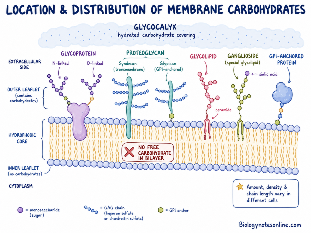
- Membrane carbohydrates are located on the extracellular leaflet of plasma membrane. Their sugar chains project towards the outside of cell.
- They are not present as free molecules inside the lipid bilayer. The hydrophobic region of membrane does not contain unattached carbohydrate.
- In glycoproteins, carbohydrate chains remain attached with membrane proteins. The attachment may be N-linked or O-linked. These chains extend from the outer cell surface.
- Membrane proteoglycans contain a core protein and long glycosaminoglycan (GAG) chains. Syndecans are present as transmembrane proteins. Glypicans remain attached by a lipid anchor.
- In glycolipids, the lipid tail remains inserted in outer layer of membrane. The carbohydrate head remains exposed towards extracellular fluid. Ceramide is commonly present as lipid part.
- Gangliosides are also present in the outer leaflet of plasma membrane. Their carbohydrate part contains one or more sialic acid molecules.
- Some surface proteins are attached with membrane by glycosylphosphatidylinositol (GPI) anchor. These proteins remain on the outer side of plasma membrane.
- Glycoproteins, glycolipids and proteoglycans together form a thick carbohydrate covering. This is called glycocalyx. It remains highly hydrated.
- The amount of membrane carbohydrate is not same in all cells. Their chain length, density and composition vary in different cell and tissue types.
Characteristics of Membrane Carbohydrates
Membrane carbohydrates have different structural and chemical properties. They remain present with membrane proteins or lipids. Some of the important characteristics are as follows-
- Membrane carbohydrates are present only on the outer surface of plasma membrane. Their carbohydrate chains face towards extracellular region. Free sugars are not present inside the hydrophobic part of lipid bilayer.
- They remain covalently attached with proteins or lipids. Carbohydrate attached with protein forms glycoproteins and proteoglycans. When attached with lipid, glycolipids are formed.
- Glycoproteins, proteoglycans and glycolipids together form an outer carbohydrate covering. This covering is called glycocalyx. It is thick, hydrated and gel-like in nature.
- Membrane carbohydrates are highly polar and hydrophilic. They attract water molecules. Many carbohydrate chains also have high negative charge due to sialic acid, glucuronic acid, iduronic acid and sulfate groups.
- Their formation is not controlled by a direct template like DNA or protein synthesis. Sugars are added one after another by specific enzymes. Trimming of sugar molecules also takes place in endoplasmic reticulum (ER) and Golgi apparatus.
- Membrane carbohydrates show high structural variation. Different monosaccharides are joined by α-glycosidic or β-glycosidic linkages. Their chain length and branching are also not same.
- N-glycans generally extend outward from the membrane surface. They may form branched and antenna-like carbohydrate chains. These are involved in receptor activity and protein folding.
- O-glycans and glycosaminoglycans (GAGs) may form long and closely packed chains. They hold water and protect the protein part. These chains also resist mechanical pressure.
- Their amount and composition vary in different cells. The arrangement is also different in tissues. Changes may occur during development, normal physiological conditions and disease.
- Membrane carbohydrates act as identification markers on cell surface. ABO blood group antigens are the common example. They are also involved in cell recognition, adhesion, immune response and pathogen attachment.
Structure and Composition of Membrane Carbohydrates
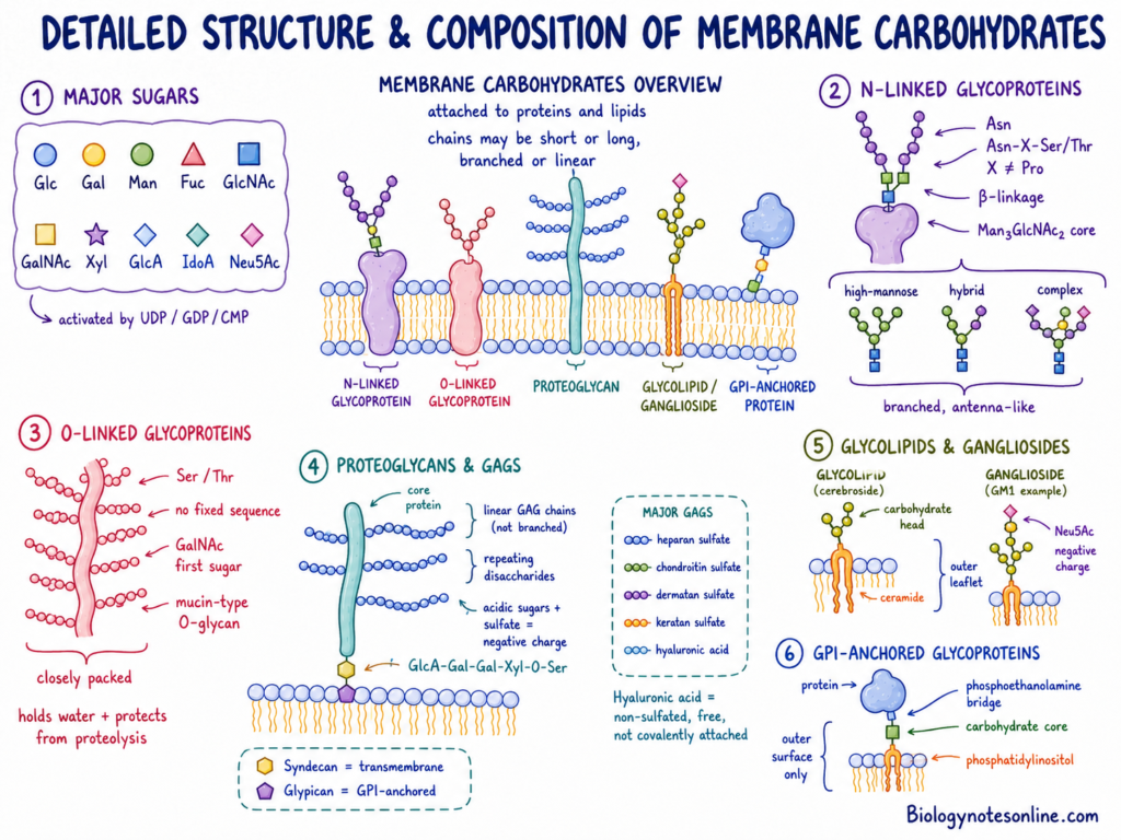
Membrane carbohydrates are formed of different monosaccharides. They remain present with proteins and lipids of plasma membrane. The chains are not same in all glycoconjugates. Some are short and branched, while long linear chains are present in proteoglycans.
Monosaccharide Building Blocks
- Major monosaccharides
- Ten major monosaccharides are used in the formation of mammalian membrane glycans. These are glucose (Glc), galactose (Gal), mannose (Man), fucose (Fuc), N-acetylglucosamine (GlcNAc) and N-acetylgalactosamine (GalNAc).
- Xylose (Xyl), glucuronic acid (GlcA) and iduronic acid (IdoA) are also found. Sialic acid or N-acetylneuraminic acid (Neu5Ac) is commonly present at the terminal end of carbohydrate chains, giving negative charge to the cell surface.
- Nucleotide donors
- Before their addition, activation of monosaccharides takes place by coupling with nucleotide donors. UDP, GDP and CMP are the major donors used for this process. Transfer of the activated sugars is carried out by different glycosyltransferase enzymes.
Structural Classes of Membrane Carbohydrates
- N-Linked Glycoproteins
- In N-linked glycoproteins, the carbohydrate is attached with amide nitrogen of asparagine (Asn). A β-glycosidic bond is present in this attachment and the asparagine generally occurs within Asn-X-Ser/Thr sequence. Here, X can be any amino acid except proline.
- The common core present in all N-linked glycans is Man₃GlcNAc₂. Addition and removal of other sugar molecules takes place in Golgi apparatus, forming high-mannose, hybrid and complex carbohydrate chains.
- Most N-linked glycans remain projected away from the protein surface. Branched antenna-like structure is commonly produced.
- O-Linked Glycoproteins
- Carbohydrate is attached with hydroxyl oxygen of serine (Ser) or threonine (Thr) in this type. No fixed amino acid sequence is required. The initial linkage is generally an α-linkage.
- N-acetylgalactosamine (GalNAc) is the first sugar present in most mucin-type O-glycans. Other sugars are then added one after another and different core structures, from Core 1 to Core 8, may be formed.
- A large number of O-linked chains are found close together in mucins. They form an extended bottle-brush type structure which covers the polypeptide part. Protection against proteolytic enzymes and physical pressure is provided by these chains.
- Proteoglycans and Glycosaminoglycans
- Proteoglycans are made up of a core protein attached with long glycosaminoglycan (GAG) chains. The chains are mainly linear and unbranched. High negative charge is present due to acidic sugars and sulfate groups.
- Repeating disaccharides form the GAG chain. One member of the disaccharide is generally an amino sugar, GlcNAc or GalNAc, while glucuronic acid (GlcA), iduronic acid (IdoA) or galactose forms the other member.
- Except hyaluronic acid, attachment of most GAGs with serine of core protein takes place through a conserved tetrasaccharide linker. The linker is GlcA-Gal-Gal-Xyl-O-Ser.
- Some of the important GAGs are heparan sulfate, chondroitin sulfate, dermatan sulfate, keratan sulfate and hyaluronic acid. Hyaluronic acid is non-sulfated and remains free, it is not covalently attached with a core protein.
- Syndecans are present as Type I transmembrane proteoglycans. In glypicans, attachment with the outer membrane surface is carried out by a GPI anchor.
- Glycolipids and Gangliosides
- Glycolipids contain a hydrophilic carbohydrate head with a hydrophobic lipid tail. Ceramide forms the lipid part in many membrane glycolipids and remains inserted within the outer leaflet of plasma membrane.
- The carbohydrate head is exposed towards extracellular fluid. In gangliosides, one or more negatively charged sialic acid (Neu5Ac) residues are present in this carbohydrate region. GM1 is one common example.
- GPI-Anchored Glycoproteins
- These are cell surface proteins attached with the outer lipid layer by a glycosylphosphatidylinositol (GPI) anchor. The anchor is formed of phosphatidylinositol and a conserved carbohydrate core.
- Joining of the protein with core glycan is carried out through a carboxyl-terminal phosphoethanolamine bridge. The complete protein part remains exposed towards extracellular surface and does not pass through the lipid bilayer.
Types of Membrane Carbohydrates
Membrane carbohydrates are found attached with proteins and lipids of plasma membrane. They occur in different forms based on the type of attachment. The following are the major types of membrane carbohydrates-
- N-Linked Glycoproteins (N-Glycans)
- In this type, carbohydrate is attached to the amide nitrogen of asparagine (Asn).
- The asparagine is present in Asn-X-Ser/Thr sequence.
- Here, X can be any amino acid except proline.
- O-Linked Glycoproteins (O-Glycans)
- These are the glycoproteins in which carbohydrate is attached to the hydroxyl oxygen of serine (Ser) or threonine (Thr).
- In collagen, it can also be attached with hydroxylysine.
- Mucin-type glycans are the common type of O-linked glycans.
- Proteoglycans and Glycosaminoglycans (GAGs)
- Proteoglycans are formed of a core protein and long carbohydrate chains.
- These carbohydrate chains are called glycosaminoglycans (GAGs).
- They are linear and highly negatively charged sugar chains.
- Some of the important GAGs are heparan sulfate, chondroitin sulfate, dermatan sulfate and keratan sulfate.
- Glycolipids and Gangliosides
- Glycolipids are formed by the attachment of carbohydrate with membrane lipid.
- Ceramide is commonly present as the lipid part.
- They are found in the outer layer of lipid bilayer.
- Gangliosides are glycolipids having one or more sialic acid residues in their carbohydrate part.
- GPI-Anchored Glycoproteins
- These are cell surface proteins attached with outer lipid layer through glycosylphosphatidylinositol (GPI) anchor.
- The GPI anchor contains a conserved carbohydrate core.
- The protein is connected with this carbohydrate core by a phosphoethanolamine bridge.
- C-Mannosylated Glycoproteins
- It is a less common type of carbohydrate attachment.
- In this type, a single mannose molecule is directly attached to the indole ring carbon of tryptophan (Trp).
- This attachment generally occurs in Trp-X-X-Trp sequence.
Formation of Membrane Glycoproteins and Glycolipids
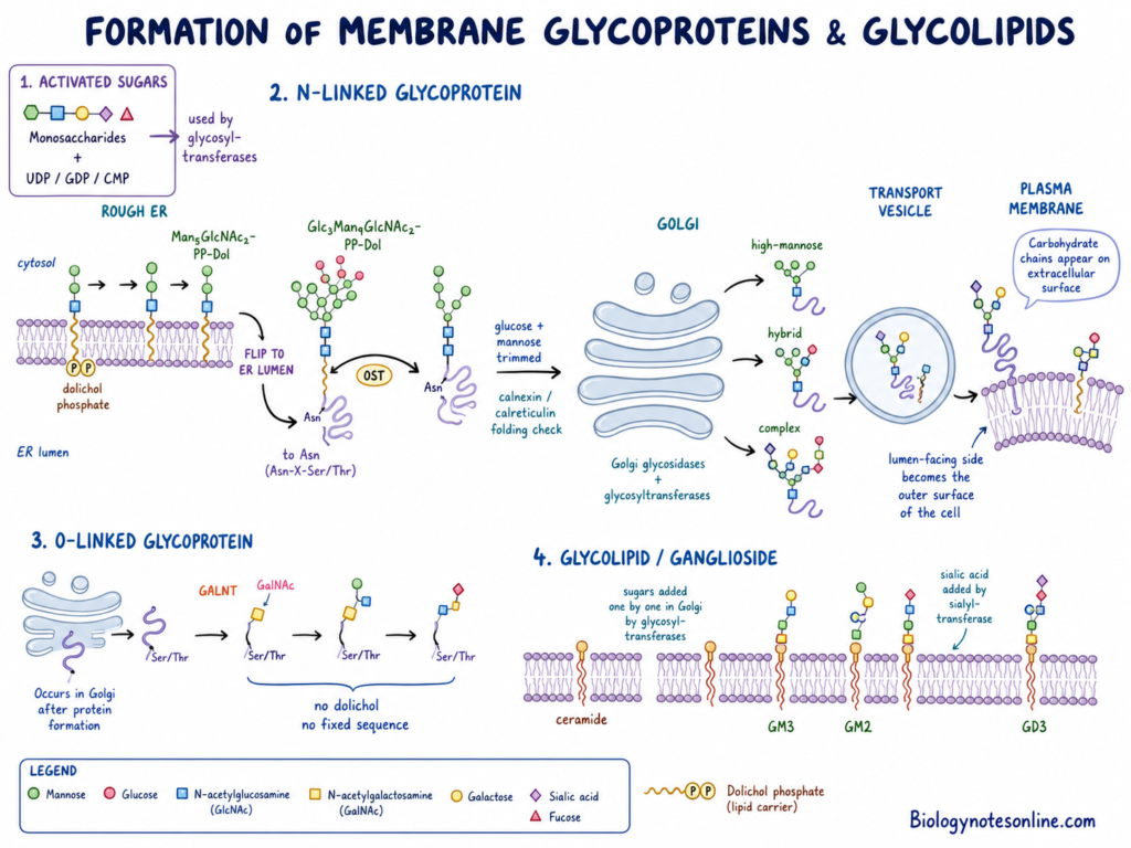
Membrane glycoproteins and glycolipids are formed in endoplasmic reticulum (ER) and Golgi apparatus. Different sugar molecules are added during this process. The formed carbohydrate chains remain attached with proteins or lipids.
Activation of Monosaccharides
- Monosaccharides are first activated in cytosol or nucleus.
- They combine with high-energy nucleotide donors.
- UDP, GDP and CMP are the major nucleotide donors.
- These activated sugars are now used by different glycosyltransferase enzymes.
Formation of N-Linked Glycoproteins
In N-linked glycosylation, carbohydrate is attached with nitrogen of asparagine (Asn). The process begins on cytosolic surface of rough endoplasmic reticulum (RER).
- Monosaccharides are first added to a lipid carrier called dolichol phosphate.
- The sugars are added one after another. A lipid-linked oligosaccharide is formed.
- A seven-sugar intermediate Man₅GlcNAc₂-PP-Dol is produced on the cytosolic side of RER.
- The formed intermediate is now transferred across the ER membrane. It reaches into the ER lumen.
- A scramblase or flippase protein is involved in this movement. Rft1 may take part in this process.
- More mannose and glucose molecules are added inside the ER lumen.
- A complete 14-sugar precursor Glc₃Man₉GlcNAc₂-PP-Dol is now formed.
- The complete oligosaccharide is transferred together to a newly forming protein. This is referred to as en bloc transfer.
- It is attached with amide nitrogen of asparagine present in Asn-X-Ser/Thr sequence.
- Enzyme involved- Oligosaccharyltransferase (OST).
- Terminal glucose and some mannose molecules are removed by ER glycosidases.
- During this process, calnexin and calreticulin are involved in checking protein folding.
- The improperly folded protein remains in ER. Properly folded glycoprotein is transferred into Golgi apparatus.
- In Golgi apparatus, some sugar molecules are removed and other sugars are added.
- Enzymes involved- Golgi glycosidases and glycosyltransferases.
- High-mannose, hybrid or complex carbohydrate chains are formed.
Formation of O-Linked Glycoproteins
O-linked glycosylation takes place in Golgi apparatus after formation of protein. Here, carbohydrate is attached with hydroxyl oxygen of serine (Ser) or threonine (Thr).
- N-acetylgalactosamine (GalNAc) is first attached with serine or threonine.
- Enzyme involved- Polypeptide GalNAc-transferase (GALNT).
- A fixed amino acid sequence is not required for this attachment.
- Other sugar molecules are now added one after another.
- Each sugar is added by a specific Golgi glycosyltransferase.
- Dolichol lipid carrier is not used in this process.
Formation of Glycolipids
Glycolipids are formed by attachment of carbohydrate with a hydrophobic lipid. Ceramide commonly acts as the lipid part.
- The ceramide backbone is initially formed in ER or cytosolic region.
- It remains inserted within the membrane.
- Further sugar addition takes place in Golgi apparatus.
- Neutral sugars are added one after another by specific glycosyltransferases.
- Sialic acid molecules are added by sialyltransferases.
- Different gangliosides are formed during this process.
- GM3, GD3 and GM2 are some examples.
Vesicular Transport and Membrane Insertion
- The formed glycoproteins and glycolipids are packed inside transport vesicles.
- These vesicles are formed from the trans-Golgi network.
- They move towards plasma membrane and fuse with it.
- The side facing vesicle lumen becomes exposed towards outside of cell.
- Due to this, carbohydrate chains remain present on the extracellular surface of plasma membrane.
Functions of Membrane Carbohydrates
Membrane carbohydrates perform different functions in cell. They are present on the outer surface of plasma membrane. Some of the important functions are as follows-
- Cell recognition– Membrane carbohydrates act as cell surface markers. They help in recognition of one cell from other cell. ABO blood group is also determined by specific carbohydrate present on red blood cells.
- Protein folding– Carbohydrate attached with newly formed protein helps in proper folding. This process takes place in endoplasmic reticulum (ER). Calnexin and calreticulin are involved in this process. They also prevent aggregation of protein.
- Cell protection– Membrane carbohydrates form a thick outer covering called glycocalyx. It protects the cell surface from mechanical and chemical injury. It also protects membrane proteins from breakdown by proteolytic enzymes.
- Cell adhesion– Membrane carbohydrates help in attachment of one cell with another cell. They also help in attachment of cell with extracellular matrix. It is involved in tissue arrangement and movement of cells.
- Cell signalling– Some membrane carbohydrates act as receptors and co-receptors. They bind with growth factors, cytokines and other signalling molecules. FGF and VEGF also bind with carbohydrate-containing receptors.
- Filtration– Membrane carbohydrates form a selective barrier on cell surface. They control movement of water, solutes and proteins. This type of function is found in vascular endothelium.
- Mechanotransduction– Membrane carbohydrates detect fluid flow and shear stress. These mechanical forces are converted into signals inside the cell. It may also increase the production of nitric oxide.
- Hydration– Glycosaminoglycans (GAGs) have high negative charge. They attract cations and water. It helps in tissue hydration, joint lubrication and shock absorption.
- Pathogen attachment– Some viruses, bacteria and toxins bind with membrane carbohydrates. They use these carbohydrates for attachment with host cell. Membrane carbohydrates also bind with lectins of immune cells.
- Reproduction– Membrane carbohydrates are involved in reproductive process. Carbohydrates on sperm surface undergo changes during its movement in reproductive tract. They also help in sperm and egg binding during fertilization.
Examples of Membrane Carbohydrates
Membrane carbohydrates are found in different forms. They remain attached with membrane proteins or lipids. Some of the important examples are as follows-
- ABO antigens– These are oligosaccharides present on red blood cell membrane. They determine the blood group. A antigen contains terminal N-acetylgalactosamine, while B antigen contains D-galactose. In O blood group, unchanged H antigen is present.
- MNS antigens– These antigens are present on glycophorins. Glycophorins are membrane glycoproteins of red blood cells. Different carbohydrate groups are found attached with them.
- GM1 ganglioside– It is a glycolipid having sialic acid. It is mainly present in neuronal cell membrane. A ceramide part remains inside the membrane and carbohydrate part remains outside.
- Other gangliosides– GM2, GM3 and GD3 are important gangliosides. One or more sialic acid molecules are present in them. They take part in cell growth and receptor function.
- Syndecans– These are membrane proteoglycans. Syndecan-1 to Syndecan-4 are the major forms. They contain heparan sulfate and sometimes chondroitin sulfate chains. These are involved in cell attachment and signalling.
- Glypicans– GPC1 to GPC6 are examples of glypicans. They are attached with outer surface of membrane by a lipid anchor. Heparan sulfate chains are present near the cell surface.
- Hyaluronic acid– It is a large non-sulfated glycosaminoglycan (GAG). It is also called hyaluronan. It binds with CD44 receptor on the cell membrane and holds large amount of water.
- DAF and CD59– Decay-accelerating factor (DAF/CD55) and CD59 are present on red blood cells. They are attached by GPI anchor. They protect the cell from complement-mediated damage.
- NCAM– Neural cell adhesion molecule (NCAM) is a surface glycoprotein. It interacts with heparan sulfate proteoglycans. It helps in attachment between cells.
- Cell-surface mucins– MUC1 and MUC16 are the common examples. They contain large number of O-linked carbohydrate chains. They form a thick covering on epithelial cells and help in hydration and protection.
- Sialyl-Lewis antigens– Sialyl-Lewis A (sLeA/CA19-9) and Sialyl-Lewis X (sLeX) are carbohydrate antigens. They are found in increased amount on some cancer cell surfaces. These antigens help in adhesion and movement of cancer cells.
- Tn antigens– Tn and Sialyl-Tn (sTn) are short O-linked carbohydrate structures. They are found on abnormal cancer cells. These are used as tumor-associated markers.
Glycoproteins vs Glycolipids
| Feature | Glycoproteins | Glycolipids |
|---|---|---|
| Core Definition | Proteins covalently attached to one or more oligosaccharide (glycan) chains. | Lipids covalently attached to a hydrophilic carbohydrate head group. |
| Non-Carbohydrate Backbone | Polypeptide chain / protein core. | Lipid anchor, primarily ceramide (forming glycosphingolipids). |
| Primary Attachment Linkages | • N-linked: β-linkage to the amide nitrogen of Asparagine (Asn). • O-linked: α-linkage to the hydroxyl oxygen of Serine (Ser) or Threonine (Thr). | β-glycosidic linkage connecting the carbohydrate head directly to the hydrophobic lipid tail. |
| Site of Biosynthesis | Initiated in the rough endoplasmic reticulum (ER) (N-linked) or Golgi apparatus (O-linked), followed by extensive Golgi remodeling. | Initiated in the cytosol/ER and completed via sequential sugar additions in the Golgi apparatus. |
| Relative Abundance | Constitute the predominant form of cell-surface glycoconjugates (over 50% of eukaryotic proteins are glycosylated). | Account for a lesser portion of total membrane-bound carbohydrates compared to glycoproteins. |
| Structural Dynamics & Conformation | Form stiff, antenna-like structures (N-glycans) or dense, extended “bottle-brush” arrays (O-glycans) projecting from the protein surface. | Amphiphilic molecules with hydrophobic lipid tails embedded in the outer membrane leaflet and sugar head groups exposed extracellularly. |
| Major Subtypes & Examples | • Cell-surface mucins (MUC1) • Adhesion molecules (NCAM, Integrins) • Transmembrane proteoglycan cores (Syndecans) | • Glycosphingolipids • Gangliosides (GM1, GM2, GM3, GD3) • Cerebrosides |
| Primary Biological Roles | Protein folding quality control, structural tissue protection, extracellular matrix anchoring, and immune signaling. | Organizing lipid microdomains (rafts), modulating membrane fluidity, cell-surface recognition, and receptor cross-talk. |
Biological and Medical Significance
Membrane carbohydrates have important role in normal body functions and disease conditions. They are involved in blood group, infection, cancer, vascular function and immune protection. Some of the important significance are as follows-
- Blood transfusion– Carbohydrates present on red blood cells determine ABO blood groups. These surface antigens are checked before blood transfusion. Wrong matching can produce severe hemolytic reaction.
- Pathogen entry– Many viruses, bacteria and parasites use membrane carbohydrates for attachment with host cells. Influenza virus binds with sialic acid. Helicobacter pylori binds with Lewis antigens. Heparan sulfate also acts as an attachment site for some viruses.
- Cancer progression– Abnormal glycosylation is commonly found in cancer cells. Increased branching, changed gangliosides and high amount of sialyl-Lewis antigens help in tumor growth and metastasis. CA19-9 and CA125 are used as tumor markers.
- Glycosylation disorders– Genetic defects in glycan-forming enzymes cause Congenital Disorders of Glycosylation (CDGs). In PMM2-CDG, phosphomannomutase 2 enzyme is affected. The formed proteins remain underglycosylated. Nervous system, heart, liver and development may be affected.
- Vascular protection– The endothelial glycocalyx protects the inner surface of blood vessels. It controls vascular permeability and blood flow. Damage of this layer is found in sepsis, diabetes, atherosclerosis and kidney disease. Tissue edema may also develop.
- Lysosomal diseases– Deficiency of lysosomal glycosidases prevents normal breakdown of glycans and glycosaminoglycans (GAGs). These substances now accumulate inside cells. Examples include Hurler syndrome, Hunter syndrome, Sanfilippo syndrome, sialidosis and mannosidosis.
- Immune regulation– Membrane carbohydrates help in immune cell recognition and leukocyte movement. They are also involved in immune surveillance. CD55/DAF and CD59 protect red blood cells from complement-mediated damage.
- Cell trafficking– Surface carbohydrate groups help leukocytes to attach with vascular endothelium. This is important during inflammation. Selectins recognize specific carbohydrate groups during this process.
References
- A higher abundance of O-linked glycans confers a selective advantage to high fertile buffalo spermatozoa for immune-evasion from neutrophils. (2020). PubMed Central (PMC). https://pmc.ncbi.nlm.nih.gov/articles/PMC7483552/
- ABO blood group antigens and differential glycan expression: Perspective on the evolution of common human enzyme deficiencies. (2023). PubMed Central (PMC). https://pmc.ncbi.nlm.nih.gov/articles/PMC9860303/
- Aberrant glycosylation as biomarker for cancer: Focus on CD43. (2014). PubMed Central (PMC). https://pmc.ncbi.nlm.nih.gov/articles/PMC3943294/
- Altered tumor-cell glycosylation promotes metastasis. (2014). PubMed Central (PMC). https://pmc.ncbi.nlm.nih.gov/articles/PMC3923139/
- BOC Sciences. (2026). N-Glycan vs O-glycan synthesis — What’s the difference? https://www.bocsci.com/glycan/resources/n-glycan-vs-o-glycan-synthesis-what-s-the-difference.html
- CDG Hub. (2026). PMM2-CDG (CDG-Ia). https://www.cdghub.com/cdg/pmm2-cdg-cdg-ia/
- Chiduza, G. N., Sakata, K., Noor, F., & Menon, A. K. (2025, December 9). Proposed mechanism for Rft1-mediated scrambling of a dolichol-linked oligosaccharide. bioRxiv. https://doi.org/10.64898/2025.12.07.692794
- Choi, S., Lee, H., Choi, J. R., & Oh, E. S. (2010). Shedding; towards a new paradigm of syndecan function in cancer. BMB Reports, 43(5), 305–312.
- Composition of the endothelial glycocalyx and its relation to its thickness and diffusion of small solutes. (2010). PubMed Central (PMC). https://pmc.ncbi.nlm.nih.gov/articles/PMC2962421/
- Creative Biolabs. (n.d.). Blood group oligosaccharides: Antigens and biosynthesis. https://www.creative-biolabs.com/glycoprotein/blood-group-oligosaccharides.htm
- Creative Biolabs. (n.d.). Different patterns of glycosylation. https://www.creative-biolabs.com/glycoprotein/different-patterns-of-glycosylation.htm
- Creative Biolabs. (n.d.). Glycosaminoglycans (GAGs): Structure, function, and pathology. https://www.creative-biolabs.com/anti-glycan-antibodies/glycosaminoglycans-gags-structure-function-pathology.htm
- Dall’Olio, F., Malagolini, N., Trinchera, M., & Chiricolo, M. (2012). Mechanisms of cancer-associated glycosylation changes. Frontiers in Bioscience, 17, 670–699. https://storage.imrpress.com/imr/journal/FBL/article/494326/1752773849086.pdf
- Dean, L. (2005). The ABO blood group. In Blood Groups and Red Cell Antigens (Chapter 5). National Center for Biotechnology Information (US). https://www.ncbi.nlm.nih.gov/books/NBK2267/
- Department of Physiology, University of Zurich. (2023). ER N-linked glycosylation. https://www.physiol.uzh.ch/en/Glycosylation/Nlinkedglycosylation/ERNlinkedglycosylation.html
- Endothelial glycocalyx. (2023). PubMed Central (PMC). https://pmc.ncbi.nlm.nih.gov/articles/PMC10214841/
- Extracellular matrix: Surface proteoglycans. (2020). PubMed Central (PMC). https://pmc.ncbi.nlm.nih.gov/articles/PMC7258702/
- Glycoconjugates: Synthesis, functional studies, and therapeutic developments. (2022). PubMed Central (PMC). https://pmc.ncbi.nlm.nih.gov/articles/PMC9674437/
- Golgi glycosylation. (2011). PubMed Central (PMC). https://pmc.ncbi.nlm.nih.gov/articles/PMC3062213/
- Goodsell, D. S. (2012, December). ABO blood type glycosyltransferases. RCSB Protein Data Bank, Molecule of the Month. https://doi.org/10.2210/rcsb_pdb/mom_2012_12
- Haslam, S. M., Freedberg, D. I., Mulloy, B., Dell, A., Stanley, P., & Prestegard, J. H. (2022). Structural analysis of glycans. In A. Varki, R. D. Cummings, J. D. Esko, et al. (Eds.), Essentials of Glycobiology (4th ed., Chapter 50). Cold Spring Harbor Laboratory Press. https://www.ncbi.nlm.nih.gov/books/NBK579945/
- Helenius, J., Ng, D., Marolda, C., Walter, P., Valvano, M., & Aebi, M. (2002). Translocation of lipid-linked oligosaccharides across the ER membrane requires Rft1 protein. Nature, 415, 447–450. https://doi.org/10.1038/415447a
- Heparan sulfate binding cationic peptides restrict SARS-CoV-2 entry. (2021). PubMed Central (PMC). https://pmc.ncbi.nlm.nih.gov/articles/PMC8308704/
- Heparan sulfate facilitates SARS-CoV-2 spike protein binding to ACE2 and potentiates viral infection. (2021). PubMed Central (PMC). https://pmc.ncbi.nlm.nih.gov/articles/PMC7844799/
- Jin, J., Fang, F., Gao, W., Chen, H., Wen, J., Wen, X., & Chen, J. (2021). The structure and function of the glycocalyx and its connection with blood-brain barrier. Frontiers in Cellular Neuroscience, 15, Article 739699. https://doi.org/10.3390/fncel.2021.739699
- King, M. W. (2026). Glycoproteins: Synthesis and clinical consequences. The Medical Biochemistry Page. https://themedicalbiochemistrypage.org/glycoproteins-synthesis-and-clinical-consequences/
- King, M. W. (2026). Glycosaminoglycans and proteoglycans. The Medical Biochemistry Page. https://themedicalbiochemistrypage.org/glycosaminoglycans-and-proteoglycans/
- Kubtan, M. A. (2025). A short review on the vascular endothelial surface layer. MOJ Surgery, 13(3), 97–100. https://doi.org/10.15406/mojs.2025.13.00301
- Lam, C., & Krasnewich, D. M. (2005, August 15; updated 2021, May 20). PMM2-CDG. In M. P. Adam, S. Bick, G. M. Mirzaa, et al. (Eds.), GeneReviews. University of Washington, Seattle. https://www.ncbi.nlm.nih.gov/books/NBK1110/
- Manon-Jensen, T., Multhaupt, H. A. B., & Couchman, J. R. (2013). Mapping of matrix metalloproteinase cleavage sites on syndecan-1 and syndecan-4 ectodomains. FEBS Journal, 280(10), 2320–2331. https://doi.org/10.1111/febs.12174
- MedlinePlus Genetics. (2010, July 1). PMM2-congenital disorder of glycosylation. U.S. National Library of Medicine. https://medlineplus.gov/genetics/condition/pmm2-congenital-disorder-of-glycosylation/
- Membrane type 1 matrix metalloproteinase mediated stromal syndecan-1 shedding stimulates breast carcinoma cell proliferation. (2010). PubMed Central (PMC). https://pmc.ncbi.nlm.nih.gov/articles/PMC2877371/
- Mesopolysaccharides: The extracellular surface layer of visceral organs. (2020). PubMed Central (PMC). https://pmc.ncbi.nlm.nih.gov/articles/PMC7498049/
- National Institutes of Health. (2020, September 15). SARS-CoV-2 may use key carbohydrate to infect cells. NIH Research Matters. https://www.nih.gov/news-events/nih-research-matters/sars-cov-2-may-use-key-carbohydrate-infect-cells
- Oh, H., Thi Thuy Tien, V., Ahmed, S., Choi, J., Ryu, K.-J., & Yang, J. (2026). Host glycan–lectin interplay in SARS-CoV-2 infection. International Journal of Molecular Sciences, 27(3), Article 1608. https://doi.org/10.3390/ijms27031608
- Proteoglycans. (1997). https://www.cryst.bbk.ac.uk/pps97/assignments/projects/emilia/Proteoglycans.HTM
- Prydz, K. (2015). Determinants of glycosaminoglycan (GAG) structure. Biomolecules, 5(3), 2003–2022. https://doi.org/10.3390/biom5032003
- Ramani, V. C., & Sanderson, R. D. (2013). Chemotherapy stimulates syndecan-1 shedding: A potentially negative effect of treatment that may promote tumor relapse. Matrix Biology, 35, 215–222. https://doi.org/10.1016/j.matbio.2013.10.005
- Role of flippases in protein glycosylation in the endoplasmic reticulum. (2016). PubMed Central (PMC). https://pmc.ncbi.nlm.nih.gov/articles/PMC4762491/
- Sonnenfeld, M. (2024). 3.2 The endomembrane system I — The endoplasmic reticulum and Golgi apparatus. In Cell & Molecular Biology. Thompson Rivers University. https://cellandmolecularbiology.pressbooks.tru.ca/chapter/3-2/
- Specific transbilayer translocation of dolichol-linked oligosaccharides by an endoplasmic reticulum flippase. (2009). PubMed Central (PMC). https://pmc.ncbi.nlm.nih.gov/articles/PMC2630073/
- The endothelial glycocalyx is hydrodynamically relevant in arterioles throughout the cardiac cycle. (2008). PubMed Central (PMC). https://pmc.ncbi.nlm.nih.gov/articles/PMC2479580/
- The endothelial glycocalyx: Composition, functions, and visualization. (2007). PubMed Central (PMC). https://pmc.ncbi.nlm.nih.gov/articles/PMC1915585/
- The relevance of the aldehyde bisulfite toluidine blue reaction and its variants in the submicroscopic carbohydrate research. (2020). PubMed Central (PMC). https://pmc.ncbi.nlm.nih.gov/articles/PMC7172417/
- The role of endothelial glycocalyx in health and disease. (2019). PubMed Central (PMC). https://pmc.ncbi.nlm.nih.gov/articles/PMC6768294/
- Thermo Fisher Scientific. (n.d.). Glycosylation. https://www.thermofisher.com/ar/en/home/life-science/protein-biology/protein-biology-learning-center/protein-biology-resource-library/pierce-protein-methods/protein-glycosylation.html
- Turning the spotlight on the oligosaccharide chain of GM1 ganglioside. (2021). PubMed Central (PMC). https://pmc.ncbi.nlm.nih.gov/articles/PMC7917043/
- UC San Diego Health. (2020, September 15). COVID-19 virus uses heparan sulfate to get inside cells [Press release]. https://health.ucsd.edu/news/press-releases/2020-09-15-covid-19-virus-uses-heparan-sulfate-to-get-inside-cells/
- Variant-specific interactions at the plasma membrane: Heparan sulfate’s impact on SARS-CoV-2 binding kinetics. (2025). PubMed Central (PMC). https://pmc.ncbi.nlm.nih.gov/articles/PMC11883730/