Contents
What is Parvovirus?
- The parvoviruses are the smallest DNA viruses (about 20 nm) and have been isolated from a variety of taxa, ranging from arthropods to humans.
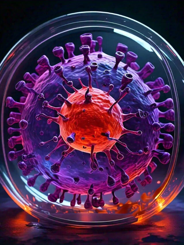
- They are icosahedral, non-enveloped, single-stranded DNA viruses.
- Human parvovirus B19 (B19) is the only known human-pathogenic parvovirus, and it demonstrates tropism towards erythroid progenitor cells.
- There are two subfamilies within the Parvoviridae family: the Parvovirinae and the Densivirinae. The latter group exclusively infects invertebrates.
- Three genera make up the family Parvovirinae: Parvovirus, Dependovirus, and Erythrovirus.
Classification of Parvovirus
- The three genera that make up the family Parvoviridae are Dependovirus, Parvovirus, and Erythrovirus.
- Dependovirus is a genus of defective poxviruses that may only replicate in conjunction with a second helper virus.
- They neither induce illness nor modify the infection produced by aiding viruses.
- These are typically discovered in conjunction with an adenovirus, and are hence referred to as adeno associated viruses. These viruses have no effect on human health.
- The genus Parvovirus contains important veterinary viruses such as the feline panleukopenia virus and the canine parvovirus.
- Erythrovirus is composed of B19, the only member of the family Parvoviridae known to cause disease in humans.
Parvovirus B19
Parvovirus B19, or B19 virus, is the causal agent of erythema infectiosum (“fifth disease”; it was fifth of the six identified exanthematous disorders of childhood), a minor viral sickness of children, as well as polyarthralgia–arthritis syndrome in immunocompetent adults.
Morphology of Parvovirus B19
B19 viruses manifest the following characteristics:
- B19 viruses are quite tiny, measuring between 18 and 26 nm in diameter.
- They possess an icosahedral capsid with no envelope.
- The length of the viral genome’s single-stranded DNA is between 4000 and 6000 nucleotides.
- Genome consists of DNA with a negative strand, but there is no virion.
- Numerous proteins are encoded by the genome, including three structural, one major nonstructural, and several minor proteins.
Viral replication
- B19 virus has a preference for (a) bone marrow cells, (b) foetal liver erythroid cells, and (c) erythroid leukaemia cells.
- The virus replicates within the nucleus.
- The single-stranded DNA genome has hairpin loops at both ends, which provide double-stranded areas for the cellular DNA polymerase to initiate synthesis of the progeny genomes.
- The viral mRNA is synthesised by the cellular RNA polymerase from the double-stranded DNA intermediate.
- The next step is the assembly of virions in the nucleus. The replication of viruses causes cell death.
Antigenic properties
- It is known that just one serotype of B19 virus exists.
Other properties
- B19 virus is highly resistant to inactivation, but formalin, beta propiolactone, and oxidising chemicals can inactivate it.
- The viruses are stable between pH 3 and 9 and can endure 30 minutes of heating at 56 degrees Celsius.
Pathogenesis and Immunity of Parvovirus B19
B19 virus exhibits tropism towards two cell types: (a) red blood cell (RBC) progenitors and (b) blood vessel endothelial cells.
Pathogenesis of parvovirus infections
- After infecting rapidly dividing erythrocyte precursors, such as bone marrow cells, foetal liver erythroid cells, and erythroid leukaemia cells, the virus kills these cells, causing aplastic anaemia.
- The infection of blood vessel endothelial cells causes erythema infectiosum.
- It has been proven that the B19 virus initially enters the body via the nasopharynx or upper respiratory tract before spreading to the blood and causing viremia.
- The infection is subsequently established when the virus infects mitotically active erythroid precursor cells in the bone marrow.
- The virus penetrates susceptible cells via the P blood antigen receptors on the progenitors of erythrocytes.
- The virus enters the nucleus of red cells, begins to replicate, and then kills the red cells.
- Due to the viruses’ destruction of erythroid precursor cells, the formation of RBCs is halted for roughly one week.
- The early phase is characterised by influenza-like symptoms induced by a high viremia.
- The viruses are transmitted by oral and respiratory secretions, and even the placenta.
- Subsequently, the development of specific antibodies against B19 virus controls viremia.
- Rash and arthralgia represent the second stage of the disease and are thought to be immune-mediated.
- This stage correlates with the departure of the B19 virus from circulation, the development of IgM and IgG antibodies specific to the B19 virus, and the synthesis of immunological complexes.
Host immunity
- The sickness has two stages: the first is a flu-like illness, and the second is the development of a rash and arthralgia.
- Immunity of the host against B19 virus infection is predominantly antibody-mediated.
- Antibodies in circulation inhibit viremia and are essential for illness resolution.
- Unknown is the role of cell-mediated immunity in providing protection against B19 virus.
Clinical Syndromes of Parvovirus B19
The B19 virus is responsible for the following clinical syndromes: (a) influenza-like illness, (b) erythema infectiosum or fifth disease, (c) infection in pregnant women, and (d) chronic B19 infection.
Flu-like illness
- Typically, B19 virus causes a flu-like disease. These are the most prevalent symptoms: malaise, headache, myalgia, and rhinorrhea.
Erythema infectiosum or fifth disease
- B19 virus is an additional culprit responsible for erythema infectiosum or fifth disease, the most prevalent childhood illness.
- The infection begins with nonspecific symptoms, followed on the fifth day by the emergence of a characteristic rash.
- A brilliant red rash appears on both cheeks, as if they had been struck.
- The rash then manifests on the trunk and progressively extends to the arms and legs.
- The ailment often resolves within one to two weeks.
Aplastic anemia: Children with chronic anaemia, such as sickle cell anaemia, thalassemia, and spherocytosis (aplastic crisis) might have transient but severe aplastic anaemia following B19 viral infection. Another severe consequence produced by B19 virus is gloves and socks syndrome. This syndrome is characterised by the appearance of erythematous exanthema on the hands and feet, with well-defined margins at the wrist and ankle joints.
Infection in pregnant women
- If the B19 virus causes reinfection in a pregnant mother who was previously infected by the same virus and is immune to it (exhibiting positive B19 antibodies), the foetus does not experience any negative effects.
- B19 virus infection is increasingly related with foetal mortality in seronegative pregnant women without immunity.
- The infection may induce severe anaemia in the foetus, which may thereafter manifest as high-output heart failure (hydrops fetalis).
- However, B19 virus does not cause congenital abnormalities in the foetus.
Chronic B19 infection
- This infection affects immunocompromised patients, including HIV patients, those undergoing immunosuppressive medicine, and transplant patients.
- Thrombocytopenia, leukopenia, and chronic anaemia are the most frequent symptoms.
Epidemiology of Parvovirus B19
Geographical distribution
- B19 virus is disseminated worldwide.
- The exact data on seropositivity in the global population is unknown.
- In the United States, over 90% of persons older than 60 are seropositive. Similar information from other regions of the globe is lacking.
Reservoir, source, and transmission of infection
- The B19 viral infection only affects humans. Only humans serve as a reservoir. Respiratory samples, which are the primary source of infection, include viruses. The infected patient is contagious for 24 to 48 hours before to the onset of prodromal symptoms and rash. The infection is spread vertically during childbirth.
- respiratory pathway through respiratory secretions
- percutaneous contact with blood and transfusion of blood or blood components (pooled RBCs, platelets, intravenous immunoglobulins, etc.).
Laboratory Diagnosis of Parvovirus B19
In early cases, the diagnosis can be made by detecting the virus in the blood, and in later cases, the antibody.
- Virus Isolation: Parvovirus B19 can be cultivated in human bone marrow or foetal liver cells for viral isolation. It can be identified by electron microscopy in the patient’s blood.
- Detection of Viral Nucleic Acid: Viral nucleic acid can be detected using either nucleic’ acid dot blot hybridization or polymerase chain reaction-based techniques.
- Antigen Detection: Using counterimmunoelectrophoresis, ELISA, or indirect immunofluorescence, antigen is detected.
- Antibody Detection: ELISA or RIA can detect IgM antibodies or a large increase in IgG antibodies. It is the most effective method.
Treatment of Parvovirus B19
No specific antiviral therapy is available for treatment of B19 virus infection.
Prevention and Control of Parvovirus B19
- There are no specific precautions available to avoid the infection. A vaccine against B19 virus is now completing phase I clinical trials.
Key Notes
- Parvoviruses are the most diminutive of DNA viruses (about 20 nm).
- There are two subfamilies within the Parvoviridae family: the Parvovirinae and the Densivirinae.
- Three genera make up the family Parvovirinae: Parvovirus, Dependovirus, and Erythrovirus.
- The Parvovirus genus consists of canine and feline parvoviruses (FPV and CPV).
- Dependoviruses require helper viral functions for reproduction, however the human adeno-associated viruses (AAV) and herpesviruses have been investigated the most.
- Erythrovirus is the only known human-infecting parvovirus (B19), along with closely related simian viruses.
- Human parvovirus B19 can cause respiratory infection accompanied by an erythematous maculopapular rash (erythema infectiosum—slapped cheek disease), joint disease, aplastic crisis in children with chronic hemolytic anaemia (sickle cell disease), nonimmune foetal hydrops following infection during pregnancy, and persistent anaemia in immunocompromised individuals.


