

Japanese encephalitis virus (JEV) is a mosquito-borne neurotropic virus that causes Japanese encephalitis (JE). JEV is the virus, while JE is the disease caused when the infection involves the central nervous system (CNS) and produces inflammation of brain.
The virus is transmitted to humans through the bite of infected mosquitoes, mainly Culex mosquitoes. It shows neurotropism, and neurons are important target cells after JEV enters the CNS. But most JEV infections are clinically inapparent. Only a small proportion of infections develop into encephalitis.
After mosquito-borne infection, JEV first replicates in peripheral tissues. The immune response can restrict the infection before important involvement of the CNS. For JE to develop, the virus has to get into the CNS and establish infection there. This neuroinvasion does not occur in most of the infections.
The exact route by which JEV gets into the brain and why this occurs only in some infected persons is still not completely understood. Once inside the CNS, neurons can be infected, together with inflammatory response in the brain producing encephalitis.
Taxonomy and Terminology
Japanese encephalitis virus belongs to the species Orthoflavivirus japonicum, genus Orthoflavivirus and family Flaviviridae. It is commonly abbreviated as JEV. Orthoflavivirus japonicum is the taxonomic species name, whereas Japanese encephalitis virus is the name of the virus.
JEV is also referred to as an “arbovirus” (arthropod-borne virus). The term “arbovirus” is not a family, genus, or any other taxonomic rank. It is an ecological description used for viruses which are biologically transmitted by blood-feeding arthropods between vertebrate hosts. In JEV, the arthropod vector is the mosquito.
Structure of Japanese Encephalitis Virus (JEV)
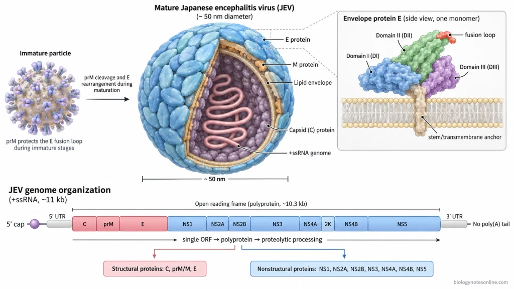
- Shape and size- Japanese encephalitis virus (JEV) is a small enveloped virus. The mature virus particle is nearly spherical, about 50 nm in diameter. Its surface proteins are arranged with icosahedral organization.
- Lipid envelope- The outermost covering is a lipid bilayer, obtained from the host cell membrane. The E and M proteins are present in this envelope. Inside it, the nucleocapsid is present.
- Envelope (E) protein- The E protein forms the major protein on surface of mature JEV. About 180 copies are present. They are closely packed and form a relatively smooth outer surface of the mature virion.E protein has three major domains, domain I (DI), domain II (DII), and domain III (DIII). DI forms the central β-barrel region. The fusion loop is present in DII, which takes part in fusion of viral and host membrane. DIII has an immunoglobulin-like structure and is involved in receptor interaction. A stem region and membrane anchor occur near its C-terminal end.
- prM and M protein- In immature JEV, precursor membrane protein (prM) is associated with the E protein. It covers the fusion loop. This prevents early or unwanted membrane fusion.During maturation, prM is cleaved by furin in the trans-Golgi region. It forms the “pr” portion and the smaller M protein. Mature JEV contains M protein instead of intact prM.
- Arrangement of E and M proteins- Mature JEV contains about 180 E and 180 M molecules. These proteins are arranged into 30 rafts on the viral surface. Three parallel E:M:M units form one raft. Most of the M protein remains below the E protein layer.
- Capsid (C) protein- The capsid protein (C) is found below the lipid envelope. It binds with the viral RNA and forms the nucleocapsid. C protein is small, about 11 kDa, and mainly α-helical. It occurs as a dimer.
- RNA genome- JEV contains a linear positive-sense single-stranded RNA (+ssRNA) genome of about 11 kb. A methylated cap is present at the 5′ end. The 3′ end does not contain a poly(A) tail.
- Genome organization- The genome contains one long open reading frame (ORF), with untranslated regions (UTRs) at its 5′ and 3′ ends. The arrangement is as follows-5′-C-prM-E-NS1-NS2A-NS2B-NS3-NS4A-2K-NS4B-NS5-3′
- Viral proteins- The single ORF is first translated into one large polyprotein. It is then cleaved by viral and host proteases. Three structural proteins are formed, C, prM/M, and E. Seven major non-structural proteins are NS1, NS2A, NS2B, NS3, NS4A, NS4B, and NS5. Structural proteins become part of the virus particle. The NS proteins mainly remain functional inside the infected cell during viral replication and processing of viral polyprotein.
- Immature JEV particle- Immature JEV does not have the same smooth surface. The prM and E proteins form projecting complexes, giving the particle a more spiky appearance. During maturation, prM is cleaved and E proteins rearrange. The surface then becomes closely packed.
Genome Structure
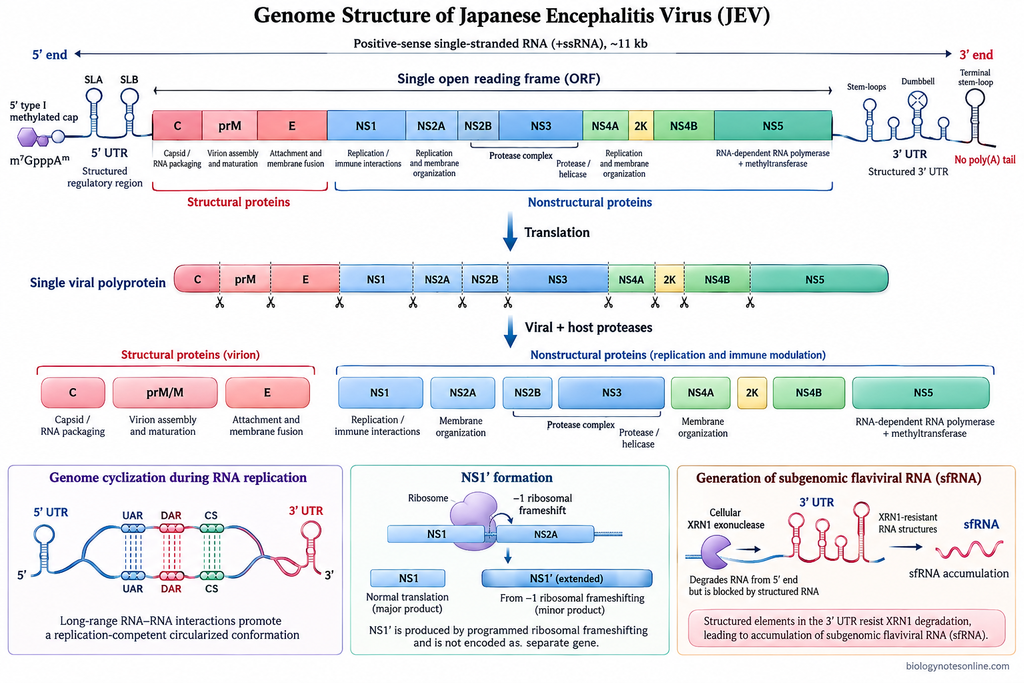
- Genome type- Japanese encephalitis virus (JEV) contains a linear positive-sense single-stranded RNA (+ssRNA) genome. It is approximately 11 kb in length. The RNA itself can function as messenger RNA after entering the host cell.
- 5′ end- A type I methylated cap structure is present at the 5′ end of JEV RNA. It is written as m⁷GpppAᵐp. The cap is involved in viral RNA translation and protects the RNA from degradation.
- 3′ end- The 3′ end has no poly(A) tail. This is a characteristic feature of the JEV genomic RNA.
- Genome organization- One long open reading frame (ORF) is present between the 5′ and 3′ untranslated regions (UTRs). The gene arrangement is as follows-5′-C-prM-E-NS1-NS2A-NS2B-NS3-NS4A-2K-NS4B-NS5-3′The structural genes are present toward the 5′ portion of ORF, followed by the non-structural genes.
- Open reading frame (ORF)- The ORF occupies most of the viral genome. In the well-characterized CNU/LP2 strain, it is 10,299 nucleotides long and codes for a polyprotein of 3432 amino acids. Genome and UTR length can show some variation among JEV strains and genotypes.
- Structural protein genes- Three structural proteins are encoded at the 5′ region of the ORF. These are capsid (C), precursor membrane (prM), and envelope (E) proteins. C protein packages the RNA, while prM and E are associated with the viral membrane and virion formation.
- Non-structural protein genes- These occur after the E gene. Seven major proteins are produced, NS1, NS2A, NS2B, NS3, NS4A, NS4B, and NS5. They mainly take part in viral RNA replication, polyprotein processing and other processes inside the infected cell.
- Polyprotein formation- JEV does not translate each major gene as a separate RNA. The single ORF is translated first into one large polyprotein. It is then cut by viral and host proteases, producing the structural and non-structural proteins.
- 5′ untranslated region (5′ UTR)- The 5′ UTR is short and highly structured. In commonly studied JEV strains it is about 95 nucleotides long. Several RNA structures are present, including stem-loop A (SLA) and stem-loop B (SLB). Some sequences from this region interact with complementary sequences near the 3′ end during genome replication.
- 3′ untranslated region (3′ UTR)- It is longer than the 5′ UTR and its length varies between JEV strains and genotypes. The region folds into several stem-loop, dumbbell and pseudoknot structures. These RNA structures take part in replication of the genome and regulation of viral RNA.The 3′ UTR contains regions that can stop the cellular XRN1 exonuclease during RNA degradation. This produces short non-coding RNAs known as subgenomic flaviviral RNAs (sfRNAs) in infected cells.
- Genome cyclization- The JEV genome can form a circularized RNA structure during replication. Complementary sequences located near the 5′ and 3′ ends pair with each other. The 5′ UAR, DAR and cyclization sequences have corresponding regions at the 3′ end.
- NS1′ formation- JEV can also produce an extended form of NS1 known as NS1′. It is formed by a −1 ribosomal frameshift near the beginning of the NS2A coding region. The frameshift adds an extra C-terminal sequence to NS1 rather than producing it as another separate gene in the genome.
Replication Cycle
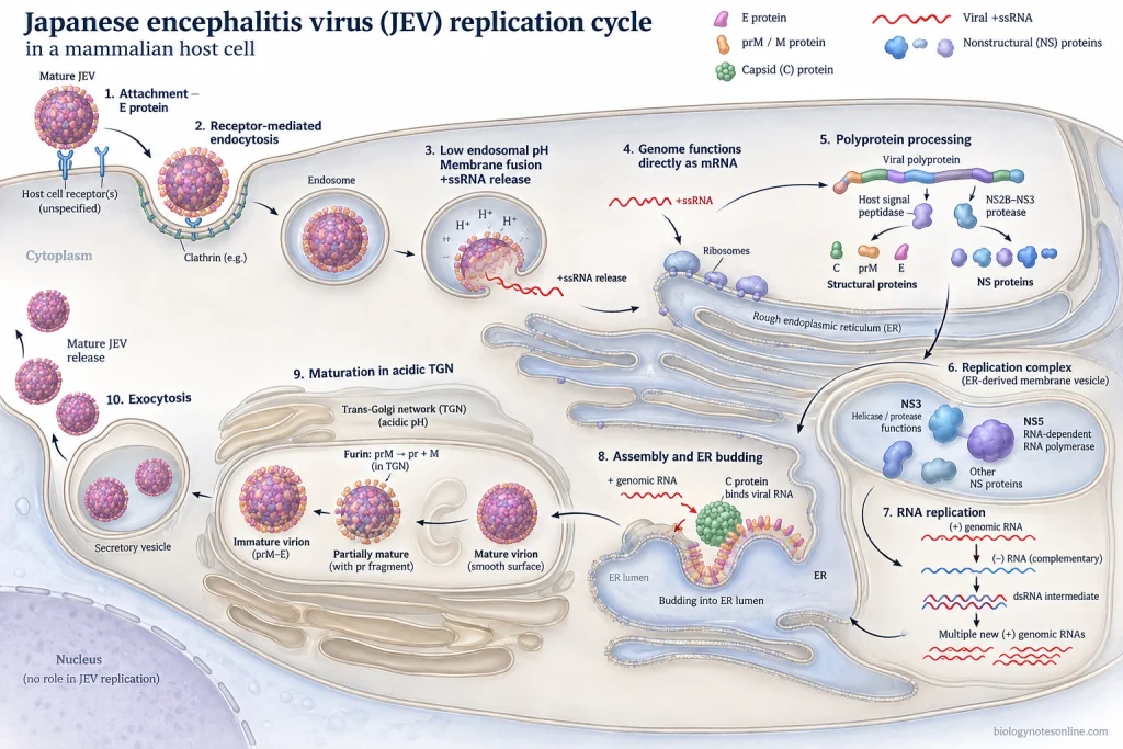
The replication of Japanese encephalitis virus (JEV) takes place mainly in the cytoplasm and on membranes derived from the endoplasmic reticulum (ER). The positive-sense RNA genome first acts as mRNA and later serves as the starting template for production of new viral RNA. The following are the major steps involved in JEV replication-
Step 1- Attachment to the host cell
JEV first attaches to the surface of a susceptible host cell. The viral envelope (E) protein is mainly involved in this process and interacts with attachment factors and receptor molecules present on the cell membrane.
No single receptor is responsible for JEV entry in all types of cells. Different host surface molecules can take part depending on the cell type.
Step 2- Endocytosis
After attachment, the virus is taken inside the cell by receptor-mediated endocytosis. Clathrin-dependent endocytosis is an important entry route reported for JEV.
The complete virion becomes enclosed inside an endosomal vesicle. It then moves through the endosomal pathway.
Step 3- Membrane fusion and uncoating
The pH gradually becomes acidic inside the endosome. This low pH changes the arrangement of E proteins present on viral envelope. The fusion loop of E protein gets exposed and inserts into the endosomal membrane.
Viral envelope then fuses with the endosomal membrane. The nucleocapsid is released and uncoating takes place, allowing the +ssRNA genome to enter into the cytoplasm.
Step 4- Translation of viral RNA
JEV RNA is positive-sense. So, after its release, the genome can directly function as mRNA.
The RNA associates with ribosomes on the rough endoplasmic reticulum (ER) and one long viral polyprotein is produced. This polyprotein contains the structural and non-structural protein sequences.
Step 5- Polyprotein processing
The newly formed polyprotein is cut into individual viral proteins. Both host and viral proteases are involved.
The NS2B-NS3 protease complex carries out several cleavages on the cytoplasmic side of ER membrane. Host signal peptidase performs other cleavages. From this processing, the structural proteins C, prM and E and non-structural proteins NS1, NS2A, NS2B, NS3, NS4A, NS4B and NS5 are formed.
Step 6- Formation of replication complex
The non-structural proteins bring about extensive changes in the ER membrane. ER-derived vesicles and membrane structures are formed, where viral RNA replication occurs.
Several NS proteins come together to form the replication complex (RC). NS5 contains the RNA-dependent RNA polymerase (RdRp) activity. NS3 has helicase activity, while NS2B acts as an important cofactor of NS3.
Step 7- Viral RNA replication
In this step, the positive-sense genomic RNA is first copied into a complementary negative-sense RNA (-RNA). A double-stranded replicative RNA form is produced during this process.
The newly formed negative strand then works as template. NS5 synthesizes many new positive-sense RNA genomes from it. These new RNAs can enter further rounds of translation or can be packed into new virus particles.
Step 8- Nucleocapsid formation and assembly
Newly synthesized genomic RNA associates with the capsid (C) protein. This forms the nucleocapsid.
Assembly occurs in association with the ER membrane. The nucleocapsid buds into the lumen of ER and gets surrounded by a host-derived lipid membrane containing prM and E proteins. At this stage the newly formed JEV particle is immature.
Step 9- Maturation of JEV
The immature virions move from the ER through the cellular secretory pathway and reach the trans-Golgi network (TGN). Their surface at this stage contains prM-E complexes.
Low pH inside the TGN causes rearrangement of these proteins. The cleavage site of prM becomes exposed. Host furin protease then cleaves prM into the “pr” portion and mature M protein.
The “pr” portion remains associated with the virion during its movement through the acidic secretory pathway. After release into the neutral extracellular environment, it dissociates from the viral surface.
Step 10- Release of mature virions
The mature JEV particles are carried to the cell surface in secretory vesicles. They are released outside the infected cell by exocytosis.
Partially mature particles containing some uncleaved prM can also be released from infected cells.
Transmission Cycle of Japanese Encephalitis Virus (JEV)
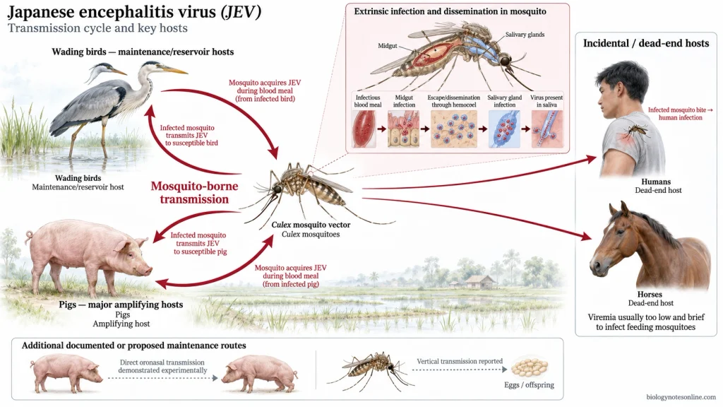
Japanese encephalitis virus (JEV) is maintained mainly in a mosquito-bird-pig transmission cycle. Culex mosquitoes act as vectors. Wild wading birds are important maintenance hosts, while pigs commonly act as amplifying hosts.
Step 1- JEV infection in birds and pigs
Wild birds, especially ardeid birds such as herons and egrets, can carry JEV and develop viraemia. They form an important natural maintenance cycle with mosquitoes.
Pigs are highly important in the domestic cycle. After JEV infection, susceptible pigs develop a relatively high viraemia for several days. This level is sufficient to infect mosquitoes feeding on them.
Step 2- Mosquito takes JEV during blood feeding
A susceptible female mosquito feeds on a viraemic bird or pig. Virus present in the blood is taken into the mosquito along with the blood meal.
Culex tritaeniorhynchus is one of the principal JEV vectors in many endemic regions. Other Culex species can also transmit the virus, depending on geographical area.
Step 3- Infection of mosquito midgut
The infected blood reaches the mosquito midgut. JEV first infects and multiplies in the midgut epithelial cells.
For further transmission, virus must cross the midgut infection and midgut escape barriers. Not every infected mosquito can complete this process.
Step 4- Dissemination inside the mosquito
After multiplication in the midgut, JEV escapes through the basal region and enters the hemocoel. From here virus spreads to other tissues.
The salivary glands are important. JEV has to infect these glands and finally enter the saliva. The period required between taking an infectious blood meal and becoming capable of transmitting virus is referred to as the extrinsic incubation period (EIP).
Step 5- Transmission during next mosquito bite
Now the infected mosquito takes another blood meal. JEV present in its saliva is introduced into the skin of a susceptible vertebrate host during feeding.
The new host may be a bird, pig, human, horse or another susceptible animal.
Step 6- Amplification in pigs
When a pig is infected, JEV multiplies and enters its bloodstream. High viraemia develops, commonly lasting for a few days. Many mosquitoes feeding on such a pig can acquire the virus.
This produces the important mosquito-pig-mosquito amplification cycle. New susceptible pigs continuously entering pig populations can also provide hosts for further virus amplification.
Step 7- Maintenance through birds
Mosquitoes also transmit JEV between susceptible and infected birds. Ardeid birds are considered important maintenance hosts and may carry the virus over wider geographical areas.
A mosquito feeding on a viraemic bird can become infected. The virus again multiplies in the mosquito and reaches its salivary glands, continuing the mosquito-bird-mosquito cycle.
Step 8- Infection of humans and horses
Humans become infected when an infectious mosquito feeds on them. Horses can be infected in the same way.
However, humans and horses generally produce low and short viraemia, which is not sufficient to infect feeding mosquitoes. They are therefore referred to as “dead-end” or incidental hosts in the normal JEV transmission cycle.
Direct transmission between pigs
JEV transmission between pigs without mosquitoes has also been demonstrated experimentally. Infected pigs can shed virus through oronasal secretions, and susceptible pigs can acquire infection through close contact or oronasal exposure.
This route can occur together with mosquito-mediated transmission. Its importance in maintaining JEV under natural field conditions is still less clearly established than the mosquito-borne cycle.
Vertical transmission in mosquitoes
JEV can also be passed from infected female mosquitoes to their offspring through eggs (vertical or transovarial transmission). This has been suggested as one possible mechanism by which virus may persist when normal mosquito-host transmission becomes reduced.
Epidemiology and Risk
- Japanese encephalitis (JE) is mainly distributed in South, Southeast and East Asia. Transmission also occurs in some parts of Western Pacific and Australia. The geographical distribution is not fixed, and JEV has been detected in areas where human JE was earlier uncommon.
- The disease burden is still considerable. Around 100,308 clinical cases were estimated for 2015, with about 25,000 deaths. The actual number may differ because laboratory diagnosis and surveillance are not equally available in all endemic areas.
- Most JEV infections are without encephalitis. Only around 1 in 200-300 infections develops into encephalitic disease. Many infections remain inapparent. Some produce only mild illness.
- Children below 15 years are commonly affected in endemic regions. This age pattern can change after long-term vaccination. In areas having good childhood vaccination, cases may occur more among unvaccinated adults and older persons.
- JE commonly shows seasonal occurrence. In temperate areas, more transmission is seen during warm and rainy months when mosquito number increases. Tropical and subtropical regions can have transmission for much longer period of the year.
- The risk is usually higher in rural agricultural areas. Irrigated rice fields provide suitable breeding sites for Culex mosquitoes. Pig rearing near human settlements also increases the opportunity for virus amplification.
- More outdoor exposure means more chance of mosquito bites. People staying outdoors during evening or night can have higher exposure in areas where JEV is circulating. Houses without mosquito screens, bed nets or other mosquito protection also increase contact with vectors.
- Unvaccinated persons are at greater risk when living in or travelling to endemic areas. Vaccination has reduced human JE greatly in several countries. But JEV still continues in its mosquito-animal transmission cycle.
- JE is rare among most travellers to Asia, usually less than 1 case per million travellers. The risk is not equal in every traveller. Long stay, rural travel and outdoor activity during transmission season can increase exposure. A short trip can also carry risk when mosquito exposure is high.
- People living or working close to rice fields, wetlands, irrigation areas and pig farms can come in contact with infected mosquitoes more frequently. Agricultural workers and people staying in rural villages may therefore receive repeated mosquito exposure. Persons remaining mainly in protected urban areas usually have less contact with these vectors.
Pathogenesis of Japanese Encephalitis
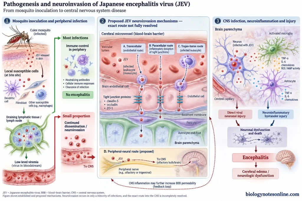
- JEV enters the body through the bite of an infected Culex mosquito. Virus is deposited into the skin with mosquito saliva. Dendritic cells, fibroblasts and other susceptible cells present around the bite site can become infected.
- The virus then moves into draining lymphatic tissues. Replication occurs in local cells and lymph nodes. From here, JEV enters the blood and a primary viraemia develops. It is usually low.
- JEV spreads through blood and lymphatic circulation into peripheral tissues. Monocytes and cells belonging to macrophage-dendritic cell lineage can also support infection. More virus may be produced during this stage.
- In most infected persons, the infection is controlled during the peripheral phase. Virus does not enter the brain. Only in a small proportion, JEV escapes this control and neuroinvasion takes place.
- How JEV exactly enters the central nervous system (CNS) is still not completely established. Different routes have been suggested. Virus may cross through infected brain endothelial cells, between endothelial cells, or can be carried by infected leukocytes (“Trojan horse”). Entry through peripheral nerves has also been proposed.
- The blood-brain barrier (BBB) normally restricts entry into brain tissue. During JEV infection, inflammatory mediators can disturb tight-junction proteins such as claudin-5, occludin and ZO-1. BBB permeability increases. But marked BBB disruption may not always be required before the virus gets into the brain.
- Once inside the CNS, JEV mainly infects neurons. Viral replication starts in these cells and infection can spread into different regions of brain. The infected neurons may be directly damaged or killed.
- Microglia and astrocytes become activated. They produce inflammatory cytokines and chemokines, including TNF-α and IL-6. Some of this response acts against viral infection. Excessive activation, however, can damage the surrounding nervous tissue.
- More inflammatory products are released as infection progresses. Matrix metalloproteinases (MMPs), cytokines, chemokines and reactive oxygen species (ROS) take part. Endothelial damage increases and the tight junctions can become further disturbed. Immune cells also move into the CNS.
- Neuronal damage is produced in more than one way. JEV replication itself causes injury to infected neurons. Activated microglia and inflammatory molecules can also damage nearby neurons which may not be directly infected.
- With extensive neuronal injury, encephalitis and cerebral edema develop. Perivascular inflammation and BBB disturbance can be seen in severe infection. Depending on the areas of CNS involved, altered consciousness, seizures, movement abnormalities and other neurological manifestations may occur.
Clinical Course and Symptoms
- Incubation period- The incubation period of Japanese encephalitis (JE) is usually 5-15 days after infection. During this period, symptoms are not seen.
- Asymptomatic infection- Most of the JEV infections remain without obvious symptoms. Some persons may develop a mild febrile illness only. Encephalitis does not develop in most infected people.
- Prodromal phase- This is the early symptomatic period of JE. Fever, headache, malaise and vomiting are common. Chills and muscle pain may occur. Anorexia, diarrhea or coryza are also seen in some patients. This phase generally lasts for a few days before neurological symptoms appear.
- Acute encephalitic phase- The illness may progress rapidly after the prodromal period. High fever continues. Confusion, altered sensorium and behavioral changes can develop when the brain becomes involved. Focal neurological deficits are also seen.
- Altered consciousness- Drowsiness and disorientation may be present initially. With severe disease, the level of consciousness becomes reduced. Stupor or coma can occur.
- Seizures- Convulsions are common, particularly in children with JE. Seizures may be focal or generalized. In severe cases they can become repeated, and status epilepticus may develop.
- Meningeal signs– Some patients also show meningitis-like features. Headache, neck stiffness, nausea, vomiting and photophobia can be present. These may occur before obvious encephalitic signs.
- Movement disorders- Tremor, rigidity, dystonia, choreoathetosis and myoclonus may occur during JE. A parkinsonian type syndrome is also described. Mask-like face, tremor, increased muscle tone and cogwheel rigidity can be seen in these patients.
- Motor weakness and paralysis- Weakness may affect only a particular part of body or become more extensive. Some patients develop acute flaccid paralysis resembling poliomyelitis, with weak muscles and reduced reflexes. Hemiplegia or quadriplegia can also occur in severe cases.
- Raised intracranial pressure- Increased intracranial pressure may develop during acute encephalitis. In severe cerebral involvement, abnormal posturing can be present. Deep coma may follow.
- Convalescent phase- In survivors, fever and acute encephalitic symptoms gradually decrease. Neurological recovery may be slow. Movement abnormalities sometimes become more noticeable when the patient starts recovering from coma.
- Neurological sequelae- Recovery is not complete in many survivors. Motor weakness, recurrent seizures, speech problems, movement disorders, cognitive impairment and behavioral changes may remain for a long period. About half of survivors have been reported with some neurological sequelae when assessed one year or more after hospital discharge.
Diagnosis
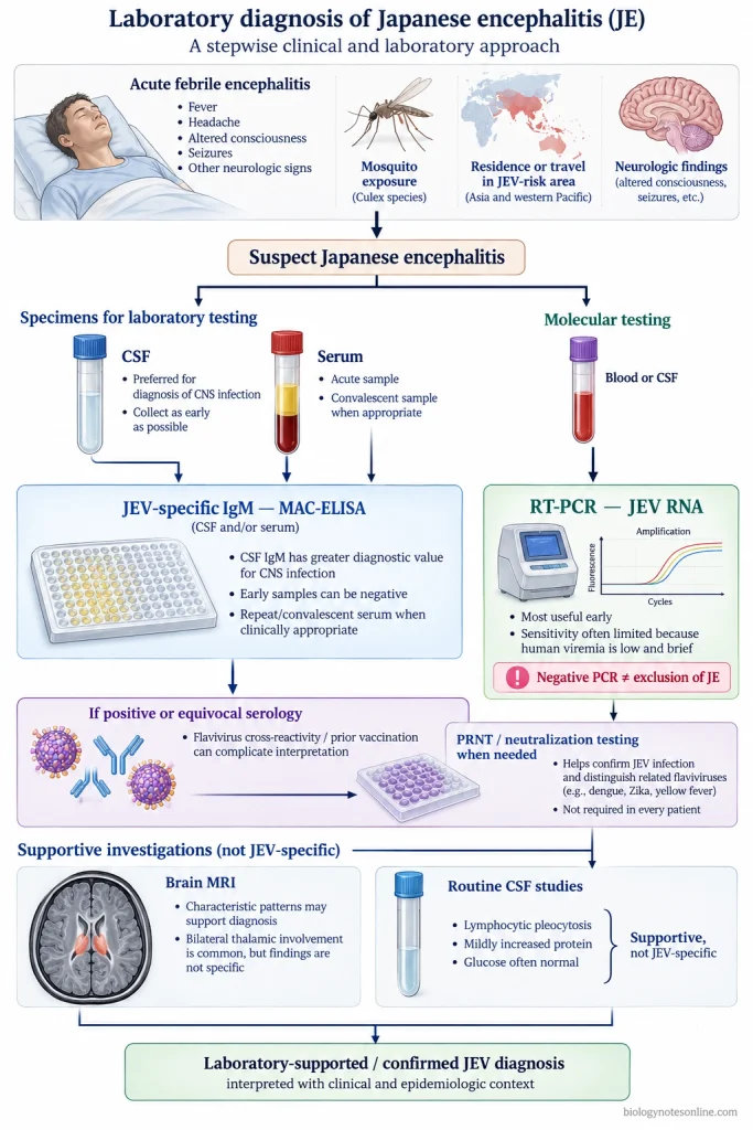
- Clinical evaluation- Japanese encephalitis (JE) is suspected in a patient with acute fever and encephalitis, mainly when there is history of mosquito exposure or residence/travel in a JEV transmission area. Altered consciousness and seizures are common findings. Movement disorders or acute flaccid paralysis may also be present. Clinical features alone are not enough for confirmation.
- CSF examination- Cerebrospinal fluid (CSF) usually shows mild to moderate lymphocytic pleocytosis. Protein may be slightly increased. Glucose generally remains normal.
- IgM capture ELISA- Detection of JEV-specific IgM antibody by MAC-ELISA is the main laboratory method used for JE diagnosis. Serum and CSF can both be tested. CSF IgM is more useful because its presence supports infection involving the central nervous system.JEV-specific IgM may be detected in CSF from about 4 days after onset of symptoms. In serum, it commonly appears around 7-8 days. If the sample is collected too early, the test can be negative. Another sample may then be required.
- Serum IgM- Presence of JEV IgM in serum indicates a recent infection. But interpretation is sometimes difficult. Antibodies against other flaviviruses can cross-react, and previous flavivirus infection or vaccination may also affect the result.
- Neutralization test- Plaque reduction neutralization test (PRNT) is used when serological findings are uncertain. It detects virus-specific neutralizing antibodies and can help separate JEV antibodies from some other cross-reacting flavivirus antibodies. A fourfold or greater rise in antibody titre between acute and convalescent serum samples may be demonstrated.
- RT-PCR- Reverse transcription PCR (RT-PCR) can detect JEV RNA in blood or CSF. It is more useful during early infection. However, JEV viraemia is usually low and remains for a short period. Viral RNA is therefore found only in a small number of clinical cases. A negative PCR does not exclude JE.
- Virus isolation- JEV can be isolated from clinical specimens, but it is rarely used for routine diagnosis. Low viraemia and short virus circulation make isolation difficult.
- MRI findings- Magnetic resonance imaging (MRI) may show lesions involving the thalamus, basal ganglia and midbrain. Bilateral thalamic lesions are commonly associated with JE. Other areas of brain can also be involved.
- Differential diagnosis- Other causes of acute encephalitis should also be considered. These include other viral encephalitides and CNS infections producing similar clinical features. In areas where several flaviviruses circulate, antibody cross-reaction can make the diagnosis more difficult.
Treatment of Japanese Encephalitis
- Specific antiviral treatment- There is no proven specific antiviral drug for Japanese encephalitis (JE). Treatment is mainly supportive and complications are managed as they appear. Dexamethasone, interferon alpha-2a, ribavirin, minocycline and intravenous immunoglobulin (IVIG) have been studied, but none has become an established specific treatment for JE.
- Hospital care- Patients with encephalitis require close hospital observation. Severe cases having coma, repeated seizures or respiratory difficulty may need treatment in an intensive care unit (ICU).
- Airway and breathing- The airway is maintained and adequate oxygenation should be provided. Patients with marked reduction of consciousness may not protect their airway properly. Assisted or mechanical ventilation can be required in severe cases.
- Seizure control- Convulsions are treated with appropriate anticonvulsant drugs. Repeated seizures need rapid control. In children, seizures may sometimes be subtle, such as eye deviation, small muscle twitching or irregular respiration, so close observation is required.
- Cerebral edema- Brain swelling and raised intracranial pressure can occur in severe JE. Intracranial pressure is controlled while maintaining adequate cerebral perfusion. Mannitol may be used when raised intracranial pressure is present.
- Fluid and electrolyte management- Adequate hydration has to be maintained. Both underhydration and overhydration should be avoided, because excess fluid may add to cerebral edema. Electrolyte and acid-base abnormalities are corrected when present.
- Fever management- High fever is controlled with antipyretic treatment and physical cooling when required. General nursing care is also important, particularly in unconscious patients.
- Aspiration and secondary infection- Patients with reduced consciousness are at risk of aspiration pneumonia. Airway care, positioning and observation for aspiration are needed. Secondary bacterial infections are treated when they occur.
- Nutrition- Nutrition and fluid intake have to be maintained during prolonged illness. If swallowing is impaired or the patient remains unconscious, feeding through a tube may be required after the patient is stabilized.
- Rehabilitation- Neurological problems may remain after the acute infection. Physiotherapy, speech therapy, cognitive and behavioral rehabilitation can be required depending on the disability. Rehabilitation may continue after hospital discharge, especially in patients having weakness, movement disorder, speech difficulty or cognitive problems.
Prevention and Control of Japanese Encephalitis
- Vaccination- JE vaccination is the main preventive measure. Inactivated Vero cell-derived vaccine, live attenuated SA14-14-2 vaccine, and live chimeric vaccine are used. Vaccination schedule varies with the vaccine.
- Routine immunization- JE vaccine is included in childhood immunization programs in endemic areas. Catch-up vaccination can also be done when vaccine is introduced. Human disease is reduced by vaccination, but JEV still circulates between mosquitoes and animal hosts.
- Mosquito bite prevention- Mosquito repellents, bed nets, screened rooms and long-sleeved clothes are used to reduce mosquito bites. Permethrin-treated clothes and nets can also be used. More protection is needed during evening and night.
- Vector control- Larvicides and insecticides are used against mosquito vectors. Breeding places are also reduced where possible. But control becomes difficult in large endemic areas because Culex mosquitoes breed in rice fields, irrigation water and other standing water.
- Environmental management- Standing water around houses should be reduced. Drainage can be improved. Irrigation-water management and changes in rice-field water practices may lower mosquito breeding.
- Pig management- Pigs are important amplifying hosts of JEV. Large pig farms can be kept away from densely populated areas to reduce mosquito contact between pigs and humans. Pig vaccination is also used in some countries.
- Surveillance- JE and acute encephalitis syndrome (AES) cases are monitored in endemic regions. Laboratory testing is used for confirmation of JEV infection. Mosquito and animal surveillance can detect virus circulation.
- Outbreak control- During outbreaks, susceptible populations may be vaccinated. Mosquito control is increased. Personal protection and case surveillance are also strengthened.
- Traveler protection- Travellers going to JE-risk areas should prevent mosquito bites. Vaccination is considered according to duration of travel, season, rural exposure and outdoor activities.
- Health education- People living in endemic areas are informed about JEV transmission, mosquito protection and vaccination.
Japanese Encephalitis at a Glance
| Feature | Key exam points |
|---|---|
| Causative agent | Japanese encephalitis virus (JEV) |
| Disease | JEV causes Japanese encephalitis (JE), an inflammatory disease of the brain |
| Taxonomy | Species: Orthoflavivirus japonicum, Genus: Orthoflavivirus, Family: Flaviviridae |
| Type of virus | Enveloped, positive-sense single-stranded RNA (+ssRNA) virus |
| Size | About 50 nm in diameter |
| Genome | Linear +ssRNA, about 11 kb, 5′ capped and no 3′ poly(A) tail |
| Genome organization | 5′-C-prM-E-NS1-NS2A-NS2B-NS3-NS4A-2K-NS4B-NS5-3′ |
| Structural proteins | C, prM/M and E |
| Major envelope protein | E protein, involved in attachment, membrane fusion and antigenicity |
| Vector | Mainly Culex mosquitoes, especially Culex tritaeniorhynchus |
| Main transmission cycle | Mosquito-bird-mosquito and mosquito-pig-mosquito cycles |
| Amplifying host | Pigs |
| Maintenance hosts | Mainly wild birds, particularly ardeid birds such as herons and egrets |
| Dead-end hosts | Humans and horses, because viraemia is generally too low to infect mosquitoes |
| Route of human infection | Bite of an infected mosquito |
| Incubation period | Usually 5-15 days |
| Most infections | Remain asymptomatic or produce mild illness |
| Encephalitis frequency | Only about 1 in 200-300 infections develops encephalitic disease |
| Major risk areas | Rural agricultural regions, rice-growing areas and places with pig rearing |
| Major risk group | Children in endemic areas, unvaccinated persons and people with repeated mosquito exposure |
| Initial pathogenesis | Virus replicates near bite site and lymphatic tissues, followed by viraemia |
| Neuroinvasion | JEV crosses into the central nervous system (CNS). Exact route is not completely established |
| Major target cells | Neurons |
| Brain injury | Produced by direct viral neuronal damage together with inflammatory response |
| Common symptoms | Fever, headache, vomiting, altered consciousness and seizures |
| Neurological signs | Tremor, rigidity, dystonia, parkinsonian features, weakness and acute flaccid paralysis |
| Severe disease | Stupor, coma, repeated seizures, cerebral edema and respiratory problems may occur |
| Main diagnostic test | Detection of JEV-specific IgM by MAC-ELISA, especially in CSF |
| CSF findings | Usually lymphocytic pleocytosis, mildly increased protein and generally normal glucose |
| PCR | RT-PCR can detect viral RNA, mainly early in infection, but sensitivity is limited by low and short viraemia |
| MRI finding | Bilateral thalamic lesions are characteristic. Basal ganglia and midbrain may also be involved |
| Treatment | No proven specific antiviral treatment. Management is mainly supportive |
| Supportive care | Airway support, seizure control, management of cerebral edema, fluids, nutrition and rehabilitation |
| Main prevention | Vaccination |
| Other prevention | Mosquito repellents, bed nets, screened rooms, protective clothing and vector control |
| Important vaccines | Inactivated Vero cell-derived vaccine, live attenuated SA14-14-2, and live chimeric vaccine |
| Public health control | Vaccination, surveillance, mosquito control, environmental management and reduction of mosquito exposure |
References
- Ashraf, U., Ding, Z., Deng, S., Ye, J., Cao, S., & Chen, Z. (2021). Pathogenicity and virulence of Japanese encephalitis virus: Neuroinflammation and neuronal cell damage. Virulence, 12(1), 968–980. https://doi.org/10.1080/21505594.2021.1899674
- Banerjee, A., & Tripathi, A. (2019). Recent advances in understanding Japanese encephalitis. F1000Research, 8, 1915. https://doi.org/10.12688/f1000research.19693.1
- Batchelor, P., & Petersen, K. (2015). Japanese encephalitis: A review of clinical guidelines and vaccine availability in Asia. Tropical Diseases, Travel Medicine and Vaccines, 1, 11. https://doi.org/10.1186/s40794-015-0013-6
- Bharucha, T., Shearer, F. M., Vongsouvath, M., Mayxay, M., de Lamballerie, X., Newton, P. N., Zitzmann, N., Gould, E., & Dubot-Pérès, A. (2020). A need to raise the bar—A systematic review of temporal trends in diagnostics for Japanese encephalitis virus infection, and perspectives for future research. International Journal of Infectious Diseases, 95, 444–456. https://doi.org/10.1016/j.ijid.2020.03.039
- Campbell, G. L., Hills, S. L., Fischer, M., Jacobson, J. A., Hoke, C. H., Hombach, J. M., Marfin, A. A., Solomon, T., Tsai, T. F., Tsu, V. D., & Ginsburg, A. S. (2011). Estimated global incidence of Japanese encephalitis: A systematic review. Bulletin of the World Health Organization, 89(10), 766–774. https://doi.org/10.2471/BLT.10.085233
- Cheng, Y., Tran Minh, N., Tran Minh, Q., Khandelwal, S., & Clapham, H. E. (2022). Estimates of Japanese encephalitis mortality and morbidity: A systematic review and modeling analysis. PLoS Neglected Tropical Diseases, 16(5), e0010361. https://doi.org/10.1371/journal.pntd.0010361
- Chin, R., & Torresi, J. (2013). Japanese B encephalitis: An overview of the disease and use of ChimeriVax-JE as a preventative vaccine. Infectious Diseases and Therapy, 2(2), 145–158. https://doi.org/10.1007/s40121-013-0018-2
- Christie, S., Chan, V., Mollayeva, T., & Colantonio, A. (2018). Systematic review of rehabilitation intervention outcomes of adult and paediatric patients with infectious encephalitis. BMJ Open, 8(5), e015928. https://doi.org/10.1136/bmjopen-2017-015928
- Gao, X., Li, X., Li, M., Fu, S., Wang, H., Lu, Z., Cao, Y., He, Y., Zhu, W., Zhang, T., Gould, E. A., & Liang, G. (2014). Vaccine strategies for the control and prevention of Japanese encephalitis in Mainland China, 1951–2011. PLoS Neglected Tropical Diseases, 8(8), e3015. https://doi.org/10.1371/journal.pntd.0003015
- Ghosh, D., & Basu, A. (2009). Japanese encephalitis—A pathological and clinical perspective. PLoS Neglected Tropical Diseases, 3(9), e437. https://doi.org/10.1371/journal.pntd.0000437
- Hernandez, R., Brown, D. T., & Paredes, A. (2014). Structural differences observed in arboviruses of the alphavirus and flavivirus genera. Advances in Virology, 2014, 259382. https://doi.org/10.1155/2014/259382
- Hills, S. L., Netravathi, M., & Solomon, T. (2023). Japanese encephalitis among adults: A review. The American Journal of Tropical Medicine and Hygiene, 108(5), 860–864. https://doi.org/10.4269/ajtmh.23-0036
- Hsieh, J. T., & St John, A. L. (2020). Japanese encephalitis virus and its mechanisms of neuroinvasion. PLoS Pathogens, 16(4), e1008260. https://doi.org/10.1371/journal.ppat.1008260
- Kumar, S., Verma, A., Yadav, P., Dubey, S. K., Azhar, E. I., Maitra, S. S., & Dwivedi, V. D. (2022). Molecular pathogenesis of Japanese encephalitis and possible therapeutic strategies. Archives of Virology, 167(9), 1739–1762. https://doi.org/10.1007/s00705-022-05481-z
- Ladreyt, H., Durand, B., Dussart, P., & Chevalier, V. (2019). How central is the domestic pig in the epidemiological cycle of Japanese encephalitis virus? A review of scientific evidence and implications for disease control. Viruses, 11(10), 949. https://doi.org/10.3390/v11100949
- Laidlow, T. A., Johnston, E. S., Zadoks, R. N., Walsh, M., Viana, M., Wiley, K. E., Singh, B. B., Baldini, F., Dhanze, H., Webb, C., & Brookes, V. J. (2025). Scoping review of Japanese encephalitis virus transmission models. Transboundary and Emerging Diseases, 2025, 9880670. https://doi.org/10.1155/tbed/9880670
- Lau, C. L., Mills, D. J., Mayfield, H., Gyawali, N., Johnson, B. J., Lu, H., Allel, K., Britton, P. N., Ling, W., Moghaddam, T., & Furuya-Kanamori, L. (2023). A decision support tool for risk-benefit analysis of Japanese encephalitis vaccine in travellers. Journal of Travel Medicine, 30(7), taad113. https://doi.org/10.1093/jtm/taad113
- Liu, H., Zhang, J., Niu, Y., & Liang, G. (2021). The 5′ and 3′ untranslated regions of the Japanese encephalitis virus (JEV): Molecular genetics and higher order structures. Frontiers in Microbiology, 12, 730045. https://doi.org/10.3389/fmicb.2021.730045
- Lord, J. S., Gurley, E. S., & Pulliam, J. R. C. (2015). Rethinking Japanese encephalitis virus transmission: A framework for implicating host and vector species. PLoS Neglected Tropical Diseases, 9(12), e0004074. https://doi.org/10.1371/journal.pntd.0004074
- Mehta, A., Singh, R., Mani, V. E., & Poddar, B. (2021). Japanese B encephalitis. Indian Journal of Critical Care Medicine, 25(Suppl. 2), S171–S174. https://doi.org/10.5005/jp-journals-10071-23843
- Money, K. M., Chauhan, L., Piquet, A. L., Tyler, K. L., & Pastula, D. M. (2023). An overview of Japanese encephalitis. The Neurohospitalist, 13(3), 328–330. https://doi.org/10.1177/19418744231164810
- Mulvey, P., Duong, V., Boyer, S., Burgess, G., Williams, D. T., Dussart, P., & Horwood, P. F. (2021). The ecology and evolution of Japanese encephalitis virus. Pathogens, 10(12), 1534. https://doi.org/10.3390/pathogens10121534
- Olie, S. E., Andersen, C. Ø., van de Beek, D., & Brouwer, M. C. (2024). Molecular diagnostics in cerebrospinal fluid for the diagnosis of central nervous system infections. Clinical Microbiology Reviews, 37(4), e00021-24. https://doi.org/10.1128/cmr.00021-24
- Park, J.-Y., Kamitani, W., Shin, H.-J., & Lee, H.-M. (2025). Japanese encephalitis virus genotype 5 infectious clone and reporter system for antiviral evaluation. Journal of Medical Virology, 97(9), e70608. https://doi.org/10.1002/jmv.70608
- Park, S. L., Huang, Y.-J. S., & Vanlandingham, D. L. (2022). Re-examining the importance of pigs in the transmission of Japanese encephalitis virus. Pathogens, 11(5), 575. https://doi.org/10.3390/pathogens11050575
- Patabendige, A., & Janigro, D. (2023). The role of the blood–brain barrier during neurological disease and infection. Biochemical Society Transactions, 51(2), 613–626. https://doi.org/10.1042/BST20220830
- Poonsiri, T., Wright, G. S. A., Solomon, T., & Antonyuk, S. V. (2019). Crystal structure of the Japanese encephalitis virus capsid protein. Viruses, 11(7), 623. https://doi.org/10.3390/v11070623
- Ricklin, M. E., García-Nicolás, O., Brechbühl, D., Python, S., Zumkehr, B., Nougairede, A., Charrel, R. N., Posthaus, H., Oevermann, A., & Summerfield, A. (2016). Vector-free transmission and persistence of Japanese encephalitis virus in pigs. Nature Communications, 7, 10832. https://doi.org/10.1038/ncomms10832
- Sahu, R. C., Suthar, T., Pathak, A., & Jain, K. (2022). Interventions for the prevention and treatment of Japanese encephalitis. Current Infectious Disease Reports, 24(11), 189–204. https://doi.org/10.1007/s11908-022-00786-1
- Savić, V., Barbić, L., Bogdanić, M., Rončević, I., Klobučar, A., Medić, A., & Vilibić-Čavlek, T. (2025). Zoonotic orthoflaviviruses related to birds: A literature review. Microorganisms, 13(7), 1590. https://doi.org/10.3390/microorganisms13071590
- Sistrom, M., Andrews, H., & Edwards, D. L. (2024). Comparative genomics of Japanese encephalitis virus shows low rates of recombination and a small subset of codon positions under episodic diversifying selection. PLoS Neglected Tropical Diseases, 18(1), e0011459. https://doi.org/10.1371/journal.pntd.0011459
- Srivastava, K. S., Jeswani, V., Pal, N., Bohra, B., Khanna, N., & Shukla, R. (2023). Japanese encephalitis virus: An update on the potential antivirals and vaccines. Vaccines, 11(4), 742. https://doi.org/10.3390/vaccines11040742
- Steffen, R., Chen, L. H., & Leggat, P. A. (2023). Travel vaccines—Priorities determined by incidence and impact. Journal of Travel Medicine, 30(7), taad085. https://doi.org/10.1093/jtm/taad085
- Upadhyay, R. K. (2013). Biomarkers in Japanese encephalitis: A review. BioMed Research International, 2013, 591290. https://doi.org/10.1155/2013/591290
- Van den Eynde, C., Sohier, C., Matthijs, S., & De Regge, N. (2022). Japanese encephalitis virus interaction with mosquitoes: A review of vector competence, vector capacity and mosquito immunity. Pathogens, 11(3), 317. https://doi.org/10.3390/pathogens11030317
- Vannice, K. S., Hills, S. L., Schwartz, L. M., Barrett, A. D., Heffelfinger, J., Hombach, J., Letson, G. W., Solomon, T., Marfin, A. A., & Japanese Encephalitis Vaccination Experts Panel. (2021). The future of Japanese encephalitis vaccination: Expert recommendations for achieving and maintaining optimal JE control. npj Vaccines, 6(1), 82. https://doi.org/10.1038/s41541-021-00338-z
- Wang, H., & Liang, G. (2015). Epidemiology of Japanese encephalitis: Past, present, and future prospects. Therapeutics and Clinical Risk Management, 11, 435–448. https://doi.org/10.2147/TCRM.S51168
- Wang, X., Li, S.-H., Zhu, L., Nian, Q.-G., Yuan, S., Gao, Q., Hu, Z., Ye, Q., Li, X.-F., Xie, D.-Y., Shaw, N., Wang, J., Walter, T. S., Huiskonen, J. T., Fry, E. E., Qin, C.-F., Stuart, D. I., & Rao, Z. (2017). Near-atomic structure of Japanese encephalitis virus reveals critical determinants of virulence and stability. Nature Communications, 8, 14. https://doi.org/10.1038/s41467-017-00024-6
- Yun, S.-I., & Lee, Y.-M. (2014). Japanese encephalitis: The virus and vaccines. Human Vaccines & Immunotherapeutics, 10(2), 263–279. https://doi.org/10.4161/hv.26902
- Yun, S.-I., & Lee, Y.-M. (2018). Early events in Japanese encephalitis virus infection: Viral entry. Pathogens, 7(3), 68. https://doi.org/10.3390/pathogens7030068