Contents
What is MHC Class I?
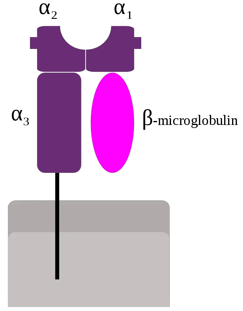
- MHC class I molecules are an essential component of the immune system found on the surface of nearly all nucleated cells in vertebrates. Alongside MHC class II molecules, they play a crucial role in antigen presentation and immune response. However, MHC class I molecules are absent on red blood cells while being present on platelets.
- The main function of MHC class I molecules is to present peptide fragments derived from proteins within the cell to cytotoxic T cells. This presentation allows the immune system to identify and target cells that display non-self antigens, triggering an immediate immune response against potential threats. The peptides presented by MHC class I molecules come from proteins found in the cytosol of the cell, leading to the pathway of MHC class I presentation being referred to as the cytosolic or endogenous pathway.
- In humans, the MHC class I molecules are encoded by the human leukocyte antigen (HLA) genes. The specific HLA genes associated with MHC class I in humans are HLA-A, HLA-B, and HLA-C. These genes encode the MHC class I molecules that are responsible for presenting antigens to cytotoxic T cells, initiating an immune response against foreign or abnormal cells.
- Understanding the role of MHC class I molecules is crucial for comprehending the mechanisms of immune recognition and response. By presenting antigens derived from within the cell, MHC class I molecules contribute to the immune system’s ability to identify and eliminate infected or mutated cells, ultimately maintaining the body’s overall health and defending against pathogens.
Functions of MHC Class I
Sure! Here’s the information about the functions of MHC Class I molecules presented in a point-by-point list format:
- MHC Class I molecules bind peptides generated from the degradation of cytosolic proteins by the proteasome.
- The MHC I:peptide complex is inserted into the external plasma membrane via the endoplasmic reticulum.
- The epitope peptide is bound to the extracellular parts of the MHC Class I molecule.
- The main function of MHC Class I is to display intracellular proteins to cytotoxic T cells (CTLs).
- This antigen presentation allows CTLs to recognize and respond to infected or abnormal cells.
- Normal cells present peptides from normal cellular protein turnover on their Class I MHC, which does not activate CTLs due to tolerance mechanisms.
- In the presence of foreign proteins, such as those during viral infections, a fraction of Class I MHC will display these peptides on the cell surface.
- CTLs specific to the MHC:peptide complex recognize and eliminate presenting cells, defending the body against pathogens.
- MHC Class I molecules can also present peptides generated from exogenous proteins through a process called cross-presentation.
- MHC Class I can serve as an inhibitory ligand for natural killer cells (NKs).
- Reduction in surface Class I MHC levels, employed by some viruses and certain tumors, activates NK cell killing.
- This mechanism allows NK cells to eliminate infected or mutated cells that evade CTL responses.
- PirB, a receptor that binds to MHC I, is involved in regulating visual plasticity in the central nervous system.
- PirB diminishes ocular dominance plasticity during the developmental critical period and adulthood.
- Mutant mice lacking PirB function exhibit enhanced plasticity in the visual cortex.
- The results suggest that PirB may modulate synaptic plasticity in the visual cortex.
What is MHC Class II?
- MHC Class II molecules are a crucial component of the immune system and belong to the major histocompatibility complex (MHC). Unlike MHC Class I molecules, which are found on most nucleated cells, MHC Class II molecules are primarily present on professional antigen-presenting cells. These include dendritic cells, mononuclear phagocytes, some endothelial cells, thymic epithelial cells, and B cells. These cells play a critical role in initiating immune responses.
- The antigens presented by MHC Class II molecules are derived from extracellular proteins, rather than cytosolic proteins as seen in MHC Class I presentation. The loading process of MHC Class II molecules involves phagocytosis. Extracellular proteins are taken up by the cell, digested within lysosomes, and the resulting peptide fragments are then loaded onto MHC Class II molecules before being transported to the cell surface.
- In humans, the MHC Class II protein complex is encoded by the human leukocyte antigen gene complex (HLA). Several HLA genes correspond to MHC Class II, including HLA-DP, HLA-DM, HLA-DOA, HLA-DOB, HLA-DQ, and HLA-DR. These genes are responsible for encoding the MHC Class II molecules that present antigens to helper T cells, facilitating the activation of immune responses.
- Mutations in the HLA gene complex can lead to a condition known as bare lymphocyte syndrome (BLS), which is a type of MHC Class II deficiency. In BLS, there is a reduced or absent expression of MHC Class II molecules, resulting in impaired immune responses. This condition can make individuals more susceptible to infections and immune-related disorders due to the compromised ability to present antigens to helper T cells.
- Understanding the role of MHC Class II molecules is crucial for comprehending the mechanisms of immune recognition and activation. By presenting antigens derived from extracellular proteins, MHC Class II molecules facilitate the communication between antigen-presenting cells and helper T cells, ultimately orchestrating effective immune responses against pathogens and foreign substances.
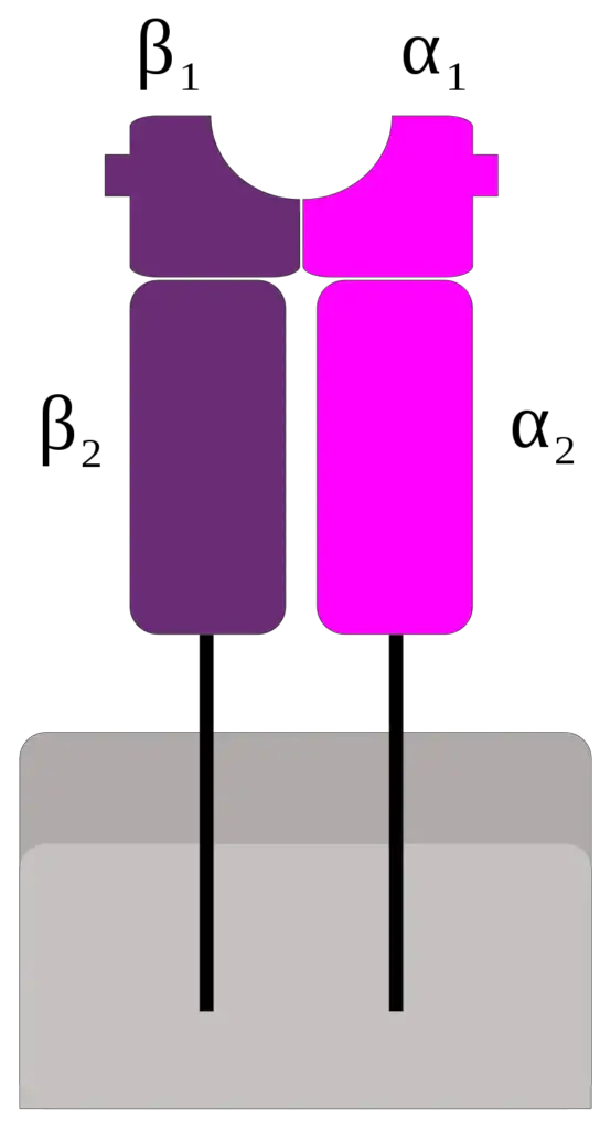
Functions of MHC Class II
The functions of MHC Class II molecules are crucial for immune recognition and response, specifically in the context of extracellular pathogens. Here are the key points about the functions of MHC Class II:
- MHC Class II molecules play a vital role in presenting peptides derived from extracellular proteins.
- The main function of Class II MHC is the presentation of antigens from extracellular pathogens, such as bacteria that may be infecting a wound or present in the bloodstream.
- Class II molecules primarily interact with immune cells, particularly T helper cells (CD4+).
- The peptide presented by MHC Class II molecules regulates how T cells respond to an infection.
- Stable binding of peptides to MHC Class II is crucial for proper immune function.
- Secure attachment of the peptide to the MHC molecule prevents detachment and degradation of the peptide.
- Without stable peptide binding, T cell recognition of the antigen, recruitment of T cells, and an appropriate immune response may be hindered.
- A proper immune response triggered by MHC Class II presentation may include localized inflammation, swelling, and recruitment of phagocytes to the site of infection.
- It can also lead to a robust antibody immune response by activating B cells.
In summary, MHC Class II molecules are responsible for presenting extracellular antigens to T helper cells, thereby regulating immune responses against pathogens. Stable peptide binding to MHC Class II is essential to ensure proper T cell recognition and subsequent immune activation, which can involve various immune mechanisms such as inflammation, phagocyte recruitment, and antibody production.
Difference between MHC Class I and MHC Class II – MHC Class I vs MHC Class II
The major difference in MHC Class I and MHC Class II (including their antigen processing pathways and presentation pathways) can be described by this chart:
| Characteristics | MHC-I molecule | MHC-II molecule |
|---|---|---|
| Distribution | Present on almost all nucleated cells including platelets. | Have a restricted tissue distribution and are chiefly found on macrophages, dendritic cells, B cells, and other antigen-presenting cells only. |
| Encoding genes | Encoded by the HLA-A, HLA-B, and HLA-C genes. | Encoded by the genes of the HLA-D region. |
| Nature of antigen presented | Antigens presented are of endogenous origin. | Antigens presented are derived from extracellular proteins. |
| Antigen | Cytosolic proteins; peptides generated within the cell or those that may enter the cytosol from phagosomes. | Peptides outside the cell, mostly internalized from the extracellular environment, such as lysosomal proteins. |
| Enzymes involved in peptide generation | Cytosolic proteasome. | Endosomal and lysosomal proteases. |
| Peptide loading of MHC | Endoplasmic reticulum. | Specialized vesicular compartment. |
| Peptide-loading complex | Includes the ER transporter associated with antigen processing (TAP1/2), tapasin, the oxidoreductase ERp57, and the chaperone protein calreticulin. | Chaperones in ER; invariant chain in ER, Golgi, and MHC Class II compartment/Class II vesicle. |
| Recognizing co-receptor | Recognized by CD8 co-receptors through the MHC Class I β2 subunit. | Recognized by CD4 co-receptors through β1 and β2 subunits. |
| Receptor T cell | Present antigens to CD8+ T cells. | Present antigens to CD4+ T cells. |
| Structure | Consists of one membrane-spanning α chain produced by MHC genes, and one β chain produced by the β2-microglobulin gene. | Consists of two membrane-spanning chains, α and β, both produced by MHC genes. |
| Building amino acids | Possess 8-10 amino acids. | Possess 13-18 amino acids. |
| Peptide binding domains | α1 and α2 are peptide binding domains. | α1 and β1 are peptide binding domains. |
| Invariant chain | No invariant chain. | Has an invariant chain. |
| Functional effect | Presence of abundant antigens targets the cell for destruction. | Presence of foreign antigens induces antibody production. |
| Detection Method | Serology. | Serology and mixed lymphocyte reaction. |
Related Posts
- Difference Between Homologous Chromosomes and Sister Chromatids
- Difference between Monocarpic and Polycarpic Plants
- Differences Between Poisonous and Non-poisonous Snakes
- Differences Between Sensitivity, Specificity, False positive, False negative
- Anabolism vs Catabolism – Differences Between Anabolism and Catabolism


