Contents
Principle of Western blotting
The principle of Western blotting involves the separation of proteins based on their size through gel electrophoresis, followed by the transfer of the separated proteins onto a solid support (blotting), and the detection of the target protein using specific antibodies (detection).
In the first step, gel electrophoresis, proteins are separated based on their electrophoretic mobility, which depends on their charge, size, and structure. The proteins are loaded onto a polyacrylamide gel and subjected to an electric field. Smaller proteins migrate faster through the gel, while larger proteins move more slowly.
Next, the separated proteins are transferred from the gel to a solid support, typically a membrane made of nitrocellulose or polyvinylidene difluoride (PVDF). This transfer, known as blotting, can be achieved by various methods, such as electroblotting or vacuum blotting. The proteins bind to the membrane through hydrophobic interactions.
After blotting, the membrane is incubated with primary antibodies specific to the target protein of interest. These antibodies recognize and bind to the target protein. Following this, secondary antibodies that are conjugated to an enzyme are added. The secondary antibody recognizes and binds to the primary antibody, forming an antibody-antigen-antibody complex.
To detect the protein bands, a substrate specific to the enzyme conjugated to the secondary antibody is added. The substrate reacts with the enzyme, resulting in the generation of a colored or luminescent product at the site of the target protein. This allows visualization of the protein bands on the membrane.
The Western blotting technique enables the identification and quantification of specific proteins in a mixture. By comparing the thickness or intensity of the bands obtained from the protein of interest with those of known standards, the amount of protein present can be estimated.
Overall, Western blotting is a valuable tool in molecular biology research for analyzing protein expression, studying protein-protein interactions, and detecting post-translational modifications of proteins.
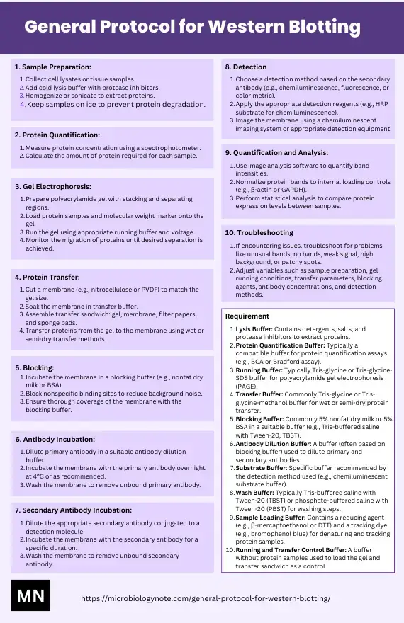
Key Solutions and Reagents Required
Lysis buffer: Radioimmunoprecipitation assay buffer (RIPA buffer)
The radioimmunoprecipitation assay (RIPA) buffer is a commonly used lysis buffer in molecular biology and biochemistry research. It is used to extract proteins from cells or tissues while maintaining their integrity and preventing their degradation. RIPA buffer contains several components that aid in protein extraction and stabilization.
The composition of RIPA buffer typically includes:
- 50 mM Tris-HCl, pH 8.0: Tris-HCl serves as a buffering agent, maintaining a stable pH during the lysis process.
- 150 mM NaCl: Sodium chloride provides the necessary ionic strength for protein solubilization and extraction.
- 1% Nonidet P-40 (NP-40) or 0.1% Triton X-100: These nonionic detergents are included to lyse the cell membranes and solubilize proteins. They disrupt lipid-protein interactions and facilitate the release of proteins into the buffer.
- 0.5% sodium deoxycholate: Sodium deoxycholate is a detergent that aids in disrupting protein-protein interactions and solubilizing membrane-associated proteins.
- 0.1% sodium dodecyl sulphate (SDS): SDS is an anionic detergent that helps solubilize and denature proteins, enabling their separation by electrophoresis.
- 1 mM sodium orthovanadate: Sodium orthovanadate is a phosphatase inhibitor that prevents the dephosphorylation of proteins during cell lysis, thereby preserving the phosphorylation state of proteins.
- 1 mM NaF: Sodium fluoride is another phosphatase inhibitor that works synergistically with sodium orthovanadate to preserve the phosphorylation status of proteins.
- Protease inhibitors tablet (e.g., Roche): These tablets contain a mixture of protease inhibitors that help prevent protein degradation by inhibiting endogenous proteases present in the cell or tissue lysate. The specific protease inhibitors included may vary depending on the manufacturer and the desired protein preservation.
RIPA buffer is typically used by adding it to cells or tissues and then lysing them through gentle agitation or sonication. The resulting lysate contains the released proteins, which can be further analyzed using techniques such as Western blotting, immunoprecipitation, or enzyme assays.
The inclusion of detergents, phosphatase inhibitors, and protease inhibitors in RIPA buffer ensures efficient protein extraction while maintaining their structural and functional integrity. It provides a suitable environment for downstream analyses by preserving protein-protein interactions, post-translational modifications, and phosphorylation states.
Loading buffer: 2x Laemmli buffer
Loading buffer is an essential component used in molecular biology experiments, particularly in protein analysis techniques like SDS-PAGE (sodium dodecyl sulfate-polyacrylamide gel electrophoresis). It aids in denaturing and preparing protein samples for separation by electrophoresis.
One commonly used loading buffer is 2x Laemmli buffer. Its composition typically includes the following components:
- 4% SDS: Sodium dodecyl sulfate (SDS) is an anionic detergent that denatures proteins by disrupting their native structure. It coats proteins with a negative charge, allowing them to separate primarily based on their molecular weight during electrophoresis.
- 10% 2-mercaptoethanol: 2-mercaptoethanol (or β-mercaptoethanol) is a reducing agent that breaks disulfide bonds in proteins, further promoting denaturation. This helps to eliminate the effects of protein folding and secondary structure during electrophoresis.
- 20% glycerol: Glycerol is a viscosity-inducing agent that provides stability to the protein samples and helps them sink into the gel wells during loading. It also aids in preventing protein aggregation and precipitation.
- 0.004% bromophenol blue: Bromophenol blue is a tracking dye that allows visual monitoring of the progress of electrophoresis. It migrates through the gel at a known rate and helps determine the migration front during electrophoresis.
- 0.125 M Tris-HCl: Tris-HCl serves as a buffering agent to maintain a stable pH during electrophoresis.
The pH of the 2x Laemmli buffer should be checked and adjusted to pH 6.8 if necessary. Proper pH ensures optimal conditions for protein denaturation and electrophoresis.
To prepare the loading buffer, the aforementioned components are mixed together in appropriate proportions and dissolved in water or a suitable buffer. The resulting buffer is typically heated at high temperature (e.g., 95°C) for a few minutes to fully denature the proteins and ensure complete reduction of disulfide bonds.
When loading protein samples onto an SDS-PAGE gel, the 2x Laemmli loading buffer is added to the protein sample, and the mixture is heated briefly to maintain protein denaturation and reduction. The heated protein-sample-buffer mixture is then loaded into the gel wells for electrophoresis.
The 2x Laemmli buffer provides denaturing conditions for proteins, enables their separation based on size, and facilitates tracking during electrophoresis. It is widely used in various protein analysis techniques, including Western blotting, protein gel electrophoresis, and protein purification.
Running buffer: Tris/Glycine/SDS
The running buffer, specifically the Tris/Glycine/SDS buffer, is a commonly used solution in SDS-PAGE (sodium dodecyl sulfate-polyacrylamide gel electrophoresis) for the separation of proteins based on their molecular weight. It provides the necessary ionic environment for protein migration during electrophoresis and maintains the stability of the gel.
The composition of the Tris/Glycine/SDS running buffer typically includes the following components:
- 25 mM Tris: Tris (tris(hydroxymethyl)aminomethane) acts as a buffering agent and helps maintain a stable pH during the electrophoresis process. The pH of the running buffer is usually adjusted to the desired value, often around pH 8.3.
- 190 mM glycine: Glycine serves as an ionic compound in the running buffer. It provides conductivity and helps establish an electric field in the gel. The glycine ions facilitate protein migration in the gel during electrophoresis.
- 0.1% SDS: Sodium dodecyl sulfate (SDS) is an anionic detergent that denatures proteins and imparts a negative charge to them. It helps to uniformly coat the proteins with a negative charge, allowing for separation based on size during electrophoresis. SDS also aids in protein solubilization and maintains a consistent environment throughout the gel.
These components are dissolved in water to prepare the Tris/Glycine/SDS running buffer. The buffer solution is usually prepared with high-quality deionized water, and its pH is adjusted as required.
During SDS-PAGE, the running buffer is poured into the electrophoresis apparatus, and the gel is submerged in the buffer. When an electric current is applied, the negatively charged proteins migrate through the gel matrix towards the positive electrode (anode). The Tris/Glycine/SDS running buffer facilitates this migration by providing the appropriate pH, ionic strength, and detergent environment required for efficient protein separation.
The Tris/Glycine/SDS running buffer is widely used in protein electrophoresis techniques, including Western blotting, native-PAGE, and protein fragment analysis. It ensures optimal separation and resolution of proteins, allowing researchers to analyze protein samples based on their molecular weight and study protein expression, interactions, and modifications.
Transfer buffer
The transfer buffer is a crucial component used in Western blotting and other protein transfer techniques. It facilitates the transfer of proteins from a polyacrylamide gel to a membrane for subsequent protein detection and analysis.
The composition of the transfer buffer typically includes:
- 25 mM Tris: Tris (tris(hydroxymethyl)aminomethane) acts as a buffering agent to maintain a stable pH during the transfer process. The pH of the transfer buffer is usually adjusted to the desired value, often around pH 8.3. Tris ensures that the transfer occurs under optimal conditions for protein stability and membrane binding.
- 190 mM glycine: Glycine serves as an ionic compound in the transfer buffer. It provides the necessary ionic strength to promote efficient protein transfer during electrophoresis. The glycine ions help maintain a stable electric field and facilitate the migration of proteins from the gel to the membrane.
- 20% methanol: Methanol is included in the transfer buffer to enhance the transfer efficiency. It improves the solubility of proteins and enhances their movement from the gel to the membrane. Methanol also aids in preventing the nonspecific binding of proteins to the gel matrix during the transfer process.
For proteins larger than 80 kDa (kilodaltons), it is recommended to include SDS (sodium dodecyl sulfate) in the transfer buffer at a final concentration of 0.1%. SDS functions as a denaturing agent, ensuring efficient transfer of large proteins by disrupting their structure and promoting their solubility. Including SDS in the transfer buffer helps prevent aggregation and allows for a more successful transfer of high-molecular-weight proteins.
To prepare the transfer buffer, the specified amounts of Tris, glycine, and methanol are dissolved in an appropriate volume of high-quality deionized water. If SDS is needed, it is added to the final buffer solution at a concentration of 0.1%.
During the transfer process, the gel containing the separated proteins is placed in a transfer apparatus or sandwiched between two membranes, with the transfer buffer surrounding it. An electric current is applied to promote the migration of proteins from the gel to the membrane. The transfer buffer provides the necessary pH, ionic strength, and denaturing conditions for efficient protein transfer.
The transfer buffer plays a vital role in Western blotting, allowing researchers to transfer and immobilize proteins on a membrane, which can then be probed with specific antibodies for protein detection and analysis. It ensures the successful transfer of proteins while maintaining their structural integrity, thus enabling accurate and reliable protein identification and quantification.
Ponceau S staining buffer
Ponceau S staining buffer is a commonly used solution in molecular biology for protein visualization and quantification. It is typically used to stain proteins on a membrane after transfer from a gel, allowing researchers to assess the success of the transfer and to verify the presence of proteins of interest.
The composition of Ponceau S staining buffer typically includes:
- 0.2% (w/v) Ponceau S: Ponceau S is a red dye that binds to proteins, resulting in a visible staining of the protein bands. It interacts with the amino acid residues present in the proteins, allowing their visualization on the membrane. Ponceau S staining is reversible, allowing the same membrane to be subsequently probed with specific antibodies for further analysis if desired.
- 5% glacial acetic acid: Glacial acetic acid is included in the staining buffer to enhance the staining process and optimize the interaction between the dye and the proteins. It helps to improve the binding of Ponceau S to the proteins, leading to more pronounced staining.
To prepare the Ponceau S staining buffer, the specified amount of Ponceau S dye is dissolved in an appropriate volume of water. Glacial acetic acid is then added to the solution to reach a final concentration of 5%.
After transferring the proteins from a gel to a membrane, the membrane is briefly immersed in the Ponceau S staining buffer. The staining time may vary but is typically short, usually ranging from a few minutes to several minutes. The membrane is then washed with water to remove excess dye and to enhance the contrast between the stained proteins and the background.
Ponceau S staining allows for a quick and visual assessment of protein transfer efficiency, as well as the overall protein pattern on the membrane. It enables researchers to determine the presence, abundance, and relative molecular weight of the transferred proteins. The stained proteins appear as red bands or spots on a white background, providing a convenient and immediate means of evaluating the success of protein transfer before proceeding to subsequent steps, such as antibody probing or other downstream analyses.
It’s important to note that Ponceau S staining is not suitable for quantifying protein levels accurately. However, it serves as a useful qualitative tool in protein analysis, offering a quick and straightforward method for initial evaluation and troubleshooting.
Tris-buffered saline with Tween 20 (TBST) buffer
Tris-buffered saline with Tween 20 (TBST) buffer is a commonly used solution in various molecular biology techniques, including Western blotting and immunohistochemistry. It is used as a washing and blocking buffer to enhance protein detection and reduce nonspecific binding.
The composition of TBST buffer typically includes:
- 20 mM Tris, pH 7.5: Tris (tris(hydroxymethyl)aminomethane) serves as a buffering agent to maintain a stable pH during the experimental procedures. The pH of the TBST buffer is typically adjusted to 7.5, which is close to physiological pH and provides optimal conditions for protein stability and interaction with antibodies.
- 150 mM NaCl: Sodium chloride (NaCl) is included in the TBST buffer to establish an appropriate ionic strength. It helps maintain proper protein solubility, stability, and interactions during antibody-based detection methods. NaCl also contributes to the removal of unbound antibodies and other nonspecific molecules during washing steps.
- 0.1% Tween 20: Tween 20 is a nonionic detergent added to TBST buffer to reduce nonspecific binding and improve antibody-antigen interactions. It helps to block and wash away any nonspecific binding sites on the membrane or other surfaces, reducing background noise and enhancing the sensitivity of protein detection.
To prepare the TBST buffer, the specified amounts of Tris and NaCl are dissolved in water, and the pH is adjusted to 7.5 using an appropriate acid or base. Tween 20 is then added to achieve a final concentration of 0.1%.
TBST buffer is primarily used for washing steps in techniques like Western blotting. After transferring proteins to a membrane and blocking nonspecific binding sites, the membrane is incubated with primary antibodies to detect specific proteins. Following the primary antibody incubation, the membrane is washed multiple times with TBST buffer to remove unbound antibodies and any other nonspecific binding molecules. The buffer helps maintain proper protein solubility, minimizes background noise, and improves the signal-to-noise ratio.
TBST buffer is also commonly used in antibody-based assays such as immunohistochemistry and immunocytochemistry, where it serves as a washing buffer to remove unbound antibodies and other reagents.
Overall, TBST buffer plays a critical role in reducing nonspecific binding, optimizing antibody-antigen interactions, and maintaining protein stability during protein detection experiments. Its composition ensures proper pH, ionic strength, and surfactant properties for efficient and specific antibody-based assays.
Tris-buffered saline with Blocking buffer
Tris-buffered saline with blocking buffer is a solution commonly used in immunoassays, such as Western blotting and immunohistochemistry, to minimize nonspecific binding and reduce background noise. It serves as a blocking agent to block nonspecific binding sites on membranes or other surfaces, preventing unwanted interactions and improving the specificity of antibody-based detection.
The composition of the blocking buffer typically includes:
- 3% bovine serum albumin (BSA): Bovine serum albumin, a protein derived from bovine blood, is included in the blocking buffer to occupy and block nonspecific binding sites on the membrane or surface. BSA serves as a proteinaceous blocking agent and helps reduce background noise caused by nonspecific interactions. It effectively binds to unoccupied sites, preventing antibodies or other proteins from binding nonspecifically and improving the signal-to-noise ratio.
The blocking buffer is prepared by dissolving the specified amount of BSA in Tris-buffered saline with Tween 20 (TBST) buffer. TBST buffer provides the necessary pH, ionic strength, and surfactant properties to optimize protein stability and antibody-antigen interactions.
To use the blocking buffer, the membrane or surface is incubated in the solution, allowing the BSA to bind and block nonspecific binding sites. This step is usually carried out prior to the incubation with primary antibodies or other detection reagents. The blocking buffer forms a protective barrier, preventing nonspecific molecules from interacting with the membrane and reducing background noise.
Blocking with BSA in TBST buffer is widely used due to the versatility and low immunogenicity of BSA. It effectively blocks a wide range of nonspecific binding sites, providing reliable and consistent results across different immunoassay techniques. The use of BSA in the blocking buffer is particularly beneficial when working with antibodies derived from species that are prone to cross-reactivity or when using complex samples that may contain abundant nonspecific binding molecules.
Overall, Tris-buffered saline with blocking buffer containing 3% BSA in TBST is a commonly employed solution to block nonspecific binding sites, reduce background noise, and improve the specificity of antibody-based detection in immunoassays. It ensures a reliable and accurate assessment of the target proteins or antigens of interest while minimizing false-positive signals.
Stripping buffer
A stripping buffer is a solution used in molecular biology techniques to remove antibodies or other proteins that have bound to a membrane, allowing for subsequent re-probing or re-use of the membrane. It effectively removes previously bound molecules, resetting the membrane for further analysis.
The composition of the stripping buffer typically includes:
- 20 ml 10% SDS: Sodium dodecyl sulfate (SDS) is a strong anionic detergent commonly used in stripping buffers. It disrupts protein-protein interactions and denatures proteins, facilitating the removal of antibodies or other proteins bound to the membrane.
- 12.5 ml 0.5 M Tris HCl, pH 6.8: Tris(hydroxymethyl)aminomethane hydrochloride (Tris HCl) acts as a buffering agent to maintain a stable pH during the stripping process. The pH of the stripping buffer is adjusted to 6.8, which is suitable for efficient antibody removal.
- 67.5 ml ultrapure water: Ultrapure water, also known as deionized water, is used as the solvent to create the buffer solution. It ensures that the buffer is free from impurities that may interfere with the stripping process.
- 0.8 ml 2-mercaptoethanol: 2-mercaptoethanol is a reducing agent that helps break disulfide bonds and further aids in the denaturation of proteins. It enhances the efficiency of stripping by reducing the stability of protein complexes.
To prepare the stripping buffer, the specified amounts of SDS, Tris HCl, and ultrapure water are mixed together. Subsequently, 2-mercaptoethanol is added to the solution.
When using a stripping buffer, the membrane with previously bound antibodies or proteins is immersed in the solution and incubated under specific conditions, such as temperature and time, to facilitate the removal of the bound molecules. The stripping buffer disrupts the interactions between antibodies/proteins and the target molecules, allowing for their detachment from the membrane.
After stripping, the membrane is thoroughly washed with an appropriate wash buffer, such as TBST (Tris-buffered saline with Tween 20), to remove any residual stripping buffer and ensure the removal of all stripped proteins.
The stripping buffer is particularly useful when performing Western blotting experiments, where it allows for the stripping of antibodies targeting a specific protein of interest, enabling the membrane to be reprobed with different antibodies for multiple protein detections.
It’s important to note that the stripping buffer may not remove all proteins completely, and some residual signal may persist. Therefore, careful optimization of the stripping conditions and subsequent antibody probing is necessary to achieve reliable and accurate results.
Overall, the stripping buffer is an essential tool in molecular biology techniques, enabling the removal of previously bound antibodies or proteins from membranes. It allows for the reuse of membranes, reduces the need for additional experiments, and enhances the versatility of protein analysis.
Western Blotting Protocol / Procedure/ Steps
1. Sample prep (based on a typical cell culture scenario)
Sample preparation is a crucial step in molecular biology experiments, including Western blotting and protein analysis. The following steps outline a typical sample preparation procedure based on a cell culture scenario:
- Start by placing the cell culture dish on ice to maintain a low temperature throughout the sample preparation process. This helps preserve the integrity of the proteins.
- Wash the cells with ice-cold Tris-buffered saline (TBS). TBS is used to rinse the cells and remove any residual culture media or other contaminants.
- Aspirate the TBS, and then add ice-cold RIPA buffer (1 ml per 100 mm dish) to the dish. RIPA buffer is a lysis buffer that helps break down the cell membrane and release the proteins of interest.
- Scrape the adherent cells off the dish using a cold plastic cell scraper. Gently transfer the cell suspension into a pre-cooled microcentrifuge tube. The cold temperature helps prevent protein degradation.
- Maintain constant agitation of the cell suspension for 30 minutes at 4°C. Agitation aids in cell lysis and ensures thorough protein extraction.
- If necessary, perform sonication on the cell suspension. Sonication involves subjecting the sample to high-frequency sound waves to further disrupt the cells and promote complete cell lysis. Sonicate the sample three times for 10-15 seconds each to ensure efficient cell lysis and shear DNA to reduce sample viscosity.
- Centrifuge the lysate at 16,000 x g for 20 minutes in a pre-cooled centrifuge at 4°C. This step separates the cellular debris and organelles from the protein-containing supernatant.
- Gently remove the centrifuge tube and place it on ice. Transfer the supernatant, which contains the protein lysate, to a fresh tube kept on ice. Discard the pellet containing the cellular debris.
- To determine the protein concentration, remove a small volume (10-20 μl) of the lysate and perform a protein assay. This step allows quantification of the protein content in the lysate.
- If necessary, aliquot the protein samples for long-term storage at -20°C. It is important to avoid repeated freeze and thaw cycles, as they can lead to protein degradation.
- Take 20 μg of each sample and add an equal volume of 2x Laemmli sample buffer. The Laemmli buffer helps denature the proteins and provides a suitable pH and reducing environment for subsequent protein analysis.
- Boil each cell lysate in the sample buffer at 95°C for 5 minutes. Heating the samples denatures the proteins, ensuring their complete unfolding and preparation for further analysis.
- Centrifuge the samples at 16,000 x g in a microcentrifuge for 1 minute to remove any insoluble particles or debris that may interfere with downstream analysis.
Following these steps, the prepared protein lysates are ready for analysis using techniques such as Western blotting or other protein assays. Proper sample preparation is essential for obtaining reliable and accurate results in protein analysis experiments.
2. Protein separation by gel electrophoresis
Protein separation by gel electrophoresis is a commonly used technique in molecular biology and biochemistry to separate proteins based on their size. The following steps outline the process:
Gel preparation
Gel preparation is an essential step in various laboratory techniques, such as gel electrophoresis, that are used to separate and analyze biomolecules like proteins and nucleic acids. The following instructions outline the process of preparing a gel using a stacking gel and separating gel.
- Start by preparing a 10% stacking gel solution. This can be done by mixing the appropriate amounts of acrylamide and bis-acrylamide with the running buffer in a flask. However, it’s important to note that the gel will not solidify until the required reagents are added, so it’s best to delay adding them until later in the process.
- Once the stacking gel solution is prepared, assemble the rack for gel solidification. This can typically be done using a gel-casting apparatus, which consists of two glass plates held together by clamps. Refer to Figure 1 for a visual representation of the setup.
- Carefully pour the stacking gel solution into the gap between the glass plates, making sure to fill it up until the level reaches the green bar or marker indicated on the apparatus (refer to Figure 2). Take caution not to overfill the gap.
- After adding the stacking gel solution, fill the remaining space at the top of the gap with distilled water (H2O). This water layer helps prevent any air bubbles from forming and ensures a smooth gel surface.
- Allow the gel to solidify for approximately 15 to 30 minutes. The stacking gel solution contains specific reagents, such as ammonium persulfate (AP) and tetramethylethylenediamine (TEMED), that initiate the gelation process. The gel should turn solid, indicating that it is ready for the next steps.
- Once the stacking gel is solidified, carefully remove the water layer from the top. It is often helpful to tilt the gel-casting apparatus and use a paper towel or similar absorbent material to absorb and remove the water.
- Overlay the solidified stacking gel with the separating gel solution. The separating gel is typically of higher percentage than the stacking gel and is poured on top of it to form a continuous gel matrix. The separating gel allows for the separation of biomolecules based on their size during electrophoresis.
- After adding the separating gel, insert a comb into the gel. The comb creates wells or indentations in the gel, which will be used to load the samples later. Ensure that the comb is inserted carefully and firmly, without creating any air bubbles.
- Allow the gel to solidify completely. The solidification time can vary but is typically around 30 minutes. To check if the gel is solidified, you can leave a small amount of gel solution in a tube and observe its solidification progress.
Electrophoresis
- Load equal amounts of protein, typically 20 μg, into the wells of a mini (8.6 x 6.7 cm) or midi (13.3 x 8.7 cm) format SDS-PAGE gel. In addition to the protein samples, molecular weight markers are also loaded onto the gel. These markers provide reference points of known molecular weights, which help in estimating the sizes of the protein bands during analysis.
- Initially, run the gel at a low voltage, typically 50 V, for 5 minutes. This step allows the proteins to concentrate in the stacking gel region, forming sharp and compact bands before they enter the separating gel.
- Increase the voltage to a higher range, usually between 100-150 V, to complete the electrophoresis run in approximately 1 hour. The voltage applied generates an electric field that causes the proteins to migrate through the gel matrix based on their size and charge.
The percentage of the gel used for separation depends on the size range of the proteins of interest. A commonly used gel type is the 4-20% gradient gel, which provides excellent separation of proteins of various sizes. This gradient gel has a lower percentage of acrylamide near the stacking gel region and gradually increases to a higher percentage in the separating gel region. This gradient allows proteins of different sizes to migrate at different rates, resulting in well-resolved bands during separation.
It’s important to note that for more specific details and recommendations regarding gel selection and optimization, referring to resources such as the Protein Blotting Guide (bulletin 2895) is advisable. These resources provide comprehensive information and guidelines for achieving optimal protein separation based on specific experimental requirements.
Overall, protein separation by gel electrophoresis is a fundamental technique used to separate proteins based on their size, enabling subsequent analysis and characterization of proteins of interest.
3. Transferring the protein from the gel to the membrane
Transferring proteins from the gel to a membrane is a critical step in Western blotting to facilitate the detection and analysis of specific target proteins. The following steps outline the process of protein transfer:
- Begin by placing the gel in 1x transfer buffer. The gel should be immersed in the transfer buffer for approximately 10-15 minutes. The transfer buffer is specifically formulated to provide the appropriate ionic conditions for efficient protein transfer.
- Assemble the transfer sandwich by layering the components in the following order: a sponge soaked in transfer buffer, a piece of filter paper, the gel containing the separated proteins, a nitrocellulose or PVDF membrane, and another piece of filter paper. It is important to ensure that there are no air bubbles trapped between the layers of the transfer sandwich. The blot (membrane) should be placed on the cathode (negative electrode), while the gel is positioned on the anode (positive electrode).
- Place the transfer sandwich into the transfer tank. To maintain a cold environment and prevent protein degradation, an ice block or other cooling methods can be added to the tank. Cooling the transfer process is especially important for heat-sensitive proteins.
- For optimal transfer efficiency, it is recommended to conduct the transfer overnight in a coldroom at a constant current of 10 mA. This slow and continuous transfer allows the proteins to migrate from the gel onto the membrane effectively.
Note: Alternative transfer conditions can be optimized depending on the size of the proteins being transferred. For example, transferring at 100 V for 30 minutes to 2 hours can be suitable for proteins of different sizes. It is essential to optimize the transfer conditions to achieve efficient and specific protein transfer for the target proteins of interest.
By following these steps, the proteins from the gel will be transferred onto the membrane, allowing subsequent detection and analysis steps, such as antibody probing and visualization of the target protein bands. Proper protein transfer ensures accurate and reliable Western blotting results.
4. Antibody incubation
Antibody incubation is a crucial step in Western blotting that involves the specific binding of antibodies to target proteins on the membrane. The following steps outline the antibody incubation process:
- After the protein transfer from the gel to the membrane, briefly rinse the membrane in water. This is followed by staining the membrane with Ponceau S solution to visually assess the transfer quality. Ponceau S staining helps confirm that the proteins are appropriately transferred and evenly distributed on the membrane.
- Rinse off the Ponceau S stain by performing three washes with TBST (Tris-buffered saline with Tween 20). TBST is a buffer solution that provides the appropriate pH and ionic conditions for antibody binding.
- Block the membrane to prevent non-specific binding of antibodies by incubating it in a blocking solution. The commonly used blocking agent is 3% BSA (bovine serum albumin) in TBST. The blocking step is performed at room temperature for 1 hour and helps saturate any remaining unbound sites on the membrane surface.
- Incubate the membrane overnight at 4°C in the primary antibody solution specific to the target protein. The primary antibody should be appropriately diluted in the blocking buffer according to the manufacturer’s recommended ratio. Incubation times may vary depending on the antibody quality and performance. Alternatively, the primary antibody may be applied for 1-3 hours at room temperature.
- After the primary antibody incubation, rinse the membrane with TBST by performing 3-5 washes of 5 minutes each. This step helps remove any unbound primary antibody and reduces background noise.
- Incubate the membrane in the secondary antibody solution conjugated with an enzyme such as HRP (horseradish peroxidase) for 1 hour at room temperature. The secondary antibody is selected to specifically bind to the primary antibody, allowing for detection of the target protein. The secondary antibody can be diluted in 5% skim milk in TBST.
- Rinse the membrane again with TBST by performing 3-5 washes of 5 minutes each. This step removes any unbound secondary antibody and helps reduce background noise.
Following these steps, the membrane is now ready for detection and visualization of the target protein bands through the enzymatic reaction catalyzed by the conjugated enzyme in the secondary antibody. The antibody incubation process ensures specific and sensitive detection of the target protein on the Western blot membrane.
5. Imaging and data analysis
Imaging and data analysis are crucial steps in Western blotting for quantifying and analyzing the protein bands of interest. The following steps outline the process of imaging and data analysis:
- Apply the chemiluminescent substrate to the blot according to the manufacturer’s recommendation. Chemiluminescent substrates are designed to react with the enzyme conjugated to the secondary antibody, generating light that is proportional to the amount of the target protein present. The substrate reaction should be carried out in a darkroom or under low light conditions to minimize background noise.
- Capture the chemiluminescent signals emitted from the blot using a CCD (charge-coupled device) camera-based imager. CCD cameras are commonly used in Western blot imaging systems due to their sensitivity and ability to capture low light signals. The camera is typically connected to a computer for image acquisition.
Note: The use of film is not recommended at this step because it has a limited dynamic range, which may result in overexposed or underexposed bands and loss of data.
- Once the chemiluminescent signals are captured, use image analysis software to quantify and analyze the band intensities of the target proteins. Image analysis software allows for accurate measurement of band intensities, background subtraction, and comparison between different samples. The software provides tools to define regions of interest (ROIs) around each band and calculates their intensity values.
By using the image analysis software, it is possible to obtain quantitative data such as band intensities, molecular weights, and sizes of the protein bands. This data can be further analyzed and compared between different samples or experimental conditions, providing valuable insights into protein expression levels, protein-protein interactions, and other parameters of interest.
Overall, imaging and data analysis play a crucial role in Western blotting, enabling the quantification and interpretation of the results obtained from the chemiluminescent signals captured on the blot. The use of specialized imaging systems and software ensures accurate and reliable data analysis, facilitating further research and understanding in the field of protein analysis and molecular biology.
6. Stripping and reprobing
Stripping and reprobing are important steps in Western blotting when it is necessary to remove the antibodies bound to the membrane and detect a different target protein or reprobe the same membrane for a loading control. The following steps outline the process of stripping and reprobing:
- Warm the stripping buffer to 50°C. The stripping buffer typically contains components that can disrupt the antibody-antigen interactions and remove the antibodies bound to the membrane.
- Add the stripping buffer to the membrane in a container designated for stripping. Ensure that the membrane is completely submerged in the buffer. Incubate the membrane at 50°C for up to 45 minutes with some agitation. This incubation time may vary depending on the stripping buffer and the strength of the antibody-antigen binding. Agitation helps in enhancing the efficiency of the stripping process.
- Rinse the blot under running water for 1 hour. Thoroughly wash the membrane to remove any residual stripping buffer and stripped antibodies.
- Transfer the membrane to a clean container and wash it 5 times for 5 minutes each with TBST (Tris-buffered saline with Tween 20). This step helps to remove any remaining contaminants and prepare the membrane for reprobing.
- Block the membrane in 3% BSA (bovine serum albumin) in TBST at room temperature for 1 hour. Blocking is necessary to prevent non-specific binding of antibodies during the reprobing process.
- Incubate the membrane overnight in the primary antibody solution against the target protein or loading control protein at 4°C. The primary antibody should be diluted in the blocking buffer according to the manufacturer’s recommended ratio.
- Rinse the blot 3–5 times for 5 minutes each with TBST to remove unbound primary antibody.
- Incubate the membrane in the HRP (horseradish peroxidase)-conjugated secondary antibody solution for 1 hour at room temperature. The secondary antibody is specific to the host species of the primary antibody and is conjugated to an enzyme, such as HRP, for signal detection.
Note: The secondary antibody can be diluted using 5% skim milk in TBST as an alternative blocking agent.
- Rinse the blot 3–5 times for 5 minutes each with TBST to remove unbound secondary antibody.
After completing these steps, the membrane is ready for detection using a suitable detection method, such as chemiluminescence or fluorescence, to visualize the target protein or loading control protein. Stripping and reprobing allow for the efficient utilization of Western blots by enabling the detection of multiple target proteins on the same membrane, saving time, and conserving sample material.
7. Imaging and data analysis
Imaging and data analysis are crucial steps in Western blotting to quantify and analyze the results obtained from the experiment. The following steps outline the process of imaging and data analysis:
- Apply the chemiluminescent substrate to the blot following the manufacturer’s suggestions. The chemiluminescent substrate reacts with the enzyme-conjugated secondary antibody bound to the target protein, resulting in the emission of light. Ensure that the substrate is evenly applied to cover the entire membrane.
- Capture the chemiluminescent signals using a CCD (charge-coupled device) camera-based imager. The CCD camera detects and captures the emitted light from the chemiluminescent reaction. It is recommended to use a CCD camera-based imager instead of film because the dynamic range of CCD cameras allows for a more accurate and quantitative detection of signals.
Note: Film-based detection systems are not recommended for chemiluminescent signals as they have a limited dynamic range and may result in saturated signals.
- Use image analysis software to read the band intensity of the loading control proteins. Load the image obtained from the CCD camera into image analysis software, which allows for quantification of the protein bands. Select the regions of interest corresponding to the loading control protein bands and measure their intensity. The intensity values reflect the amount of loading control protein present in each lane.
- Use the loading control protein levels to normalize the target protein levels. Normalize the intensity values of the target protein bands by dividing them with the corresponding intensity values of the loading control protein bands. This normalization corrects for any variations in protein loading or transfer efficiency across the different samples.
By normalizing the target protein levels to the loading control protein levels, the data analysis accounts for any discrepancies that may have occurred during the experimental process. The normalized data can then be statistically analyzed, compared between different samples or conditions, and presented in graphical form to draw conclusions about the relative expression levels or changes in the target protein of interest.
Theory
Sample Preparation for Western Blot:
In Western blot analysis, cell lysates are commonly used as samples for protein analysis. The goal of protein extraction is to collect all the proteins present in the cell cytosol. To prevent protein denaturation, the extraction process should be performed at a cold temperature, and protease inhibitors should be added. However, when working with tissue samples that possess a higher degree of structure, additional mechanical methods such as homogenization or sonication are required to extract the proteins effectively.
Once the proteins are extracted, it is crucial to determine their concentration accurately. This information is essential for ensuring that the samples are compared on an equivalent basis. Protein concentration can be measured using a spectrophotometer, which allows the researcher to determine the mass of the protein being loaded into each well based on the relationship between concentration, mass, and volume.
After determining the appropriate sample volume, the sample is diluted into a loading buffer. The loading buffer contains glycerol, which helps the samples sink easily into the wells of the gel. It also contains a tracking dye, typically bromophenol blue, which allows the researcher to monitor the progress of separation during electrophoresis. The diluted sample is then heated to denature the higher order structure while retaining sulfide bridges. Denaturing the protein structure ensures that the negative charge of amino acids is not neutralized, enabling the proteins to move in an electric field during electrophoresis.
It is crucial to include positive and negative controls when preparing the samples. Positive controls involve using a known source of the target protein, such as purified protein or a control lysate. These controls help confirm the identity of the protein and validate the activity of the antibody. Negative controls, such as a null cell line like β-actin, are also used to confirm that the staining is not nonspecific.
Gel Electrophoresis:
Western blot analysis involves using two types of agarose gel: a stacking gel and a separating gel. The stacking gel is slightly acidic (pH 6.8) and has a lower acrylamide concentration, resulting in a porous gel that separates proteins poorly but allows for the formation of thin, well-defined bands. The separating gel, on the other hand, is basic (pH 8.8) and has a higher polyacrylamide content, creating narrower gel pores. This separation gel facilitates the separation of proteins based on their size, with smaller proteins traveling more easily and rapidly through the gel than larger proteins.
When loading the proteins onto the gel, they acquire a negative charge due to denaturation through heating. Consequently, they migrate toward the positive electrode when a voltage is applied. The gel is typically prepared by pouring it between two glass or plastic plates using the specified solution. The protein samples and a marker are loaded into the wells, while the empty wells are filled with sample buffer. The gel is then connected to a power supply and allowed to run. It is crucial to set the voltage appropriately to prevent overheating and distortion of the protein bands.
Blotting:
After the proteins are separated in the gel, they need to be transferred to a membrane for further analysis. The transfer is accomplished using an electric field oriented perpendicular to the gel’s surface, causing the proteins to migrate out of the gel and onto the membrane. The membrane is positioned between the gel surface and the positive electrode in a sandwich-like configuration, which includes fiber pads (sponges) at each end and filter papers to protect the gel and blotting membrane. It is important to ensure close contact between the gel and the membrane for clear imaging, and the membrane should be placed correctly to allow the negatively charged proteins to migrate from the gel to the membrane. This transfer process is known as electrophoretic transfer and can be conducted in semi-dry or wet conditions. Wet conditions are generally preferred as they minimize the risk of drying out the gel and are more suitable for larger proteins.
The membrane, acting as a solid support, is a critical component of the Western blotting process. There are two types of membranes commonly used: nitrocellulose and PVDF. Nitrocellulose is preferred for its high affinity for proteins and retention abilities. However, it is brittle and does not allow for easy reprobing of the membrane. In contrast, PVDF membranes provide better mechanical support and allow for reprobing and storage. However, PVDF membranes tend to have higher background noise, so careful washing is essential.
Washing, Blocking, and Antibody Incubation:
Blocking is a crucial step in Western blotting as it prevents nonspecific binding of antibodies to the membrane. Blocking is typically performed using 5% BSA or nonfat dried milk diluted in a wash buffer such as TBST (Tris-buffered saline with Tween). Nonfat dried milk is often preferred due to its availability and cost-effectiveness. However, the choice of blocking solution should consider compatibility with the detection labels used. For example, BSA blocking solutions are preferred with biotin and alkaline phosphatase (AP) antibody labels and antiphosphoprotein antibodies, as milk contains casein, a phosphoprotein, and biotin, which can interfere with the assay results. It is often advantageous to incubate the primary antibody with BSA, as higher amounts of primary antibody are typically required compared to the secondary antibody. Placing the antibody in a BSA solution allows for its reuse if the initial blot does not yield satisfactory results.
The concentration of the antibody should follow the manufacturer’s instructions and can be diluted in a wash buffer such as PBS or TBST. Washing steps are crucial to minimize background noise and remove unbound antibodies. However, the membrane should not be left to wash for an excessively long time, as this can reduce the signal.
Detection:
The membrane is detected using a labeled antibody, often conjugated with an enzyme such as horseradish peroxidase (HRP). The enzyme generates a detectable signal corresponding to the position of the target protein. This signal is captured on a film, typically developed in a dark room.
Quantification:
It is important to note that Western blot data is typically considered semi-quantitative rather than providing an absolute measure of protein quantity. This is due to variations in loading and transfer rates between samples in separate lanes and on different blots. To enable more precise comparisons, these differences need to be standardized. Additionally, the signal generated by detection is not linear across the concentration range of the samples, so it should not be used to model the concentration directly.
Troubleshooting for Western Blotting
1. Unusual or Unexpected Bands:
- Protease degradation: Use a fresh sample kept on ice or consider changing the antibody.
- Bands too high: Reheat the sample to break the quaternary protein structure.
- Blurry bands: Run the gel at a lower voltage, ensure proper transfer sandwich preparation, and check for air bubbles during transfer.
- Non-flat bands: Optimize the gel to fit the sample size.
- White (negative) bands: Decrease the protein or antibody concentration.
2. No Bands:
- Improper antibody: Use appropriate primary or secondary antibodies.
- Low antibody concentration: Ensure the antibody concentration is sufficient for detection.
- Low or absent antigen concentration: Confirm the presence of the antigen by using another source.
- Prolonged washing: Optimize the washing time.
- Contaminated buffers: Use new and non-contaminated buffers to avoid sodium azide interference.
3. Faint Bands or Weak Signal:
- Low antibody or antigen concentration: Increase the antibody or antigen concentration.
- Increase exposure time: Adjust the exposure time to enhance band visibility.
- Nonfat dry milk masking antigen: Use BSA or decrease the amount of milk used.
4. High Background:
- High antibody concentration: Decrease the antibody concentration to prevent binding to PVDF membranes.
- Old buffers: Use fresh buffers to reduce background noise.
- Increased washing time: Optimize the washing time to minimize background.
- High exposure: Check different exposure times to find the optimum duration.
5. Patchy or Uneven Spots on the Blot:
- Improper transfer: Ensure there are no air bubbles between the gel and the membrane during transfer.
- Use a shaker for incubation: Maintain even agitation during the incubation steps.
- Thorough washing: Properly wash the blot to remove background noise.
- Antibodies binding to blocking agents: Try a different blocking agent or filter the blocking agent to remove contaminants.
- Aggregation of the secondary antibody: Centrifuge and filter the secondary antibody to remove aggregates.
FAQ
What is Western blotting and why is it used?
Western blotting is a widely used laboratory technique for detecting and analyzing specific proteins in a sample. It involves the separation of proteins by gel electrophoresis, transfer to a membrane, and subsequent detection using specific antibodies. Western blotting is used to study protein expression, protein-protein interactions, and protein modifications.
What are the main steps involved in the Western blotting protocol?
The main steps in Western blotting include protein extraction, gel electrophoresis, protein transfer to a membrane, blocking to prevent nonspecific binding, incubation with primary and secondary antibodies, and detection of the target protein bands.
How do I prepare my protein samples for Western blotting?
Protein samples can be prepared by lysing cells or tissues using appropriate lysis buffers. The lysates are typically supplemented with protein inhibitors to prevent degradation. After lysis, the protein concentration can be determined using methods such as the Bradford or BCA assay.
What is the purpose of running a gel in Western blotting?
Running a gel is an essential step in Western blotting for separating proteins based on their molecular weight. Sodium dodecyl sulfate polyacrylamide gel electrophoresis (SDS-PAGE) is commonly used, where proteins are denatured and loaded onto the gel. Electrophoresis separates the proteins based on size, creating distinct bands that can be transferred to a membrane for further analysis.
How do I transfer proteins from the gel to a membrane?
Protein transfer is typically done using a method called electroblotting. The gel and membrane are sandwiched together and placed in a transfer apparatus. An electric current is applied, causing the proteins to migrate out of the gel and onto the membrane. Common transfer methods include wet transfer using a buffer system or semi-dry transfer using a special apparatus.
What blocking agents can I use to prevent nonspecific binding in Western blotting?
Common blocking agents used in Western blotting include nonfat dry milk, bovine serum albumin (BSA), and casein. These blocking agents are added to the membrane to occupy nonspecific binding sites, preventing antibodies from binding nonspecifically and reducing background signal.
How do I choose the appropriate primary antibody for my Western blot experiment?
The choice of primary antibody depends on the specific target protein you want to detect. Considerations include antibody specificity, sensitivity, and the host species of the primary antibody. Consult the literature or antibody databases for validated antibodies or perform pilot experiments to select the most suitable primary antibody.
How do I select the secondary antibody for Western blotting?
Secondary antibodies are chosen based on the host species of the primary antibody. Secondary antibodies are conjugated to detection molecules such as enzymes (e.g., horseradish peroxidase) or fluorophores. Select a secondary antibody that recognizes the host species of the primary antibody and is compatible with your detection method.
What detection methods can be used for visualizing protein bands in Western blotting?
Common detection methods include chemiluminescence, fluorescence, and colorimetric detection. Chemiluminescent detection using substrates like enhanced chemiluminescence (ECL) provides sensitive and quantitative results. Fluorescent detection allows multiplexing and quantification of multiple targets simultaneously. Colorimetric detection uses enzyme-substrate reactions to generate a visible color change.
How can I analyze and quantify the results of my Western blot experiment?
Analysis and quantification of Western blot results can be performed using various software or imaging systems. Bands can be quantified by measuring their intensity using densitometry. Normalization with loading controls, such as housekeeping proteins like β-actin or GAPDH, helps account for variations in sample loading. Statistical analysis can be performed to determine significant differences between experimental groups.
References
- Mahmood T, Yang PC. Western blot: technique, theory, and trouble shooting. N Am J Med Sci. 2012 Sep;4(9):429-34. doi: 10.4103/1947-2714.100998. PMID: 23050259; PMCID: PMC3456489.
- Burnette, W. N. (1981). “Western blotting”: electrophoretic transfer of proteins from sodium dodecyl sulfate-polyacrylamide gels to unmodified nitrocellulose and radiographic detection with antibody and radioiodinated protein A. Analytical Biochemistry, 112(2), 195-203.
- Towbin, H., Staehelin, T., & Gordon, J. (1979). Electrophoretic transfer of proteins from polyacrylamide gels to nitrocellulose sheets: procedure and some applications. Proceedings of the National Academy of Sciences, 76(9), 4350-4354.
- Sambrook, J., & Russell, D. W. (2001). Western blotting. Molecular Cloning: A Laboratory Manual (3rd ed., Vol. 1), Cold Spring Harbor Laboratory Press.
- Harlow, E., & Lane, D. (1999). Antibodies: A Laboratory Manual (2nd ed.). Cold Spring Harbor Laboratory Press.
- Kurien, B. T., & Scofield, R. H. (2015). Western blotting. Methods in Molecular Biology (Clifton, N.J.), 1312, 183-189.
- Towbin, H. (1992). Western blotting and protein detection. Methods in Enzymology, 182, 499-507.
- Gallagher, S. R. (2012). One-dimensional SDS gel electrophoresis of proteins. Current Protocols in Molecular Biology, Chapter 10, Unit 10.1.
- Towbin, H., Gordon, J., & Lightheart, P. (1993). Immunoblotting and dot immunobinding—current status and outlook. Journal of Immunological Methods, 157(1-2), 1-20.
- Ausubel, F. M., Brent, R., Kingston, R. E., Moore, D. D., Seidman, J. G., Smith, J. A., & Struhl, K. (Eds.). (2002). Current Protocols in Molecular Biology. John Wiley & Sons.
- Winer, J., Jung, C. K., Shackel, I., & Williams, P. M. (1999). Development and validation of real-time quantitative reverse transcriptase-polymerase chain reaction for monitoring gene expression in cardiac myocytes in vitro. Analytical Biochemistry, 270(1), 41-49.
- https://www.abcam.com/protocols/general-western-blot-protocol
- https://www.bio-rad.com/webroot/web/pdf/lsr/literature/Bulletin_6376.pdf
Related Posts
- DNA Library – Types, Construction, Applications
- Emulsion PCR – Principle, Procedure, Advantages, Limitations, Uses
- TaqMan Probe – Definition, Principle, Applications
- Differences Between Sensitivity, Specificity, False positive, False negative
- Protein Synthesis (Translation)- Definition, Steps, Sites, Machinery


