
Simple Staining – Procedure, Principle, Result
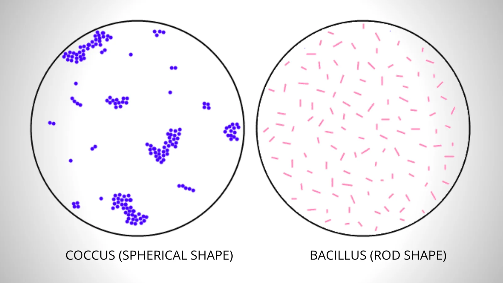
The main purpose of simple staining is to determine the cell shape, size, and arrangement of bacterial cells.
The main purpose of simple staining is to determine the cell shape, size, and arrangement of bacterial cells.
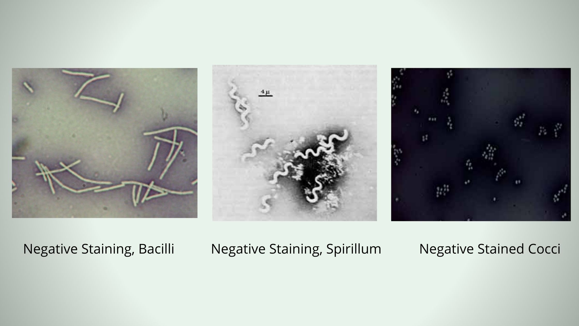
In negative staining method, an acidic dye is used known as India Ink or Nigrosin. When the bacterial cells are exposed to this stain, due to the presence of acidic nature it readily gives up a hydrogen ion (proton) and the chromophore. As a result, the dye becomes negatively charged, now the bacterial cell surface deflects the stain.
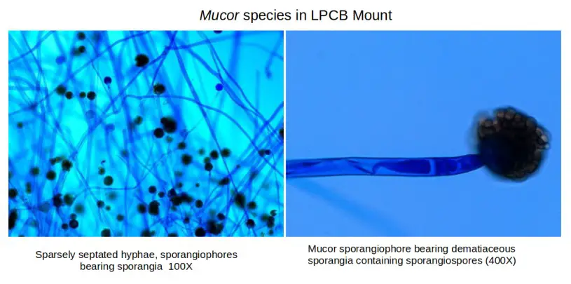
The lactophenol cotton blue (LPCB) wet mount preparation is the most used method for staining and viewing fungus, and its preparation is straightforward. The formulation contains the following ingredients: As a mounting medium and staining agent, lactophenol cotton blue solution is used to prepare slides for microscopic study of fungus. Elements of fungi are dyed … Read more
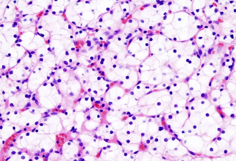
What is Hematoxylin and Eosin (H&E) Staining? Hematoxylin and Eosin (H&E) staining is a commonly used histological staining technique that is used to visualize the structure of cells and tissues in a sample. The staining is performed by first staining the tissue with hematoxylin, a basic dye that stains acidic structures such as the cytoplasm … Read more

The term Giemsa stain originated from a name of German chemist and bacteriologist Gustav Giemsa. He apply this stain with a combination of reagents to detect the presence of malaria parasites. This stain is used for nucleic acid staining and histopathological diagnosis of malaria and other parasites. Giemsa Stain is a types of Romanowsky stains … Read more

Gram-staining used to differentiate between Gram-Positive and Gram-Negative Bacteria.
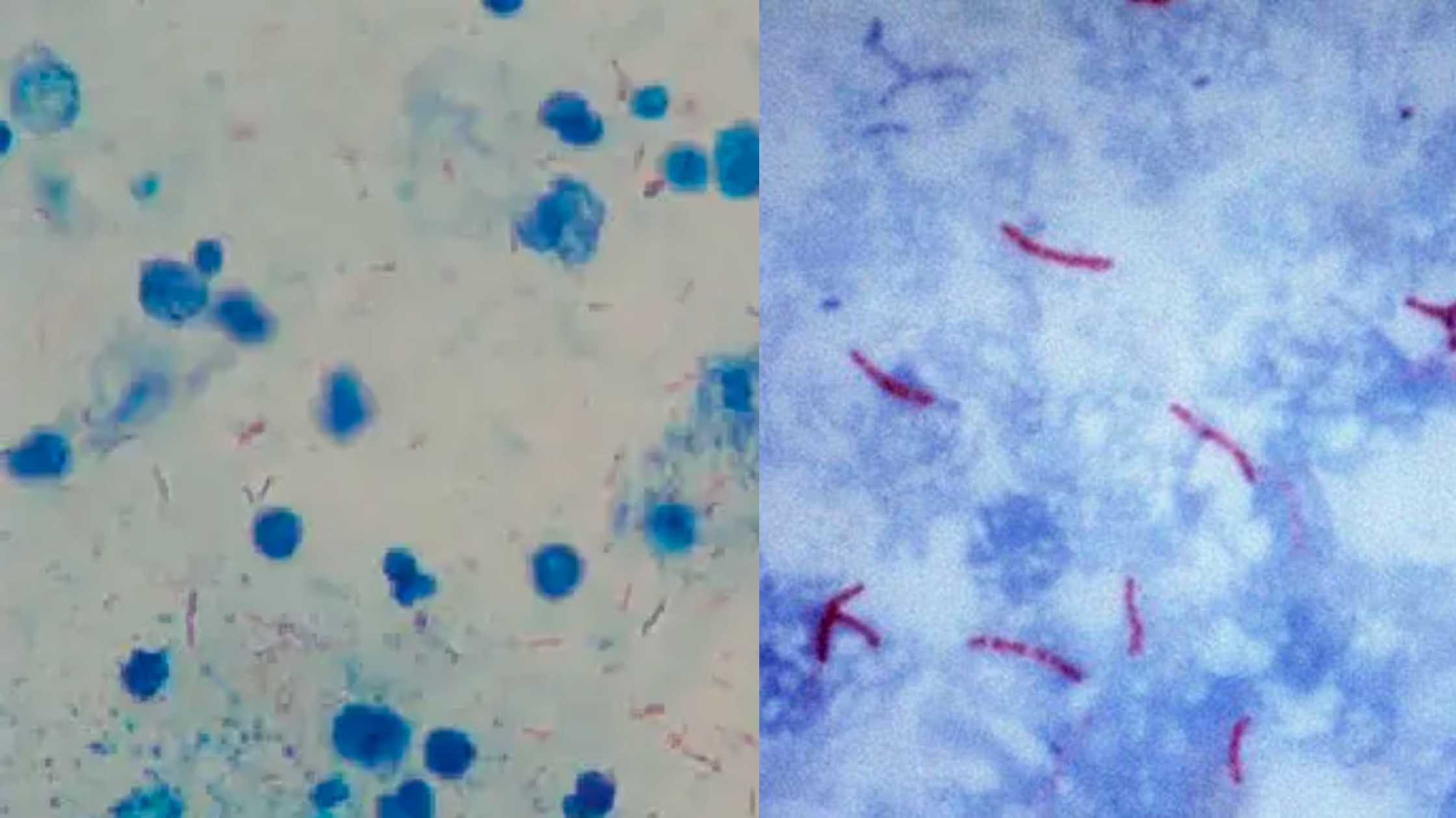
What is Acid Fast Stain? Objective of Acid Fast Stain Principle of Acid Fast Stain The principle of the Acid Fast Stain revolves around the unique characteristics of certain bacterial cells, particularly their resistance to conventional staining methods due to the presence of mycolic acid in their outer membrane. This expository explanation aims to elucidate … Read more
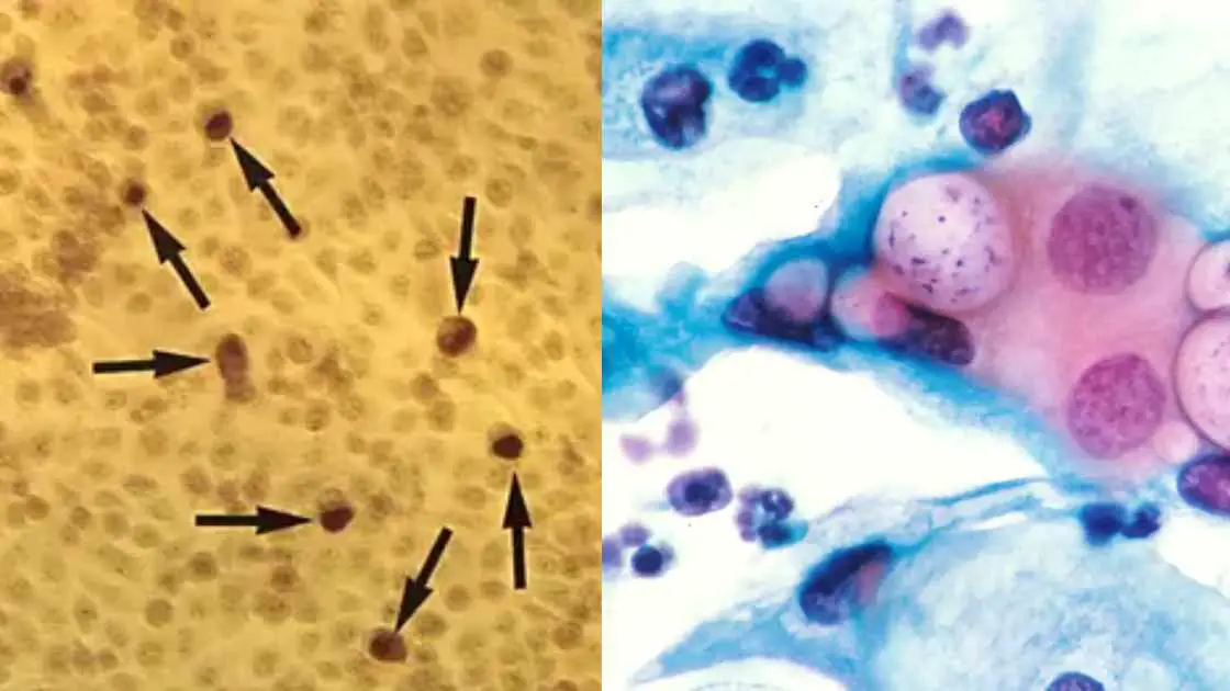
What is Chlamydia? Distribution of Chlamydia species Chlamydia species have a wide distribution across the world, and their prevalence varies in different regions. The family Chlamydiaceae, which includes the genus Chlamydia, is believed to have originated from the Order Chlamydiales approximately seven million years ago. However, evidence of Chlamydia trachomatis infections has been found dating … Read more
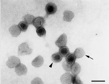
Negative staining is a method used to visualize viruses and other small particles under a microscope. In this method, a drop of the sample is placed on a flat surface and a small amount of a negatively charged stain is added to the sample. The stain surrounds the virus particle, but does not adhere to … Read more
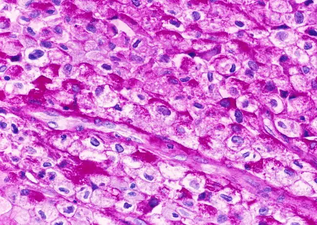
What is Periodic Acid-Schiff (PAS) Staining? Periodic Acid-Schiff (PAS) staining is a laboratory staining technique used to detect the presence of specific sugars and carbohydrates in tissue samples. The PAS stain highlights the presence of sugars, glycoproteins, and mucopolysaccharides in biological tissues and cells. The staining process is often used in the diagnosis of diseases … Read more
We've detected that you are using AdBlock Plus or some other adblocking software which is preventing the page from fully loading.
We don't have any banner, Flash, animation, obnoxious sound, or popup ad. We do not implement these annoying types of ads!
We need money to operate the site, and almost all of it comes from our online advertising.
Please add biologynotesonline.com to your ad blocking whitelist or disable your adblocking software.
