Contents
Wright Stain
- Wright’s stain is a modified Romanowsky stain. In hematology laboratory, it is used for the staining of peripheral blood smear, bone marrow aspirates, and urine samples.
- This stain is consists of eosin (red) and methylene blue dyes.
- Wright Stain was first devised by James Homer Wright in 1902, that’s why its called Wright Stain.
- This stain can easily distinguish between blood cells, that’s why it is used for differential white blood cell counts.
- Some examples of related stains are the buffered Wright stain, the Wright-Giemsa stain (a mixture of Wright and Giemsa stains), and the buffered Wright-Giemsa stain.
Wright Stain Principle
Wright Stain is a type of polychromatic staining solution which is a mixture of methylene blue and eosin. The methylene blue and eosin is a basic and acidic dye, these help to induce multiple colors when applied to cells. As its a methanol-based stain, thus it does not require any special fixation step prior to staining, the methanol works as a fixative and also as a solvent. This fixative agent does not alter the cell and helps them to get attached to the glass slide.
The acidic dye (eosin) of the Wright Stain will help to stain the basic component of white cells (i.e. hemoglobin, eosinophilic granules, and cytoplasm) from orange to pink color, these are known as eosinophilic or acidophilic. While the basic dyes (methylene blue) of the Wright Stain helps to stain the acidic components (e.g. nucleus with nucleic acid) as a result it becomes blue to purple shades, these are known as basophilic. The neutral components of the cell are stained by both the dyes of wright stain and result in variable colors.
Reagents Required
Staining Solution
- Wright’s stain powder – 1.0 gm
- Glycerol – 50.00 ml
- Methanol, absolute – 50.00 ml
Stock stain solution
- Acetone – 4.00 ml
- Phosphate buffer(1/15M,pH 6.5) – 3.00 ml
- Distilled water – 2.00 ml
- Both (a) and (b) are mixed in coplin jar – 31.00 ml
Composition of Phosphate buffer (1/15M, pH 6.5)
- Potassuum dihydrogen phosphate, anhydrous – 0.663 gm
- Disodium phosphate ,anhydrous – 0.256 gm
- Distilled water – 100.00 ml
Type of specimen
Clinical samples: In this experiment Blood sample is used as a specimen.
Wright Stain Procedure
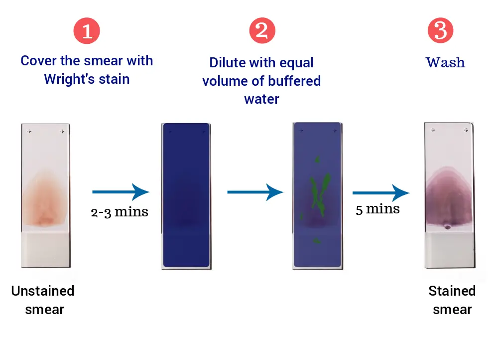
- Make a blood film on a clean microscopic slide and allow it to air dry.
- Place the smear on a staining rack, smear side will facing upwards.
- Flood the smear with the stain for 3 minutes (fixation), covering the slide completely. The stain will fix and partially stains the smear.
- Wait for 2-3 minutes.
- Gently add buffer of the same quantity as the stain, and mix by blowing gently on the surface. Leave for 5 minutes.
- Keep the slide horizontally and flooded it with distilled water until the thinner parts of the film are pinkish red.
- Air-dry the slide at room temperature.
- Now, the slide is ready to observe under the microscope.
Results of Wright Stain
- The basic component of the cell will appear in orange to pink color.
- The acidic components of the cell will appear in blue to purple shades.
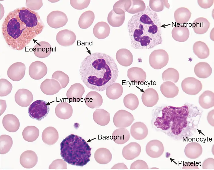
| Cells | Result |
|---|---|
| Erythrocytes | Yellowish-red |
| Neutrophils | Nucleus: Dark purple Granules: Reddish ileac granules Cytoplasm: Pale pink |
| Eosinophils | Nucleus: Blue Granules: Red to orange red Cytoplasm: Blue |
| Basophils | Nucleus: Purple to dark blue Granules: Dark purple |
| Lymphocytes | Nucleus: Dark purple Cytoplasm: Sky blue |
| Monocytes | Nucleus: Dark purple Cytoplasm: Mosaic pink and blue |
| Platelets | violet to purple granules |
Limitations of Wright Stain
- To maintain the morphology of cells, films must be fixed without delay and the films should never be left unfixed for more than a few hours.
- Methanol used as a fixative should be completely water-free. As little as 1% water may affect the appearance of the films and a higher water content causes gross changes.
- The red cells will also be affected by traces of detergent on inadequately washed slides.
- Sometimes when thick films are stained they become overlaid by a residue of stain or spoil by the envelopes of the lysed red cells.
Quality Control of Wright Stain
- Appearance: Ink blue coloured solution
- Clarity: Clear without any particles.
- Microscopic Examination: Blood staining is carried out and staining characteristics are observed under microscope by using oil immersion lens.
Storage and Shelf Life of Wright Stain
- Store between 10 – 300C in a tightly closed container and away from bright light.
- Use before the expiry date on the label.
- On opening, the product should be properly stored in a dry ventilated area protected from extremes of temperature and sources of ignition.
- Seal the container tightly after use.
Application of Wright stain
- Wright’s Stain is used as a staining solution for blood films.
- Used to stain urine samples.
- Used to stain bone marrow aspirates.
- It is used to stain chromosomes for the diagnosis of syndromes and diseases.
- Used for the diagnosis of interstitial nephritis or urinary tract infection.
- Used for differential white blood cell counts.

