- Among the Zygomycetes fungi, the genus Rhizopus contains some of the best-known and most-studied species. Typically, these fungi are saprophytes and can be found living on deceased and decaying organic matter, such as leaves or soil.
- They are widespread and can be found in a variety of niches, ranging from balmy and humid Southern Asia to colder Northern Europe.
- Rhizopus oryzae is the most described member of the Rhizopus genus. Not only has this species garnered considerable research interest, but it is also utilised in large-scale industrial applications.
- One traditional use of these fungi is in tempe, a cuisine made from fermented soybeans by R. oryzae or the related Rhizopus microsporus that has been consumed by millions of people in Southeast Asia since the 15th century.
- In addition, numerous other edibles and beverages are processed with Rhizopus species. Current research on Rhizopus has two primary foci: The production of organic acids, specifically L-lactic acid and fumaric acid, is a major focus.
- Rhizopus also possesses an impressive array of enzymes, which is the subject of the second concentrate. Phytases, amylases, pectinases, cellulases, proteases, and cellulases are prominent examples.
- Rhizopus has also been studied for purposes such as the treatment of industrial effluent derived from organic sources and the production of animal feed.
- The Rhizopus genus is not only recognised for its positive qualities. It is also a documented cause of food spoilage, especially of crops, resulting in enormous economic losses during storage and transport.
- Rhizopus stolonifer is a prominent example of the pathogen that causes Rhizopus-soft rot. This species is capable of spoiling food even after preventative treatment, as its enzymes are exceptionally heat resistant and remain active even after 40 minutes at 100 degrees Celsius.
- Some Rhizopus isolates are also recognised as opportunistic pathogens that can cause zygomycosis in immunocompromised individuals.
Contents
Habitat of Rhizopus spp
Rhizopus spp. is a genus of fungi found in numerous habitats, including soil, decomposing plant matter, and food. Additionally, they are prevalent in indoor environments, such as homes and businesses. Rhizopus spp. can thrive in a variety of environments, including warm and humid conditions as well as cooler and drier conditions. Additionally, they can tolerate a wide spectrum of pH levels.
These are some of the most common habitats for Rhizopus spp.
- Soil: Rhizopus spp. are prevalent soil fungi. They aid in decomposing organic matter and returning nutrients to the soil.
- Decaying plant matter: Rhizopus spp. are also commonly found on decaying plant matter, including leaves, fruits, and vegetables. They aid in decomposition and return this matter to the soil.
- Food: Rhizopus spp. can thrive on bread, fruits, vegetables, and dairy products, among others. This fungus can cause food spoilage, so it is essential to store food adequately to prevent its growth.
- Indoor environments: Rhizopus spp. can also thrive indoors. Typically, they inhabit moist environments, such as restrooms and kitchens. In addition to carpets and furniture, they are also found in other areas of the residence.
Rhizopus spp. can be annoying, but they can also be dangerous. Some Rhizopus spp. species can cause infections in both humans and animals. These infections can be severe, and in some instances, fatal. It is essential to be aware of the potential dangers associated with Rhizopus spp. and to take measures to prevent the fungus’s growth.
Features of Rhizopus
- Mycelia is their branching body structure.
- The preponderance of rhizopus are decomposers, or saprobic.
- They primarily consume decomposing organisms or organic matter.
- They reproduce by producing spores.
- They are utilised in the industrial production of biotin, intoxicating beverages, etc.
- They are a dark greyish brown hue.
- The height of rhizoids is 10 mm.
- Sporangia are 100 micrometre in diameter.
Morphology of Rhizopus spp
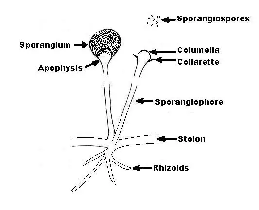
The morphology of Rhizopus spp. is characterized by the following features:
- Mycelium: Rhizopus spp. mycelium is a network of branching, thread-like hyphae. The hyphae are non-septate, which indicates that they lack cross-walls. This enables the hyphae to rapidly grow and spread.
- Stolons: Long, horizontal hyphae that grow along the surface of a substrate are stolons. They aid in affixing the fungus to the substrate and facilitate the transport of nutrients.
- Rhizoids: Rhizoids are slender, root-like hyphae that develop within the substrate. They assist the fungus in adhering to the substrate and absorbing nutrients.
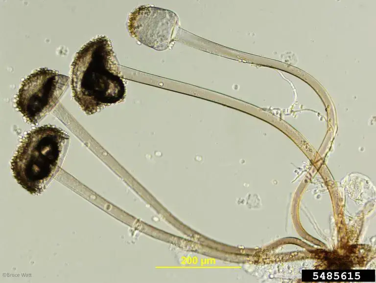
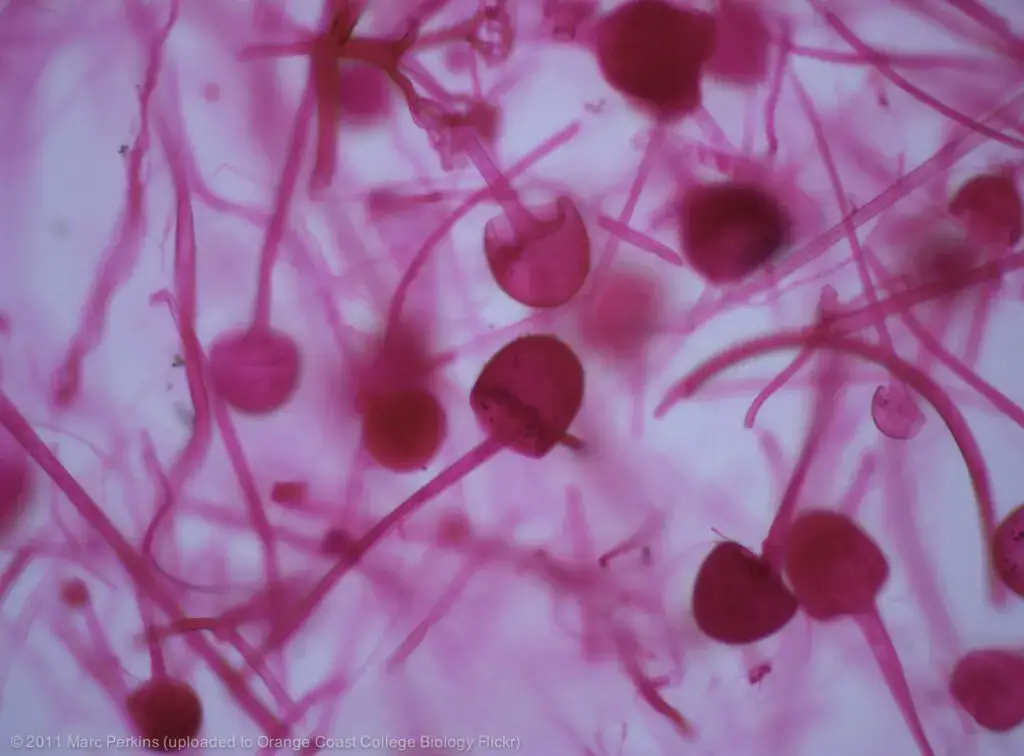
- Sporangiophores: Sporangiophores are hyphae that are erect and bear sporangia. Sporangia are spherical, spore-containing structures. The spores are dispersed by air currents after being discharged from the sporangia.
- Spores: The spores of Rhizopus spp. are produced by mitosis and are asexual. They are haploid, which means they have half as many chromosomes as the progenitor cell. The particles develop into new hyphae.
- Aseptate hyphae: Rhizopus spp. hyphae are not partitioned into cells by septa. This facilitates the quick transport of nutrients and other substances throughout the mycelium.
- Sporangiospores: Sporangiospores are generated by Rhizopus spp. as asexual spores. They are carried by air currents and are capable of germinating into new hyphae.
- Zygospores: Rhizopus spp. produce zygospores, which are sexual spores. When two hyphae of opposite mating types come into contact, they form. Zygospores have robust walls and can withstand extreme conditions. When environmental conditions improve, zygospores germinate to form new mycelium.
Cultural Characteristics of Rhizopus spp
Rhizopus spp. are filamentous fungi that can be grown in culture to observe their cultural characteristics. These characteristics can provide important information for the identification and classification of the fungus. Some of the cultural characteristics of Rhizopus spp. include:
- Growth rate: Rhizopus spp. grow rapidly on suitable media, with visible growth often observed within 24 to 48 hours.
- Colony morphology: The colonies of Rhizopus spp. are fast-growing, cottony, and often fluffy in texture. They are typically white or gray at first, but can darken to shades of gray or brown with age. The colony surface is often covered with sporangia and sporangiophores.
- Sporulation: Rhizopus spp. produce sporangia and sporangiophores on the surface of the colony. The sporangia are usually black and spherical, while the sporangiophores are long and branched.
- Substrate utilization: Rhizopus spp. are able to grow on a variety of organic substrates, including fruits, vegetables, grains, and decaying plant matter.
- Temperature tolerance: Rhizopus spp. can grow over a wide range of temperatures, from 15°C to 40°C, with the optimal growth temperature around 30°C.
- Humidity: Rhizopus spp. require high humidity to grow.
- pH: Rhizopus spp. grow best at a pH of 5.5 to 6.5.
- Oxygen requirement: Rhizopus spp. are facultative anaerobes, meaning they can grow in the presence or absence of oxygen.
- Odor: Rhizopus spp. colonies emit a distinctive odour that is typically described as fragrant or musty.
Culture media used for the growth of Rhizopus spp
Rhizopus spp. can be cultured on a variety of different media, depending on the specific research or diagnostic application. Some commonly used media for the growth of Rhizopus spp. include:
- Potato Dextrose Agar (PDA): PDA is a general-purpose medium that supports the growth of a wide variety of fungi, including Rhizopus spp. It contains potato infusion, dextrose, and agar, which provide a nutrient-rich environment for fungal growth.
- Sabouraud Dextrose Agar (SDA): SDA is another general-purpose medium commonly used for the growth of fungi, including Rhizopus spp. It contains peptone, dextrose, and agar, and has a low pH that inhibits the growth of bacteria.
- Malt Extract Agar (MEA): MEA is a medium that is particularly useful for the cultivation of Zygomycetes such as Rhizopus spp. It contains malt extract, peptone, and agar, and has a slightly acidic pH.
- Czapek-Dox Agar (CDA): CDA is a medium that is often used for the isolation and enumeration of fungi in soil and plant material. It contains sodium nitrate, potassium chloride, magnesium sulfate, ferrous sulfate, dextrose, and agar.
- Rose Bengal Agar: This is a selective medium used for the isolation of Rhizopus spp. It contains rose bengal, peptone, dextrose, and agar.
Here are some growing suggestions for Rhizopus spp. on culture media:
- Before using the media, sterilise it. This can be accomplished via autoclaving or chemical disinfectant.
- Inoculate the medium with a small amount of inoculum from Rhizopus spp. This is accomplished by removing mycelium from a colony onto the medium.
- The media should be incubated at the appropriate temperature. Rhizopus spp. flourish best at temperatures between 25 and 30 degrees Celsius.
- Observe the fungus’s growth over time. Within a few days, Rhizopus spp. will grow swiftly and form colonies.
Pathogenesis of Rhizopus spp
Rhizopus spp. are opportunistic pathogens that can cause a wide range of infections in humans, particularly in individuals with compromised immune systems, uncontrolled diabetes, or those who have suffered from trauma or burns. The pathogenesis of Rhizopus spp. infections is complex and involves multiple virulence factors, including:
- Adhesion: Rhizopus spp. are able to adhere to host tissues using surface proteins and other adhesins. Once attached, the fungus can begin to invade the host tissues. Rhizopus spp. produce a variety of adhesion-promoting molecules. These molecules include adhesins, which are proteins that bind to specific receptors on host cells, and extracellular matrix (ECM)-degrading enzymes, which enable the fungus to invade host tissues by degrading the ECM.
- Invasion of host tissues/Enzymatic activity: Rhizopus spp. produce a range of hydrolytic enzymes, including proteases, lipases, and chitinases, that can break down host tissues and extracellular matrix components.
- Iron acquisition: Rhizopus spp. are able to secrete siderophores, which are small molecules that can scavenge iron from host tissues and other sources.
- Mycotoxin production: Some strains of Rhizopus spp. can produce mycotoxins such as rhizoxin and fumonisin, which can have toxic effects on host tissues.
- Immune evasion: Rhizopus spp. are able to evade the host immune system by producing a thick, branched hyphal cell wall that is difficult for immune cells to penetrate.
Infections caused by Rhizopus spp. typically begin with the inhalation of fungal spores, which can lead to pulmonary infection. In immunocompromised patients, the fungus can then spread to other tissues and organs, causing invasive disease. In addition to pulmonary infection, Rhizopus spp. can cause a range of other clinical syndromes, including rhinocerebral, cutaneous, and gastrointestinal infections.
Overall, the pathogenesis of Rhizopus spp. infections is multifactorial and involves a range of virulence factors that allow the fungus to invade and damage host tissues, evade the host immune system, and cause disease in susceptible individuals.
The following infections can be caused by Rhizopus spp. in humans and animals:
- Rhinocerebral mucormycosis: This is an infection of the airway, sinuses, and brain known as rhinocerebral mucormycosis. It is the most prevalent form of human mucormycosis.
- Pulmonary mucormycosis: This is a lung infection called pulmonary mucormycosis. It is the second most prevalent form of human mucormycosis.
- Cutaneous mucormycosis: This is a cutaneous infection. It is the third most prevalent form of human mucormycosis.
- Disseminated mucormycosis: This infection has spread to other regions of the body, including the kidneys, liver, and bones. This is the worst form of mucormycosis.
Clinical manifestation of mucormycosis/zygomycosis
- Rhinocerebral mucormycosis: The most prevalent form of mucormycosis. It is an infection of the sinuses, brain, and airways. Among the symptoms of rhinocerebral mucormycosis are:
- Nasal congestion
- Sinus pain
- Headache
- Facial swelling
- Black eschars on the skin
- Eye pain
- Vision loss
- Pulmonary mucormycosis: The second most prevalent form of mucormycosis. This condition is an infection of the airways. The following are symptoms of pulmonary mucormycosis:
- Fever
- Cough
- Dyspnea
- Hemoptysis
- Cutaneous mucormycosis: The third most prevalent form of mucormycosis. The condition is an infection of the epidermis. The following are symptoms of cutaneous mucormycosis:
- Black eschars on the skin
- Pain
- Swelling
- Redness
- Disseminated mucormycosis: This is the worst type of mucormycosis. It is an infection that has migrated to other organs, including the kidneys, liver, and bones. These are the symptoms of disseminated mucormycosis:
- Fever
- Weight loss
- Fatigue
- Multiple organ failure
Laboratory Diagnosis of Mucormycosis (Rhizopus spp)
The laboratory diagnosis of mucormycosis involves a combination of clinical evaluation, imaging studies, and laboratory testing to identify the causative fungus. Here are some of the laboratory tests that may be used to diagnose mucormycosis caused by Rhizopus spp:
- Microscopic examination: A direct microscopic examination of clinical specimens such as tissue biopsy samples, bronchoalveolar lavage, or cerebrospinal fluid can be used to identify the characteristic hyphae of Rhizopus spp. These hyphae are broad, non-septate, and have right-angle branching.
- Culture: Fungal cultures can be used to isolate and identify the causative organism. Rhizopus spp. grow rapidly on Sabouraud agar, and typically produce white, cottony colonies that can become black and necrotic over time.
- Imaging studies: Imaging studies, such as computed tomography (CT) scans and magnetic resonance imaging (MRI) scans, can assist in determining the infection’s severity. CT scans are beneficial for visualising the sinuses and lungs in particular. MRI scans detect microscopic lesions more sensitively than CT scans.
- Histopathology: Tissue biopsy samples can be examined under a microscope after staining with hematoxylin and eosin or periodic acid-Schiff stain to identify the characteristic hyphae of Rhizopus spp. Histopathological examination can also help identify tissue necrosis and invasion.
- Molecular testing: Molecular techniques, such as polymerase chain reaction (PCR), can be utilised to identify the infectious agent. PCR is a technique for amplifying the DNA of a particular organism. The DNA of Rhizopus spp. can be identified using PCR from tissue samples, respiratory secretions, and blood.
- Laboratory tests/Serology: Antibodies to Rhizopus spp. are detectable in the blood of some mucormycosis patients. Serology is not a reliable diagnostic test, however. Laboratory tests, such as blood cultures and fungal cultures, can assist in identifying the infectious agent. In mucormycosis (Rhizopus spp), blood cultures are frequently negative, so fungal cultures are more important. Typically, fungi are grown on Sabouraud dextrose agar (SDA) and incubated at 37 degrees Celsius for five to seven days. Colonies of Rhizopus spp. range in colour from white to grayish-brown and have a cottony or frothy appearance.
Rhizopus life cycle/life cycle of rhizopus
Rhizopus reproduces via all three mechanisms, i.e., vegetative, asexual, and sexual.
1. Vegetative Reproduction
Under this mode of reproduction, tiny fragments are formed on the surface of the rhizopus body, and the stolon may break into two or more than two small units, each of which is capable of growing as a mother mycelium, if the stolon breaks accidentally.
2. Asexual Mode of Reproduction
It occurs either through the formation of sporangiospores or chlamydospores.
A. By Sporangiospore Formation
Sporangiospores are produced at the apex of sporangiophores, which are sporangia of aerial mycelium. They originate from favorable conditions.
- Sporangiophores originate from the upper surface of the rhizoid node.
- As nuclei and cytoplasm move apically, the apical portion swells, producing sporangium.
- The cytoplasm of sporangium differentiates into a peripheral region with a greater number of nuclei and a central columella region with fewer nuclei and more vacuoles.
- Sporangiospores develop inside sporangium. They have multiple nuclei and are immobile.
- After sporangium maturation, its wall ruptures and sporangiospores emerge as a powdery mass.
- After acquiring the proper environment and substrate, each spore germinates into a new mycelium.
B. Formation of Chlamydospores
This type of axeual reproduction involves the development of zoospores, aplanospores, hypnospores, or a palmella stage. During unfavorable conditions, such as a lack of sustenance and water, the protoplasm develops thick, nutrient-rich walls. Then, the chlamydospore separates from the vegetative hyphae and enters the quiescent phase; when the chlamydomonas receives sufficient moisture, the chlamydospores form a germ tube, which leads to the formation of a new thallus.
3. Sexual reproduction
Sexual reproduction is accomplished through the fusion of two compatible hyphae. The majority of Rhizopus species (R. stolonifer) are heterothallic, meaning that their + and – mating strains have distinct mycelium. This species is homothallic.
- The compatible hyphae assemble. In each mycelia, a microscopic protrusion develops. It is called progametangia.
- Nuclei and cytoplasm migrate to the apical region and make contact with progametangia.
- By forming septa, the apical region is separated from the remainder of the hyphae. This is referred to as gametangia
- Gametangia conjugate to produce a multinucleated structure
- Plasmogamy is followed by karyogamy, which produces a diploid (2n) zygote. It is called zygospore. The remaining unpaired nuclei degenerate.
- Zygospores grow in size, thicken their walls, and become resistant to adverse environmental conditions. When favorable conditions are present, zygospores germinate.
- The inner wall of the zygospore transforms into germ sporangiophore and germ sporangium forms apically.
- Meiosis takes place, and haploid meiospores are created. After the rupture of the germ sporangium wall, they emerge and develop into new mycelia.
Rhizopus treatment/Treatment of Mucormycosis
The treatment of mucormycosis caused by Rhizopus spp. involves a combination of antifungal therapy, surgical debridement, and management of underlying risk factors. Here are some of the treatments that may be used:
- Antifungal therapy: Treatment with antifungal medications is critical for the management of mucormycosis. Liposomal amphotericin B is the preferred antifungal agent, and high doses (5-10 mg/kg/day) are usually required for several weeks. Posaconazole and isavuconazole are alternative antifungal agents that may be used in patients who are unable to tolerate amphotericin B or who have a contraindication to its use. Antifungal medications used to treat mucormycosis include:
- Amphotericin B
- Posaconazole
- Isavuconazole
- Voriconazole
- Surgical debridement: Surgical removal of necrotic tissue is a key component of the management of mucormycosis. This can help to control the spread of the infection and improve outcomes. Debridement should be performed as soon as possible, and may require multiple procedures.
- Control of underlying risk factors: Control of underlying risk factors such as uncontrolled diabetes, neutropenia, and immunosuppression is critical for the prevention and management of mucormycosis.
- Supportive care: Supportive care measures such as hydration, electrolyte balance, and management of organ dysfunction are important for improving outcomes in patients with mucormycosis.
Industrial uses of Rhizopus/Economic Importance of Rhizopus
- Production of enzymes: Rhizopus oryzae produces a wide array of enzymes that are utilised in numerous industries. Amylases are used to convert carbohydrates into sugars, while proteases and lipases are used to degrade proteins and lipids, respectively. These enzymes are utilised in the food and beverage industry as well as the pharmaceutical industry.
- Fermented foods and beverages: Rhizopus oryzae is utilised to produce numerous fermented foods and beverages, including tempeh, koji, and sake. Tempeh is a traditional Indonesian dish prepared from fermented soy beans and Rhizopus oryzae. Koji is a Japanese cuisine consisting of rice fermented with Rhizopus oryzae. Sake is a Japanese fermented rice-based alcoholic beverage.
- Biofuels: Rhizopus oryzae can be utilised to produce biofuels including ethanol and biodiesel. Ethanol is an intoxicant that can be utilised as a fuel in automobiles and other vehicles. Biodiesel is a fuel that can be produced from either vegetable oils or animal lipids.
- Bioremediation: Rhizopus oryzae can be utilised for bioremediation of contaminated soil and water. Bioremediation is the procedure of removing pollution using living organisms. Rhizopus oryzae can be utilised to degrade pollutants in soil and water, thereby protecting the environment.
- Biomaterials: Rhizopus oryzae can be utilised to create biomaterials such as plastics and composites. Biomaterials are substances derived from biological organisms. Rhizopus oryzae can be used to produce robust and durable plastics and composites.
- Production of organic acids: Rhizopus spp. are used for the commercial production of organic acids such as lactic acid, fumaric acid, and gluconic acid. These organic acids are used in the food, pharmaceutical, and chemical industries.
Important Species of Rhizopus
There are many important species of Rhizopus, but some of the most notable include:
- Rhizopus oryzae: This species is commonly used for the production of tempeh, a traditional soybean-based food product. It is also an opportunistic pathogen that can cause mucormycosis in immunocompromised individuals.
- Rhizopus stolonifer: This species is a common postharvest pathogen of fruits and vegetables, and is responsible for the soft rot of these crops. It is also a common cause of mucormycosis in humans, particularly in individuals with uncontrolled diabetes.
- Rhizopus microsporus: This species is an important pathogen of animals, particularly in pigs, and can cause fatal infections such as rhinocerebral mucormycosis. It is also used for the commercial production of fumaric acid.
- Rhizopus oligosporus: This species is commonly used for the production of tempeh in Indonesia, and is known for its ability to produce a high-quality product with a desirable texture and flavor.
- Rhizopus arrhizus: This species is an opportunistic pathogen that can cause mucormycosis in immunocompromised individuals. It is also used for the commercial production of organic acids such as lactic acid and fumaric acid.
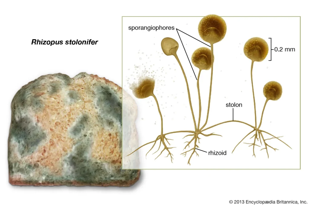
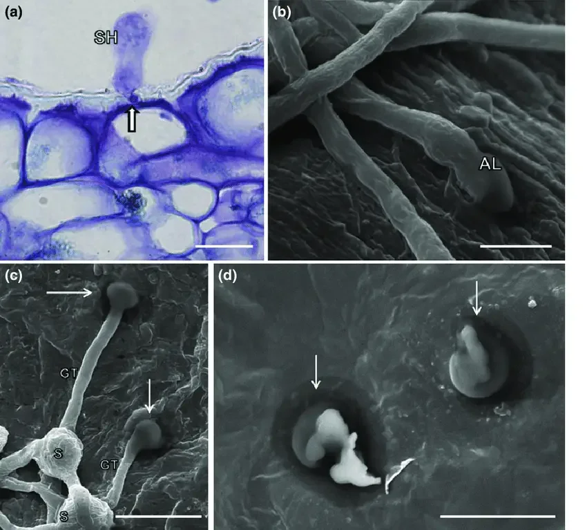
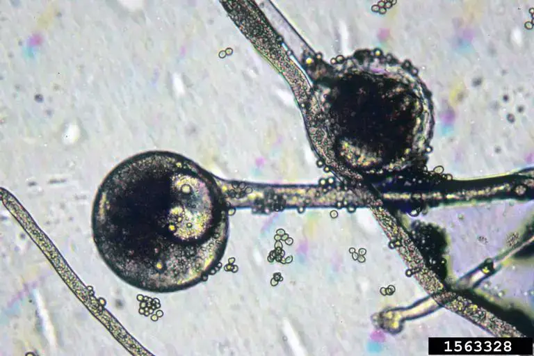
Rhizopus under microscope
FAQ
What is Rhizopus?
Rhizopus is a genus of filamentous fungi that belongs to the class Zygomycetes. These fungi are commonly found in soil, decaying organic matter, and on plants. Some species of Rhizopus have important industrial and agricultural applications, while others are opportunistic pathogens that can cause serious infections in humans and animals.
What are the industrial uses of Rhizopus?
Rhizopus spp. have several industrial uses due to their ability to produce various enzymes and organic acids. These fungi are used for the commercial production of organic acids such as lactic acid, fumaric acid, and gluconic acid. They are also used for the production of enzymes such as lipases, proteases, and amylases, which have various applications in the food, feed, detergent, and textile industries. In addition, Rhizopus spp. are used for the traditional fermentation of soybeans to produce tempeh, a protein-rich food product that is widely consumed in Southeast Asia.
What are the medical uses of Rhizopus?
Rhizopus can cause serious infections in immunocompromised individuals, particularly those with uncontrolled diabetes. In some cases, these infections can be fatal. Treatment of Rhizopus infections usually involves antifungal medication and surgical intervention, if necessary.
What are the environmental uses of Rhizopus?
Rhizopus spp. are used for bioremediation of contaminated soils and water bodies due to their ability to degrade various organic pollutants. Some strains of Rhizopus spp. also have biocontrol potential against plant pathogenic fungi, and are used as biopesticides in the agriculture industry.
How does Rhizopus reproduce?
Rhizopus reproduces both sexually and asexually. Sexual reproduction occurs through the formation of zygospores, which are formed by the fusion of haploid hyphae from different mating types. Asexual reproduction occurs through the production of sporangia, which contain spores that can germinate and form new colonies.
What are the symptoms of Rhizopus infection?
Rhizopus infections can cause a range of symptoms depending on the site of infection. Common symptoms include fever, headache, facial pain, nasal congestion, and skin lesions. In severe cases, Rhizopus infection can cause tissue necrosis and invasive disease.
How is Rhizopus infection treated?
Treatment of Rhizopus infection usually involves antifungal medication, such as amphotericin B, posaconazole, or isavuconazole. In severe cases, surgical intervention may be necessary to remove infected tissue.
What are the different species of Rhizopus?
There are several species of Rhizopus, some of which are pathogenic to humans and animals, while others have important industrial and agricultural applications. Some of the common species include Rhizopus oryzae, Rhizopus stolonifer, Rhizopus microsporus, Rhizopus oligosporus, and Rhizopus arrhizus.
How can I prevent Rhizopus infection?
Preventing Rhizopus infection involves maintaining good hygiene practices and avoiding exposure to contaminated soil and decaying organic matter. Individuals with uncontrolled diabetes should take steps to manage their blood sugar levels to reduce the risk of infection.
What are the future research directions for Rhizopus?
Future research directions for Rhizopus include developing novel strains with improved properties for industrial and agricultural applications, understanding the genetic basis of pathogenesis in Rhizopus, and developing new treatments for Rhizopus infections.
References
- Lennartsson, P. R., Taherzadeh, M. J., & Edebo, L. (2014). Rhizopus. Encyclopedia of Food Microbiology, 284–290. doi:10.1016/b978-0-12-384730-0.00391-8
- Morales-Franco B, Nava-Villalba M, Medina-Guerrero EO, Sánchez-Nuño YA, Davila-Villa P, Anaya-Ambriz EJ, Charles-Niño CL. Host-Pathogen Molecular Factors Contribute to the Pathogenesis of Rhizopus spp. in Diabetes Mellitus. Curr Trop Med Rep. 2021;8(1):6-17. doi: 10.1007/s40475-020-00222-1. Epub 2021 Jan 22. PMID: 33500877; PMCID: PMC7819772.
- Ibrahim AS, Spellberg B, Walsh TJ, Kontoyiannis DP. Pathogenesis of mucormycosis. Clinical infectious diseases. 2012 Apr 1;54(suppl_1):S16-22. doi: 10.1093/cid/cir866.
- Ribes JA, Vanover-Sams CL, Baker DJ. Zygomycetes in human disease. Clinical microbiology reviews. 2000 Oct;13(4):236-301. doi: 10.1128/CMR.13.4.236-301.2000.
- Lee MH, Jang YS, Kim KJ, Kim SW. Rhizopus oligosporus-mediated solid-state fermentation of soybean for production of soy sauce. Applied and Environmental Microbiology. 2003 Nov 1;69(11):5864-7. doi: 10.1128/AEM.69.11.5864-5867.2003.
- Srimanote P, Chitlaru T, Lipovčić N, Angsuthanasombat C, Vasil ML, Tshuva EY, Bayer EA. A novel Rhizopus oryzae alpha-glucosidase has enhanced stability and specificity for maltose. Biochimica et Biophysica Acta (BBA)-Proteins and Proteomics. 2008 Sep 1;1784(9):1285-94. doi: 10.1016/j.bbapap.2008.05.008.
- Pal AK, Singh P, Jain S, Nigam VK. Lactic acid production from wheat bran by Rhizopus oryzae under solid-state fermentation. Journal of industrial microbiology & biotechnology. 2006 Mar 1;33(3):233-7. doi: 10.1007/s10295-005-0056-2.
- Chaturvedi V, Chaturvedi S. Zygomycosis in India: the rapid emergence of Apophysomyces species in humans. The Open Mycology Journal. 2008;2:29-37. doi: 10.2174/1874437000802010029.
- Kwon-Chung KJ, Bennett JE. Medical mycology. Lea & Febiger; 1992.
- Radhakrishnan VV, Kumutha K. Rhizopus–A review. Journal of Pharmaceutical Sciences and Research. 2012;4(12):2291-5. https://jpsr.pharmainfo.in/Documents/Volumes/vol4Issue12/jpsr04121217.pdf.
- Singh P, Dutt D, Tyagi VV. Enzymatic saccharification of sweet sorghum bagasse and ethanol production by simultaneous saccharification and fermentation using a thermotolerant Rhizopus fungal strain. Energy. 2014 Jan 1;66:182-90. doi: 10.1016/j.energy.2013.11.003.
- Choudhary J, Singh R, Yadav R, Kumar R, Kumar R, Kumar P, et al. Plant growth-promoting potential of endophytic fungi isolated from Solanum surattense Burm. F. Journal of Applied Microbiology. 2016 Sep;121(3):766-77. doi: 10.1111/jam.13180.
- http://en.fungaleducation.org/rhizopus-spp/
Related Posts
- Basidiomycetes – Life cycle, Characteristics, Significance, Mycelium and Examples
- Deuteromycetes – Reproduction, Characteristics, Classification and Examples
- Yeast – Structure, Reproduction, Life Cycle and Uses
- Candida albicans – Morphology, Life Cycle, Pathogenesis, Treatment
- Hyphae – Definition, Types, Structure, Production, Functions, Examples




Excellent Article, Excellent Blog , Excellent Site ✅✅✅