

Loa loa is a filarial nematode that causes loiasis in humans. It is also known as the African eye worm.
- It is mainly found in West and Central Africa. The infection is common in rainforest and swamp forest regions.
- The parasite is transmitted by the bite of day-biting flies of genus Chrysops.
- The major vectors are Chrysops silacea and Chrysops dimidiata.
- During blood meal, the infected fly releases third-stage larvae on the skin.
- The larvae enter through the bite wound. Then it moves into the subcutaneous tissue.
- In the subcutaneous tissue, the larvae develop into adult worms after several months.
- The adult worms move continuously under the skin and connective tissues.
- The female worm is larger than male worm. Female measures about 40-70 mm, while male measures about 20-34 mm.
- The outer cuticle of adult worm contains irregular elevations. These elevations are called bosses.
- Adult worms can remain in the human body for about 15-20 years.
- The female worms release sheathed larvae called microfilariae into the blood.
- The microfilariae show diurnal periodicity. It is mostly found in peripheral blood during daytime.
- The highest number of microfilariae is found between 10:00 AM and 2:00 PM.
- During night, the microfilariae move into the blood vessels of lungs.
- Loa loa does not contain the endosymbiotic bacteria Wolbachia.
- The main clinical features are Calabar swelling and movement of worm across the eye.
- Calabar swelling is a temporary subcutaneous swelling. It is generally seen near the joints of arms and legs.
- The movement of adult worm under the conjunctiva gives the name African eye worm.
- Some infected persons do not show any symptoms. This is mostly seen in persons living for long time in endemic areas.
- Travellers and non-immune persons may show severe itching, high eosinophil count and increased IgE level.
- In heavy infection, kidney disease, heart involvement and encephalopathy may develop.
- Diagnosis is carried out by detection of microfilariae in thick blood smear.
- The blood sample is generally collected between 10:00 AM and 2:00 PM.
- Direct observation of adult worm in the eye can also be used for diagnosis.
- qPCR and antibody detection tests are also used.
- Diethylcarbamazine (DEC) is the main drug used for treatment of loiasis.
- In patients with high microfilarial count, DEC can cause severe encephalopathy.
- In such cases, the number of microfilariae is first reduced by albendazole or apheresis. After this, DEC treatment is started.
What is Loa loa?
Loa loa is a tissue-dwelling filarial nematode that causes loiasis in humans. It is transmitted by arthropod vector and is commonly known as the African eye worm.
Humans are the main natural host of this parasite. The adult worms live and move through the subcutaneous tissues and connective fascia under the skin.
The adult worm may also migrate across the subconjunctiva of eye. This movement is temporary and generally remains for few hours to few days. Loiasis is different from river blindness which is caused by Onchocerca volvulus.
Classification of Loa loa
The biological classification of Loa loa is as follows-
- Kingdom: Animalia
- Phylum: Nematoda (Nemathelminthes)
- Class: Chromadorea (Secernentea)
- Order: Rhabditida (Spirurida)
- Superfamily: Filarioidea
- Family: Onchocercidae
- Genus: Loa
- Species: Loa loa
Morphology of Loa loa
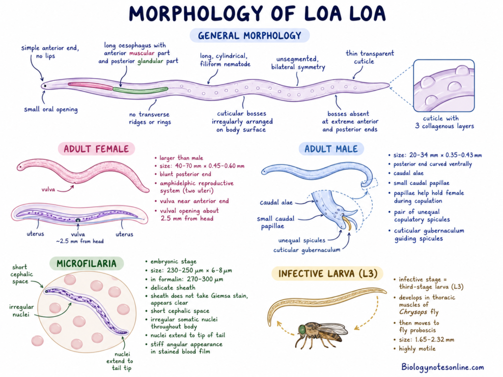
General Morphology
- Loa loa is a long, cylindrical and filiform nematode.
- The body is unsegmented and shows bilateral symmetry.
- The outer body is covered by a thin, flexible and transparent cuticle.
- The cuticle is made up of three collagenous layers.
- The important feature of adult Loa loa is the presence of cuticular bosses.
- These are irregularly arranged rounded elevations present on the body surface.
- Cuticular bosses are absent at the extreme anterior and posterior ends.
- Under Scanning Electron Microscope (SEM), the bosses are seen as rounded tubercles, dome-shaped bosses and ellipsoid elevations.
- Transverse cuticular ridges or rings are absent.
- The anterior end is simple and lips are absent.
- A small oral opening is present at the anterior end.
- The oesophagus is long. It is divided into an anterior muscular part and posterior glandular part.
Adult Female
- The adult female is larger than the male worm.
- It measures about 40-70 mm in length and 0.45-0.60 mm in width.
- The posterior end is blunt.
- The reproductive system is amphidelphic with two uteri.
- The vulval opening is located near the anterior end.
- It is present about 2.5 mm from the head.
Adult Male
- The adult male measures about 20-34 mm in length and 0.35-0.43 mm in width.
- The posterior end is curved towards the ventral side.
- Caudal alae and small caudal papillae are present.
- The caudal papillae help in holding the female during copulation.
- A pair of unequal copulatory spicules are present.
- A cuticular gubernaculum is also present. It guides the spicules during copulation.
Microfilariae
- The microfilariae are the embryonic stage of Loa loa.
- It measures about 230-250 µm in length and 6-8 µm in width in stained blood smear.
- In formalin-fixed condition, it may measure about 270-300 µm.
- The microfilaria is covered by a delicate sheath.
- The sheath does not take Giemsa stain. It appears as a clear area around the body.
- A short cephalic space is present.
- The somatic nuclei are arranged irregularly throughout the body.
- The nuclei extend continuously up to the tip of tail.
- In stained blood film, the microfilariae generally show a stiff and angular appearance.
Infective Larva
- The third-stage larva (L3) is the infective stage of Loa loa.
- It develops in the thoracic muscles of Chrysops fly.
- After development, the larva moves towards the proboscis of the fly.
- It measures about 1.65-2.32 mm in length.
- The larva is highly motile.
Key Identification Features of Loa loa
| Category | Identification feature | Description |
|---|---|---|
| Adult worm | Cuticular bosses | Irregularly arranged rounded elevations are present on the cuticle. These are absent at the extreme head and tail. It is the important identifying feature of adult Loa loa. |
| Adult worm | Transverse ridges | Transverse cuticular ridges or rings are absent. |
| Adult worm | Head structure | The anterior end is simple and smooth. Lips are absent. |
| Adult female | Size | The adult female measures about 40-70 mm in length and 0.45-0.60 mm in width. The posterior end is blunt. |
| Adult male | Size | The adult male measures about 20-34 mm in length and 0.35-0.43 mm in width. It is smaller than the female worm. |
| Adult male | Tail structure | The posterior end is coiled towards the ventral side. Caudal alae, sessile caudal papillae and two unequal copulatory spicules are present. A gubernaculum guides the spicules. |
| Microfilariae | Dimensions | It measures about 230-250 µm in length and 6-8 µm in width in stained blood film. In 2% formalin, it measures about 270-300 µm. |
| Microfilariae | Sheath | A delicate sheath is present around the microfilaria. It does not take Giemsa stain and appears as a clear halo. |
| Microfilariae | Somatic nuclei | The nuclei are crowded and irregularly arranged. It extends continuously up to the tip of tail. |
| Microfilariae | Posture | In stained blood film, the microfilariae show a stiff and angular appearance. |
| Microfilariae | Diurnal periodicity | The microfilariae are present in peripheral blood during daytime. The highest number is found between 10:00 AM and 2:00 PM. During night, it remains in pulmonary capillaries. |
| Biological feature | Wolbachia | Loa loa does not contain the endosymbiotic bacteria Wolbachia. |
| Clinical feature | Eye worm migration | The live adult worm may be seen moving below the conjunctiva of eye. Direct observation or removal of worm is a characteristic feature of loiasis. |
| Clinical feature | Calabar swelling | Temporary, non-pitting subcutaneous swellings are formed. These are caused by hypersensitivity reaction against the migrating adult worm. |
| Laboratory feature | Specific antibody | Parasite-specific IgG4 antibody against recombinant Ll-SXP-1 can be detected. |
| Laboratory feature | Molecular detection | Circulating cell-free parasite DNA can be detected by LL2643 repeat qPCR. |
Habitat, Hosts and Vector of Loa loa
Habitat of Loa loa
- Loa loa is mainly found in the rainforest and swamp forest regions of West and Central Africa.
- It is also present in forest-savannah border areas.
- The vector flies generally breed in muddy soil of rainforest floor.
- The adult flies are mostly present in the high forest canopy.
- In human body, the adult worms live in the subcutaneous tissues.
- They also move through subcutaneous fat and intermuscular connective tissues.
- The adult worm may temporarily migrate through the subconjunctival space of eye.
- The microfilariae are present in peripheral blood during daytime.
- The maximum number is generally found between 10:00 AM and 2:00 PM.
- During night, the microfilariae move into the pulmonary capillaries of lungs.
- Sometimes microfilariae may also be found in spinal fluid, urine and sputum.
- In the vector fly, microfilariae first enter the midgut.
- Then it moves into the haemocoel and thoracic flight muscles.
- In the thoracic muscles, the larvae develop through L1, L2 and infective L3 stage.
- The infective larvae finally move towards the proboscis of the fly.
Hosts of Loa loa
- Humans are the main definitive host of Loa loa.
- Humans also act as the natural reservoir of infection.
- In human host, the adult worms mature, mate and produce microfilariae.
- Some non-human primates can also carry the parasite.
- Baboons, mandrills, drills and monkeys are used as experimental hosts.
Vector of Loa loa
- Loa loa is transmitted by day-biting tabanid flies of genus Chrysops.
- These flies are also known as deer flies, mango flies or mangrove flies.
- The main vector species are Chrysops silacea and Chrysops dimidiata.
- Chrysops distinctipennis may also act as a vector in Central Africa.
- Chrysops atlanticus has been used as a vector in experimental studies.
Geographic Distribution and Epidemiology of Loa loa
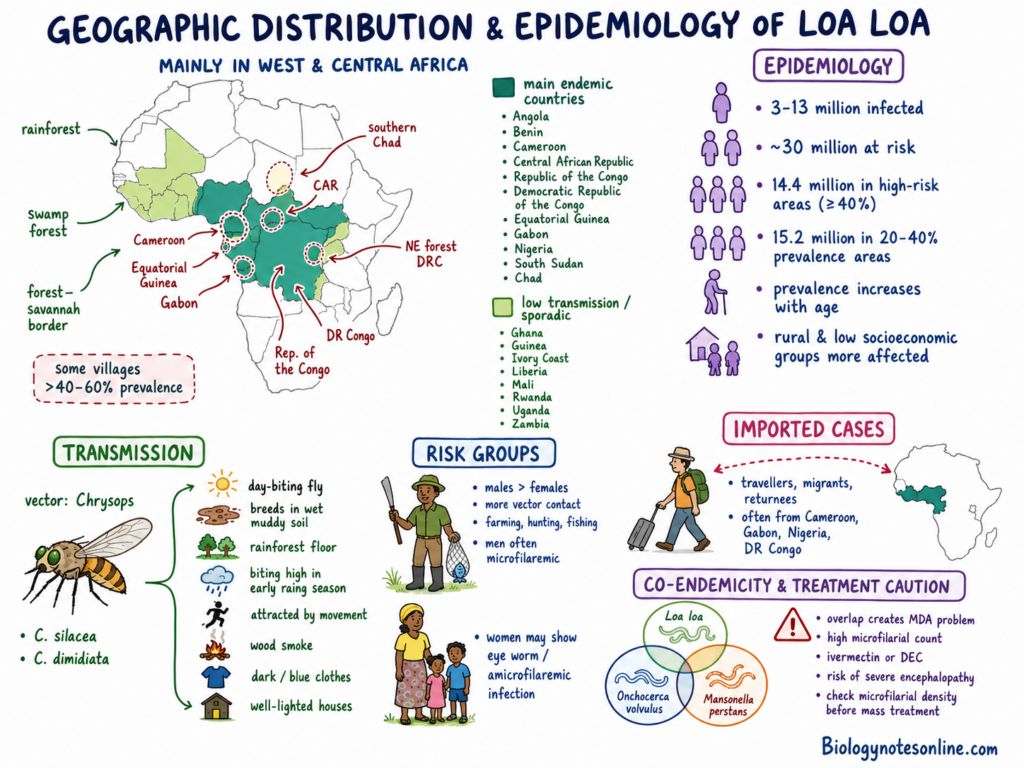
Loa loa is mainly distributed in the equatorial regions of West and Central Africa. It is common in rainforest, swamp forest and forest-savannah border areas.
The infection is mainly found in Angola, Benin, Cameroon, Central African Republic, Republic of the Congo, Democratic Republic of the Congo, Equatorial Guinea, Gabon, Nigeria, South Sudan and Chad. Low transmission or sporadic cases are also found in Ghana, Guinea, Ivory Coast, Liberia, Mali, Rwanda, Uganda and Zambia.
About 3-13 million people are infected with Loa loa. Nearly 30 million people live in the areas where transmission may occur. About 14.4 million people live in high-risk regions with prevalence of 40% or more. Another 15.2 million people live in areas having prevalence between 20% and 40%.
The major endemic regions are present in Cameroon and Democratic Republic of the Congo. These countries contain a large part of the population at risk. High infection is also found in Equatorial Guinea, Gabon, southern Cameroon, Central African Republic, Republic of the Congo and southern Chad.
Another important endemic region is present in the northeastern forest areas of Democratic Republic of the Congo. In some rural forest villages, prevalence may be more than 40-60%.
The infection is more common in males than females. This is mainly due to increased contact with the vector during farming, hunting and fishing. Men commonly show microfilariae in blood, while women may show amicrofilaremic or eye worm infection.
The prevalence increases with age because of repeated exposure to infected flies. Rural populations and persons with low socioeconomic condition are more affected.
Transmission occurs through day-biting flies of genus Chrysops. The main vector species are Chrysops silacea and Chrysops dimidiata. These flies breed in wet muddy soil of rainforest floor.
The biting activity is generally high during the early rainy season. The flies are attracted by movement, wood fire smoke, dark or blue clothes and well-lighted houses.
Imported cases are found among travellers, migrants and persons returning from endemic regions. Most of these infections are acquired from Cameroon, Gabon, Nigeria and Democratic Republic of the Congo.
The distribution of Loa loa overlaps with Onchocerca volvulus and Mansonella perstans in many regions. This creates a major problem during mass drug administration.
In persons with high microfilarial count, treatment with ivermectin or diethylcarbamazine (DEC) may produce severe encephalopathy. Therefore, microfilarial density should be determined before mass treatment in co-endemic regions.
How is Loiasis Transmitted?
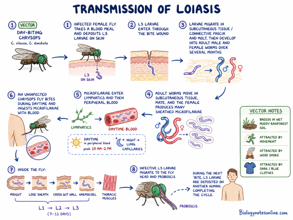
- Loiasis is transmitted to humans through the bite of infected day-biting Chrysops flies. Chrysops silacea and Chrysops dimidiata are the main species involved in the transmission.
- The female fly deposits the third-stage infective larvae (L3) on the skin while taking the blood meal. From the skin, larvae enter through the wound produced by the bite.
- After entering into human body, the larvae migrate in the subcutaneous tissues and connective fascia. Here, they undergo molting and gradually develop into the adult male and female worms, which may take several months.
- The adult worms move freely in the subcutaneous tissues. After mating, female worms produce large numbers of sheathed microfilariae, which enter the lymphatic vessels and later appear in the peripheral blood.
- The microfilariae have a diurnal periodicity, therefore they are found in the peripheral circulation during daytime. Their number is highest between 10:00 AM and 2:00 PM, which corresponds with the biting period of the vector fly.
- At night, most of the microfilariae leave the peripheral circulation and remain in the capillary vessels of lungs.
- When an uninfected Chrysops fly takes blood from the infected person during daytime, microfilariae are ingested along with the blood. In the midgut of fly, they lose the sheath and pass through the gut wall into the haemocoel.
- From the haemocoel, microfilariae migrate to the thoracic flight muscles. In this place, the first-stage (L1), second-stage (L2) and finally the infective third-stage larvae (L3) are developed within about 7-12 days.
- The fully developed infective larvae then migrate towards the head and proboscis of the fly. They are deposited on the skin when the fly again takes blood meal from a human host.
- The vector flies mostly breed in the wet and muddy soil present in rainforest areas. Movement of persons, wood fire smoke and dark or blue clothes commonly attract these flies.
Life Cycle of Loa loa
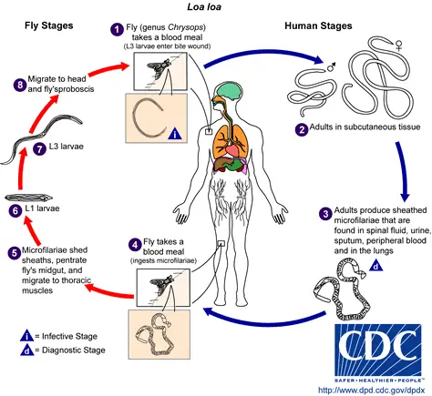
The life cycle of Loa loa is completed in humans and day-biting Chrysops fly. Humans act as the definitive host while the fly acts as the intermediate host and vector.
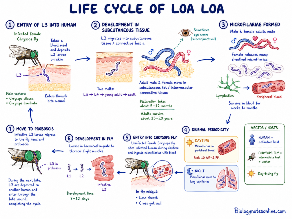
1. Entry of infective larva into humans
- During blood meal, the infected female Chrysops fly deposits third-stage infective larvae (L3) on the skin.
- The main vectors are Chrysops silacea and Chrysops dimidiata.
- The larvae enter into the human body through the bite wound.
2. Development in subcutaneous tissues
- After entry, the L3 larvae migrate into the subcutaneous tissues and connective fascia.
- In this place, the larvae undergo two molts. The L3 larva changes into L4 larva and later into young adult worm.
- The complete development into mature adult worm takes about 5-12 months.
- Adult male and female worms move through the subcutaneous fat and intermuscular connective tissues.
- Sometimes, the adult worm moves through the subconjunctival tissue of eye.
- The adult worms may remain alive in human body for about 15-20 years.
3. Formation of microfilariae
- The adult male and female worms mate in the subcutaneous tissues.
- After fertilization, the female worm releases large number of sheathed embryos called microfilariae.
- These microfilariae enter into the lymphatic vessels and later reach the peripheral blood.
- They may remain alive in the blood for several weeks to months.
4. Diurnal periodicity
- The microfilariae show diurnal periodicity.
- During daytime, they remain in large number in the peripheral blood.
- The highest number is generally found between 10:00 AM and 2:00 PM.
- During night, the microfilariae move from peripheral blood and remain in the capillary beds of lungs.
5. Entry into Chrysops fly
- A female Chrysops fly takes the microfilariae while feeding on an infected human.
- The microfilariae enter into the midgut of fly along with the blood meal.
- Inside the midgut, they lose their protective sheath and cross the gut wall.
6. Development in the vector
- After crossing the gut wall, the larvae enter into the haemocoel and migrate towards the thoracic flight muscles.
- In the thoracic muscles, the larvae undergo further development.
- First-stage larva (L1), second-stage larva (L2) and infective third-stage larva (L3) are formed.
- This development generally takes about 7-12 days.
7. Migration towards proboscis
- The infective L3 larvae leave the thoracic muscles and move towards the head and proboscis of fly.
- These larvae remain near the proboscis until the next blood meal.
- During feeding on another human, the larvae are deposited on the skin and enter through the bite wound.
Infective and Diagnostic Stages of Loa loa
Infective Stage of Loa loa
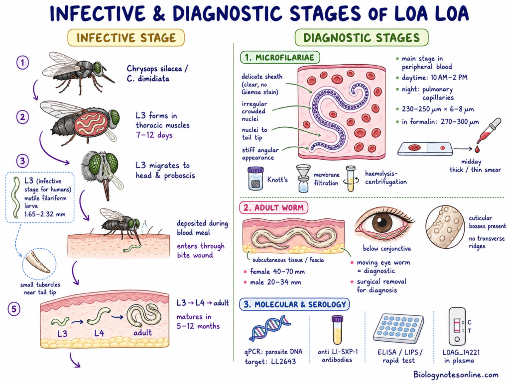
- The third-stage larva (L3) is the infective stage of Loa loa for humans.
- It is a motile filariform larva measuring about 1.65-2.32 mm in length. Small tubercles are present near the tip of tail.
- The infective larvae are developed in the thoracic flight muscles of Chrysops silacea and Chrysops dimidiata.
- Development from microfilariae to infective L3 larva takes about 7-12 days inside the fly.
- After development, the L3 larvae migrate towards the head and proboscis of fly.
- During blood meal, the larvae are deposited on the human skin. They enter into the body through the bite wound.
- In the subcutaneous tissues, L3 larva changes into L4 larva and later forms the adult worm.
- The development into mature adult worm takes about 5-12 months.
Diagnostic Stages of Loa loa
1. Microfilariae
- The microfilariae are the main diagnostic stage found in the peripheral blood.
- They are present in higher number during daytime, mainly between 10:00 AM and 2:00 PM.
- During night, the microfilariae remain in the pulmonary capillaries.
- It measures about 230-250 µm in length and 6-8 µm in width in stained blood smear.
- In 2% formalin, the length may increase up to 270-300 µm.
- The microfilariae are covered by a delicate sheath. The sheath does not take Giemsa stain and appears as a clear space.
- The somatic nuclei are irregular and crowded. They extend continuously up to the tip of tail.
- In stained blood film, the microfilariae generally show a stiff and angular appearance.
- Detection is carried out by thick or thin blood smear collected during midday.
- Knott’s concentration method, membrane filtration and haemolysis-centrifugation methods are also used.
2. Adult Worm
- The adult worm is another diagnostic stage of Loa loa.
- It lives and migrates in subcutaneous tissues, connective fascia and below the conjunctiva of eye.
- The important identifying feature is the presence of irregularly arranged cuticular bosses.
- Transverse cuticular ridges are absent.
- The adult female measures about 40-70 mm in length and the male measures about 20-34 mm in length.
- Direct observation of moving worm below the conjunctiva can confirm the infection.
- Surgical removal of the worm from eye or subcutaneous tissue is also used for diagnosis.
3. Molecular and Serological Markers
- qPCR is used for detection of circulating cell-free parasite DNA.
- The LL2643 repeat sequence is commonly used as the molecular target.
- Antibodies against recombinant Ll-SXP-1 can be detected by ELISA, LIPS or rapid diagnostic test.
- The microfilarial secretory protein LOAG_14221 may also be detected in plasma.
Pathogenesis and Disease Mechanism of Loa loa
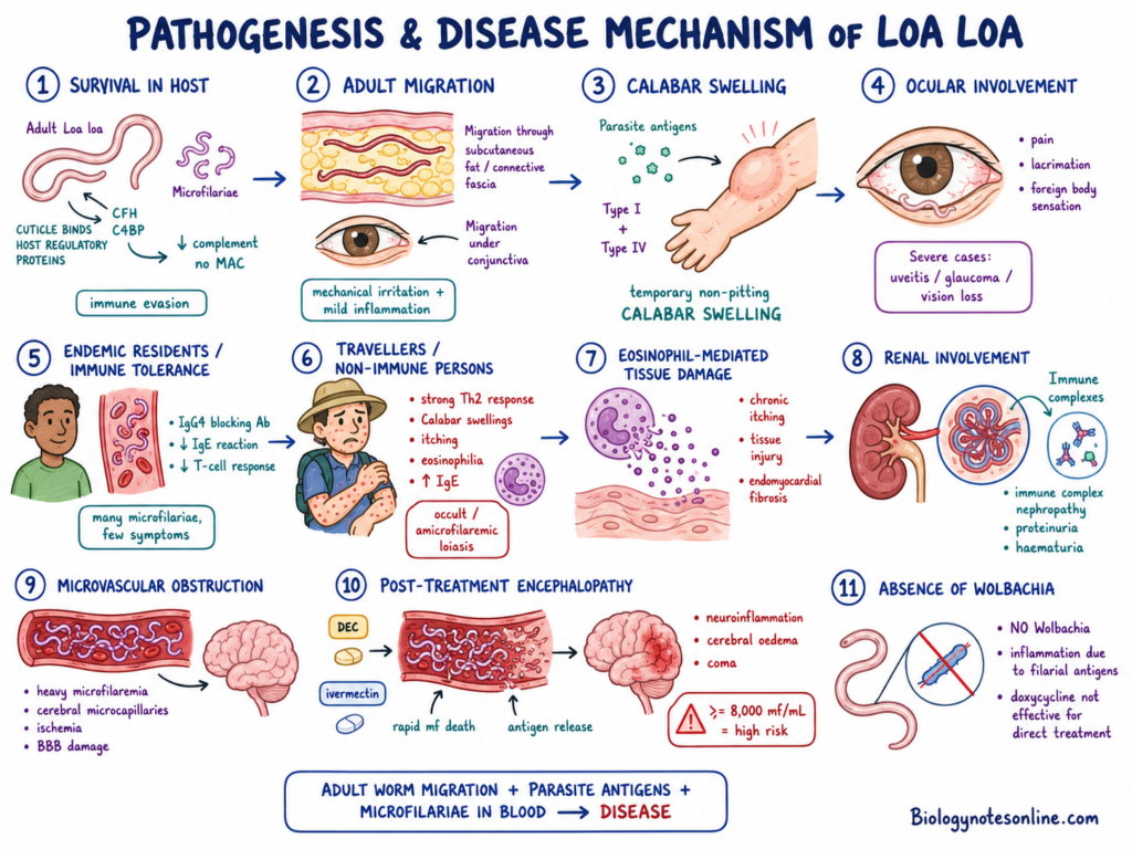
The pathogenesis of Loa loa mainly occurs due to migration of adult worms, release of parasitic antigens and presence of microfilariae in blood. The disease changes are produced in the following steps-
Step 1- Survival inside the human host
Adult worms and microfilariae can survive inside the human body for many years.
The parasite binds host regulatory proteins such as Complement Factor H (CFH) and C4b-binding protein (C4BP) on its outer cuticle.
These proteins reduce the activity of complement system. Formation of Membrane Attack Complex (MAC) is prevented and the parasite escapes complement-mediated destruction.
Step 2- Migration of adult worms
Adult worms continuously move through subcutaneous fat, connective tissues and intermuscular fascia.
During this process, local mechanical irritation and mild inflammatory changes are produced.
The worms may also migrate through the subconjunctival tissue of eye.
Step 3- Formation of Calabar swelling
Migrating worms release different somatic and excretory antigens into the surrounding tissues.
These antigens produce hypersensitivity reaction. Type I and Type IV reactions may be involved.
This results in temporary, non-pitting subcutaneous swelling known as Calabar swelling.
The swellings are mainly seen on limbs near the joints. It appears suddenly and disappears after some time.
Step 4- Ocular involvement
When adult worm passes through the subconjunctival space, local pain, lacrimation and foreign body sensation are produced.
Conjunctival irritation and inflammatory reaction may also occur.
Sometimes the worm may enter the anterior chamber of eye. In such cases, uveitis, hypopyon, cataract, glaucoma and loss of vision may develop.
Step 5- Immune response in endemic persons
Persons living for long time in endemic regions may develop immune tolerance against the parasite.
In these persons, high level of IgG4 antibody acts as blocking antibody and reduces the IgE-mediated reaction.
T-cell response against filarial antigens also becomes low.
As a result, large number of microfilariae may be present in blood but the person shows few or no symptoms.
Step 6- Immune response in travellers
Travellers and non-immune persons do not have early immune tolerance against the parasite.
A strong Th2 immune response is produced in these persons.
Frequent Calabar swellings, intense itching, eosinophilia and increased IgE level are commonly seen.
Microfilariae may be absent or remain in low number in blood. This is referred to as occult or amicrofilaremic loiasis.
Step 7- Eosinophil-mediated tissue damage
The parasitic antigens produce continuous stimulation of eosinophils.
Activated eosinophils release their granule contents into the tissues.
Long-term eosinophilic reaction may produce chronic itching, tissue injury and endomyocardial fibrosis.
Step 8- Renal involvement
Circulating parasitic antigens combine with antibodies and form immune complexes.
These immune complexes may be deposited in the renal glomeruli.
Inflammatory nephropathy is produced, which may cause proteinuria and haematuria.
Step 9- Microvascular obstruction
In heavy infection, very large number of microfilariae may be present in blood.
The microfilariae can obstruct small blood vessels, mainly cerebral microcapillaries.
This may produce focal ischemia, damage of blood-brain barrier and neurological changes.
Step 10- Post-treatment encephalopathy
Severe reaction can occur when diethylcarbamazine (DEC) or ivermectin is given to patients having high microfilarial count.
These drugs cause rapid destruction of a large number of microfilariae.
During this process, large amount of filarial antigens are released into blood.
Severe neuroinflammatory reaction, cerebral oedema, coma and fatal encephalopathy may occur.
The risk becomes higher when the microfilarial density is 8,000 mf/mL or more.
Step 11- Absence of Wolbachia
Loa loa does not contain the endosymbiotic bacterium Wolbachia.
The inflammatory changes are mainly caused by filarial antigens and not by bacterial products.
Therefore, anti-Wolbachia drugs such as doxycycline are not effective for direct treatment of Loa loa.
Clinical Forms and Symptoms of Loa loa
The clinical condition of Loa loa infection varies among infected persons. Some persons remain asymptomatic, while others develop allergic, ocular, neurological and systemic symptoms.
Clinical Forms
- Patent loiasis– Microfilariae are present in the peripheral blood. It is commonly found in long-term residents of endemic regions, and many infected persons may remain asymptomatic.
- Occult loiasis– Adult worms are present but microfilariae are not detected in peripheral blood. This form is more common in travellers and non-immune persons.
- Eye worm form– Adult worm is seen moving below the conjunctiva of eye. A previous history of eye worm migration may also be present.
- Combined form– Eye worm migration and circulating microfilariae are both present. This is also referred to as EWMF infection state.
Cardinal Symptoms
- Calabar swelling– Temporary and non-pitting swelling is formed under the skin.
- It is commonly present on arms and legs near the joints.
- The swelling generally remains for 1-3 days.
- It is formed due to hypersensitivity reaction against the migrating adult worm.
- Eye worm migration– Adult worm moves across the bulbar conjunctiva of eye.
- Pain, irritation, foreign body sensation and lacrimation may occur.
- Swelling around the eye and eyelid may also be present.
- Pruritus– Generalized itching is commonly found, mainly in travellers and occult loiasis.
- Urticaria– Allergic wheals or rashes may develop due to hypersensitivity reaction.
Neurological Symptoms
- Paresthesia– Tingling and numbness may occur in some infected persons.
- Limb weakness– Temporary weakness or paralysis of limbs may sometimes develop.
- Headache– Severe or repeated headache may occur during symptomatic infection.
- Fatigue– Generalized weakness and tiredness are commonly found.
- Spontaneous encephalopathy– A very high number of microfilariae may obstruct cerebral capillaries.
- Confusion and other neurological changes may develop.
- Severe condition may result in coma.
- Post-treatment encephalopathy– It may occur after treatment with diethylcarbamazine (DEC) or ivermectin in persons having high microfilarial count.
- Rapid killing of microfilariae causes release of large amount of filarial antigens.
- Cerebral oedema, coma and sometimes death may occur.
Ocular Complications
- Conjunctival changes– Migration of adult worm may produce redness, chemosis and swelling of eyelid.
- Intraocular infection– Adult worm may rarely enter the anterior chamber or vitreous body of eye.
- Acute uveitis and hypopyon may develop.
- Cataract and glaucoma are also found.
- Severe inflammation may produce permanent loss of vision.
Chronic Systemic Complications
- Cardiac changes– Persistent eosinophilic reaction may damage the heart tissues and produce endomyocardial fibrosis.
- Renal changes– Antigen-antibody complexes may be deposited in renal glomeruli.
- Proteinuria and haematuria may occur.
- Chronic nephropathy may develop.
- Myalgia– Recurrent muscle pain may occur near the site of worm migration.
- Arthralgia– Joint pain may be present, mainly near the affected areas.
- Lymphadenopathy– Mild enlargement of lymph nodes may be found.
- Subcutaneous abscess– Localized abscess may develop around dying adult worms.
- Splenic nodules– Small nodular changes may sometimes be present in spleen.
Laboratory Findings
- Eosinophilia– Marked increase in peripheral eosinophil count is commonly found in symptomatic infection.
- High IgE– Total serum IgE level is generally increased.
- IgG4 antibodies– Parasite-specific IgG4 antibodies may be present in chronic infection.
Diagnosis of Loiasis
The diagnosis of loiasis is based on history, clinical features and detection of microfilariae or adult worm. Different laboratory methods are also used.
Clinical Examination
- Travel history– History of residence or travel in endemic regions of West and Central Africa is noted.
- Eye worm history– Previous or present movement of adult worm below the conjunctiva is an important finding.
- Calabar swelling– Recurrent, temporary and non-pitting swelling near the joints may indicate loiasis.
- Pruritus– Generalized itching and urticaria are commonly found in symptomatic persons.
- Eosinophilia– Marked increase in peripheral eosinophil count supports the diagnosis.
- RAPLOA– It is a questionnaire method used for recording the history of eye worm migration in endemic regions.
Blood Smear Examination
- Blood timing– Blood is collected between 10:00 AM and 2:00 PM because microfilariae show diurnal periodicity.
- Sample type– Capillary blood or EDTA venous blood can be used for examination.
- Thick smear– Thick blood smear is used for detecting microfilariae.
- Thin smear– Thin blood smear is used for studying the morphology of microfilariae.
- Staining– Blood films are generally stained with Giemsa stain or hematoxylin and eosin.
- Microfilarial size– The microfilariae measure about 230-250 µm in length and 6-8 µm in width.
- Sheath– A delicate sheath is present, but it does not take Giemsa stain.
- Tail nuclei– Somatic nuclei extend continuously up to the tip of the tapered tail.
- Posture– The microfilariae commonly show a stiff and angular appearance in stained film.
Microfilarial Quantification
- MFD count– Microfilarial density is expressed as microfilariae per millilitre of blood (mf/mL).
- Low density– A count below 8,000 mf/mL is considered as low microfilaremia.
- High density– A count between 8,000 and 19,999 mf/mL is considered as high microfilaremia.
- Hyper density– A count of 20,000 mf/mL or more is referred to as hyper-microfilaremia.
- Treatment safety– Microfilarial count is determined before giving DEC or ivermectin.
- LoaScope– It is a mobile phone-based video microscope used for rapid counting of moving microfilariae.
- Repeat testing– More than one midday sample may be examined because the count can vary between samples.
Concentration Methods
- Knott’s method– Blood is mixed with 2% formalin and centrifuged for concentrating the microfilariae.
- Hemolysis method– Red blood cells are lysed by saponin or formalin, followed by centrifugation.
- Membrane filtration– Hemolysed blood is passed through a membrane filter for collecting microfilariae.
- Low infection– These methods are useful when microfilariae are present in low number.
Adult Worm Detection
- Direct observation– A live adult worm may be seen moving below the bulbar conjunctiva.
- Surgical removal– Adult worm can be removed from the eye or subcutaneous tissue under local anaesthesia.
- Cuticular bosses– Irregular rounded elevations on the cuticle are the important identifying feature.
- Female worm– Adult female measures about 40-70 mm in length.
- Male worm– Adult male measures about 20-34 mm in length.
- Head structure– The anterior end is simple and lips are absent.
- Male tail– The male has a coiled posterior end with caudal papillae and unequal spicules.
Serological Tests
- Ll-SXP-1 test– IgG4 antibodies against recombinant Ll-SXP-1 can be detected.
- ELISA– ELISA is used for detection of antifilarial antibodies.
- LIPS test– Luciferase Immunoprecipitation System is used for detection of anti-**Ll-SXP-1 antibodies.
- Rapid test– Lateral flow rapid diagnostic tests may also be used.
- Cross-reaction– Serological tests may react with other filarial and helminth infections.
- Past infection– Antibody tests cannot clearly differentiate past infection from active infection.
- False negative– Heavy microfilaremia may sometimes produce false-negative serological results.
Molecular Tests
- qPCR– Quantitative PCR is used for sensitive detection of Loa loa DNA.
- LL2643 target– The repeated LL2643 genomic sequence is commonly used as the molecular target.
- ccfDNA– Circulating cell-free parasite DNA can be detected in plasma or urine.
- Occult loiasis– Molecular tests are useful when microfilariae are not detected in blood.
- Other targets– LL20, mitochondrial 12S rDNA, Cox1 and ITS1 may also be used.
- LAMP– Loop-mediated isothermal amplification is used for rapid detection of parasite DNA.
- Secretory marker– The protein LOAG_14221 may be detected in plasma and is related with microfilarial density.
- Stool PCR– Stool-based PCR is not useful for diagnosis of loiasis.
Diagnostic Forms
- Patent loiasis– Microfilariae are detected in midday peripheral blood.
- Occult loiasis– Adult worm infection is present but microfilariae are not found in blood.
- Eye worm form– Present or previous subconjunctival migration of adult worm is found.
- Combined form– Eye worm migration and circulating microfilariae are both present.
Differential Diagnosis
- Onchocerciasis– Microfilariae are unsheathed and mainly found in skin snips. The tail tip is free from nuclei.
- Wuchereria infection– Microfilariae show nocturnal periodicity and the tail tip is free from nuclei.
- Brugia infection– Microfilariae show nocturnal periodicity and two terminal nuclei are present in the tail.
- Mansonella perstans– Microfilariae are unsheathed, non-periodic and have a blunt tail.
- Mansonella streptocerca– Microfilariae are present in skin and have a bent tail.
- Dirofilaria– Adult worms have a thick striated cuticle and cuticular bosses are absent.
- Other eye worms– Thelazia and Gnathostoma may also produce ocular infection.
Treatment and Management of Loiasis
The treatment of loiasis depends on the number of microfilariae present in blood. The microfilarial count is checked before starting the treatment because rapid killing of large number of microfilariae may produce severe encephalopathy.
Pre-Treatment Evaluation
- Blood collection– Blood sample is collected during daytime, mainly between 10:00 AM and 2:00 PM.
- Microfilarial count– The number of microfilariae per millilitre of blood (mf/mL) is determined by blood smear or LoaScope.
- Co-infection screening– The patient is examined for infection with Onchocerca volvulus and lymphatic filariasis before starting the drug.
- Medical supervision– Treatment should be carried out under a physician having experience in tropical and filarial diseases.
Diethylcarbamazine Treatment
- Drug of choice– Diethylcarbamazine (DEC) is the main drug used for treatment of loiasis. It acts against both microfilariae and adult worms.
- Indication– DEC is generally given when the microfilarial count is below 8,000 mf/mL.
- Dose– DEC is given orally at 8-10 mg/kg/day in three divided doses for 21 days.
- Treatment course– One or two courses may be required depending upon the response of patient.
- Treatment response– Disappearance of symptoms, reduction of eosinophilia and absence of microfilariae indicate response to treatment.
- Contraindication– DEC should not be given in patients having Onchocerca volvulus infection.
- It may produce severe ocular inflammation.
- A strong Mazzotti reaction may also develop.
Heavy Microfilarial Infection
- High parasite count– DEC or ivermectin should not be directly given when the microfilarial count is 8,000 mf/mL or more.
- Encephalopathy risk– Rapid destruction of microfilariae releases large amount of parasitic antigens.
- Cerebral oedema, coma and fatal encephalopathy may occur.
- The risk becomes higher in persons having more than 20,000 mf/mL.
- Albendazole– It is used to slowly reduce the microfilarial count before starting DEC.
- The usual dose is 200 mg twice daily for 21 days.
- It reduces the production of microfilariae by acting slowly on adult female worms.
- Apheresis– Microfilariae may be removed directly from blood by plasmapheresis or cytapheresis.
- It is carried out in specialized hospital centres.
- DEC is started after the count is reduced below the safer level.
Refractory Infection
- Treatment failure– Some patients may remain infected after two complete courses of DEC.
- Alternative drug– Albendazole at 200 mg twice daily for 21 days may be used in these cases.
- Co-infection treatment– In patients having light Loa loa infection with onchocerciasis, ivermectin may be used first for treatment of O. volvulus under medical supervision.
Doxycycline
- Wolbachia absence– Loa loa does not contain the endosymbiotic bacteria Wolbachia.
- No direct action– Doxycycline is not effective for direct treatment of Loa loa infection.
- Co-endemic use– It may be used for treatment of onchocerciasis in areas where Loa loa is also present.
Surgical and Symptomatic Treatment
- Worm removal– Adult worm moving below the conjunctiva may be removed surgically with forceps under local anaesthesia.
- Subcutaneous worm– A visible or palpable worm present under the skin may also be removed.
- Antihistamines– These are used for reducing itching, urticaria and allergic symptoms produced during treatment.
- Corticosteroids– These may be given during the first few days of treatment for controlling severe inflammatory reaction.
Prevention
- DEC prophylaxis– Long-term travellers to highly endemic areas may receive DEC 300 mg once weekly under medical advice.
- Insect repellent– DEET-based repellents are used for preventing the bite of Chrysops flies.
- Protective clothing– Long sleeves, full trousers and permethrin-treated clothes are used in forest areas.
- Vector habitat– Contact with swamp forest, muddy stream borders and other fly breeding places should be reduced.
- Fly attractants– Wood smoke, movement and dark or blue clothes commonly attract Chrysops flies.
Prevention of Loiasis
- DEC prophylaxis– Diethylcarbamazine (DEC) may be given at 300 mg once weekly for long-term travellers and persons living in highly endemic areas.
- Insect repellent– Repellents containing DEET are applied on exposed parts of skin.
- Long sleeves– Full-sleeved shirts are used to reduce the contact of flies with skin.
- Long trousers– Full trousers are used while visiting forest and swamp areas.
- Permethrin clothes– Clothes treated with permethrin provide additional protection against Chrysops flies.
- Light clothes– Light-coloured clothes are preferred because the flies are attracted towards blue and dark colours.
- Forest avoidance– Muddy rainforest floors and swamp forest regions should be avoided.
- River areas– Shaded places near forest rivers and streams should be avoided.
- Wood smoke– Open wood fire and wood smoke should be avoided because it attracts the vector flies.
- Daylight exposure– Outdoor forest activities should be reduced during the main biting period.
- Peak hours– The risk of fly bite is higher between 10:00 AM and 4:00 PM.
- Rainy season– Extra protection is required during the early months of rainy season when the vector population is high.
- Indoor screens– Window and door screens can reduce the entry of flies into houses.
- Bed nets– Bed nets provide limited protection because Chrysops flies mainly bite during daytime.
- Vector habitat– Contact with bogs, wet soil and muddy breeding places should be reduced.
- No vaccine– No approved vaccine is available for prevention of Loa loa infection.
- Vector control– Large-scale control of Chrysops flies is difficult because the breeding areas are widely distributed in rainforest regions.
Loiasis vs Onchocerciasis
Here is a detailed comparative overview of Loiasis and Onchocerciasis:
| Feature / Criterion | Loiasis (Loa loa) | Onchocerciasis (Onchocerca volvulus) |
|---|---|---|
| Common Name | African eye worm | River blindness |
| Insect Vector | Day-biting tabanid flies / deer flies (Chrysops silacea, Chrysops dimidiata) | Blackflies (Simulium species) |
| Adult Worm Habitat | Subcutaneous tissues, intermuscular fascia, and subconjunctival space of the eye | Fibrous subcutaneous nodules (onchocercomata) |
| Microfilariae (mf) Location | Peripheral blood stream (during daylight hours) and pulmonary capillaries (at night) | Dermis of the skin, lymphatics, and ocular tissues |
| Microfilarial Periodicity | Strict diurnal periodicity (peaking in peripheral blood between 10:00 AM and 2:00 PM) | Non-periodic (present continuously in skin tissues) |
| Microfilaria Sheath & Tail | Sheathed; column of somatic nuclei extends continuously to the absolute tip of the tail | Unsheathed; column of nuclei does not extend to the tip of the tail |
| Adult Cuticular Morphology | External cuticle contains irregularly spaced elevations called bosses; lacks transverse ridges | External cuticle features prominent transverse annular ridges (rugae) and striae |
| Wolbachia Endosymbiont | Absent (Loa loa does not harbor Wolbachia) | Present (obligate symbiont essential for adult worm survival/fertility) |
| Primary Clinical Features | Calabar swellings (transient angioedema), subconjunctival worm migration across the eye, severe pruritus | Severe pruritic onchodermatitis, skin depigmentation (“leopard skin”), subcutaneous nodules, sclerosing keratitis, chorioretinitis, permanent blindness |
| Primary Diagnostic Methods | Microscopic identification of microfilariae in midday blood draws (10 AM–2 PM), direct visualization of migrating eye worm, ccfDNA qPCR, or LoaScope | Skin snip biopsy (microfilariae emerging in saline), palpation/biopsy of nodules, or slit-lamp eye examination |
| First-Line / Curative Drug | Diethylcarbamazine (DEC) (kills both microfilariae and adult worms, indicated when MFD < 8,000 mf/mL) | Ivermectin (microfilaricide) and/or Doxycycline (anti-Wolbachia macrofilaricide) |
| Major Treatment Hazards & Conflicts | Fast-acting drugs (DEC/ivermectin) in heavy infections (≥8,000 mf/mL) trigger fatal encephalopathy. Anti-Wolbachia doxycycline is ineffective for Loa loa. | DEC is strictly contraindicated in onchocerciasis because it causes severe ocular Mazzotti reactions and blindness. |
Loiasis vs Lymphatic Filariasis
| Feature / Criterion | Loiasis (Loa loa) | Lymphatic Filariasis |
|---|---|---|
| Causal Agent(s) | Loa loa (African eye worm) | Wuchereria bancrofti, Brugia malayi, or Brugia timori |
| Primary Insect Vector(s) | Day-biting tabanid flies / deer flies (Chrysops silacea, Chrysops dimidiata) | Mosquitoes (Culex, Anopheles, Aedes, Mansonia, Coquillettidia) |
| Anatomical Habitat of Adults | Subcutaneous tissues, intermuscular connective fascia, and subconjunctival space of the eye | Lymphatic vessels and regional/inguinal lymph nodes |
| Microfilarial Periodicity | Strict diurnal periodicity (circulates in peripheral blood during daylight hours; peaks 10:00 AM–2:00 PM) | Predominantly nocturnal periodicity (peaking in peripheral blood between 10:00 PM and 2:00 AM) |
| Microfilarial Morphology | Sheathed; column of somatic nuclei extends continuously all the way to the absolute tip of the tail | Sheathed; somatic nuclei do not extend to the tip of the tail (W. bancrofti) or specific terminal nuclei are present (B. malayi) |
| Wolbachia Endosymbiont | Absent (Loa loa completely lacks Wolbachia bacteria) | Present (obligate endosymbiont required for worm survival and reproduction) |
| Primary Clinical Features | Calabar swellings (transient angioedema), visible subconjunctival worm migration (“eye worm”), intense pruritus | Acute adenolymphangitis, filarial fever; chronic lymphedema, elephantiasis of limbs, and hydrocele (testicular swelling) |
| Key Diagnostic Methods | Microscopic identification of microfilariae in midday blood samples (10 AM–2 PM), direct visual observation of migrating adult worm, ccfDNA qPCR (LL2643), or LoaScope | Microscopic identification of microfilariae in night blood samples (10 PM–2 AM), circulating filarial antigen (CFA) detection, or “filarial dance sign” on ultrasound |
| Primary Treatment | Diethylcarbamazine (DEC) (kills microfilariae and adult worms; used when MFD < 8,000 mf/mL). If MFD ≥ 8,000 mf/mL, pre-treat slowly with albendazole or apheresis to prevent fatal encephalopathy. | Diethylcarbamazine (DEC), Albendazole, or Ivermectin. Doxycycline is effective as an anti-Wolbachia macrofilaricide (4–6 weeks). |
| Efficacy of Doxycycline | Ineffective for Loa loa because the parasite lacks Wolbachia. | Highly effective; destroys endosymbiotic Wolbachia, sterilizing and slowly killing adult worms. |
Microfilarial Laboratory Identification & Comparison
| Diagnostic Feature | Loa loa | Wuchereria bancrofti | Brugia malayi | Mansonella perstans | Mansonella streptocerca |
|---|---|---|---|---|---|
| Sheath | Sheathed (does not retain Giemsa stain) | Sheathed | Sheathed | Unsheathed | Unsheathed |
| Primary Specimen / Location | Blood (peripheral bloodstream) | Blood (peripheral bloodstream) | Blood (peripheral bloodstream) | Blood (peripheral bloodstream) | Skin (dermis / skin snip biopsies) |
| Periodicity | Diurnal (peaks 10:00 AM – 2:00 PM) | Nocturnal (peaks 10:00 PM – 2:00 AM; diurnal/subperiodic in South Pacific) | Nocturnal (after 8:00 PM) | Non-periodic (circulates day and night) | Non-periodic |
| Tail Shape | Tapered | Tapered to a point | Tapered with a characteristic terminal constriction/swelling | Bluntly rounded | Blunt / Hooked (often bent like a “shepherd’s crook”) |
| Arrangement of Terminal Nuclei | Column of nuclei extends continuously all the way to the absolute tip of the tail | Tail tip is free of nuclei (nuclei do not extend to the end) | Two distinct terminal nuclei separated at the tip of the tail | Somatic nuclei extend all the way to the tip of the tail | Somatic nuclei extend all the way to the tip of the tail |
| Geographic Context | Rainforests and swamp forests of West and Central Africa | Global tropics/subtropics: Sub-Saharan Africa, Southeast Asia, India, Pacific Islands, Caribbean, South America | Asia & Pacific: Southeast Asia, India, China, Pacific Islands | West/Central Africa and Central/South America | Rainforests of West and Central Africa, and Uganda |
FAQ
What disease does Loa loa cause?
Loa loa is the parasitic roundworm responsible for loiasis (commonly referred to as African eye worm disease), a neglected tropical filarial infection endemic to West and Central Africa.
Why is it called the African eye worm?
It earned the common name African eye worm because adult worms famously migrate beneath the clear subconjunctiva of the human eye, creating a visible, moving thread across the sclera.
What is the vector?
The parasite is transmitted by day-biting tabanid flies (commonly known as deer flies or mango flies) belonging to the genus Chrysops, primarily Chrysops silacea and Chrysops dimidiata.
What is the infective stage?
The third-stage infective larva (L3) is the obligate infective stage for humans. L3 larvae develop inside the fly vector, migrate to its proboscis, and enter the human body through the fly’s bite wound.
What is the diagnostic stage?
The primary diagnostic stages are sheathed microfilariae (embryonic larvae) circulating in the blood and adult worms observed migrating beneath the ocular conjunctiva or surgically extracted from tissue.
Why is blood collected during the daytime?
Blood collection must take place between 10:00 AM and 2:00 PM (peaking near noon) because Loa loa microfilariae display diurnal periodicity. They circulate in peripheral blood during daylight hours to align with vector feeding patterns and retreat to the vascular beds of the lungs at night.
What are Calabar swellings?
Calabar swellings are localized, non-pitting subcutaneous angioedemas (fluid swellings) that spontaneously appear and regress over several days, predominantly on the limbs near joints. They represent localized hypersensitivity reactions to allergens released by migrating adult worms.
Can it cause blindness?
Subconjunctival migration across the eye surface is painful but rarely causes permanent tissue damage. However, in rare cases where an adult worm penetrates the anterior chamber or vitreous inside the eye, it can cause severe intraocular inflammation, uveitis, secondary cataracts, glaucoma, and permanent vision loss.
Can it spread between people?
No, loiasis cannot spread directly from person to person. It strictly requires an intermediate Chrysops fly vector to ingest microfilariae from an infected host and allow them to develop into infective L3 larvae before transmitting them to another individual.
Can an eye worm be removed?
Yes. An adult worm actively migrating beneath the bulbar conjunctiva can be safely extracted or removed with forceps by a clinician after applying local anesthesia to paralyze the worm.
Does eye-worm removal cure loiasis?
No. Surgical removal extracts only that single migrating adult worm. Other adult parasites and thousands of circulating microfilariae remain elsewhere in the body, requiring systemic antiparasitic medication for complete cure.
What is the drug of choice?
The primary drug of choice is diethylcarbamazine (DEC) because it kills both circulating microfilariae and adult worms, resulting in complete cure for most patients.
Why can ivermectin be dangerous?
When patients harbor high microfilarial densities (≥8,000 to >20,000 mf/mL), rapid destruction of larvae by ivermectin (or DEC) causes a massive release of foreign filarial antigens. This can trigger severe neuroinflammatory reactions, blood-brain barrier disruption, coma, and fatal post-treatment encephalopathy.
Can blood microscopy be negative?
Yes. Up to 70% of infected individuals—particularly non-endemic travelers and expatriates—have occult (amicrofilaremic) loiasis, where adult worms reside in tissues but microfilariae are cleared or absent from peripheral blood. These cases require molecular tests (such as ccfDNA qPCR) or serological assays for confirmation.
How can infection be prevented?
Loiasis can be prevented by taking weekly prophylactic DEC (300 mg once a week) when traveling in hyperendemic zones, applying DEET insect repellent, wearing long-sleeved clothing treated with permethrin, avoiding dark or blue fabrics that attract flies, and staying clear of wood smoke and fly breeding habitats.
References
- Boussinesq, M., Gardon, J., Gardon-Wendel, N., & Chippaux, J. P. (2003). Loa loa encephalopathy temporally related to ivermectin administration reported from onchocerciasis mass treatment programs from 1989 to 2001: Implications for the future. Filaria Journal, 2(Suppl 1), S8.
- Burbelo, P. D., Ramanathan, R., Klion, A. D., Iadarola, M. J., & Nutman, T. B. (2008). Rapid, novel, specific, high-throughput assay for diagnosis of Loa loa infection / Discovery of specific antigens that can predict microfilarial intensity in Loa loa infection. Journal of Clinical Microbiology, 46(7), 2298–2304. https://doi.org/10.1128/JCM.00490-08
- Centers for Disease Control and Prevention. (2024, June 13). Clinical treatment of loiasis. U.S. Department of Health and Human Services. https://www.cdc.gov/filarial-worms/hcp/clinical-care/loiasis.html
- Centers for Disease Control and Prevention. (2024). DPDx – Diagnostic procedures: Blood specimens. U.S. Department of Health and Human Services. https://www.cdc.gov/dpdx/diagnosticprocedures/blood/specimencoll.html
- Centers for Disease Control and Prevention. (2024). DPDx – Loiasis. U.S. Department of Health and Human Services. https://www.cdc.gov/dpdx/loiasis/index.html
- Dieki, R., Nsi-Emvo, E., & Akue, J. P. (2022). The human filaria Loa loa: Update on diagnostics and immune response. Research and Reports in Tropical Medicine, 13, 41–54. https://doi.org/10.2147/RRTM.S355104
- Drame, P. M., et al. (2024). A novel, highly sensitive nucleic acid amplification test assay for the diagnosis of loiasis and its use for detection of circulating cell-free DNA. PubMed Central.
- Drugs for Neglected Diseases initiative. (2025). Fusidic Acid Macrofilaricide Evaluation (FAME). DNDi. https://dndi.org/global-networks/fusidic-acid-macrofilaricide-evaluation-fame/
- Eballe, A. O., Epée, E., Koki, G., Owono, D., Mvogo, C. E., & Bella, A. L. (2008). Intraocular live male filarial Loa loa worm. Clinical Ophthalmology, 2(4), 965–967. https://doi.org/10.2147/co.s3877
- Esum, M. E., et al. (2020). Generation of Loa loa infective larvae by experimental infection of the vector, Chrysops silacea. PLOS Neglected Tropical Diseases, 14(8), e0008415. https://doi.org/10.1371/journal.pntd.0008415
- Ferris, H. (2026). Loa loa. Nemaplex, University of California, Davis. http://nemaplex.ucdavis.edu/taxadata/G989s1.aspx
- Forrer, A., Obie, E. D., Abong, R. A., Ekanya, R., Njouendou, A. J., Nji, T. M., Amuam, A., Eyong, E. M., Ndzeshang, B. L., Nkimbeng, D. A., Fombad, F. F., Teghen, S., Suireng, A., Ashu, E. E., Hamill, L., Enyong, P., Turner, J. D., Wanji, S., & Taylor, M. J. (2026). Efficacy of test and treat with doxycycline on palpable nodules, microfilarial load and Wolbachia density in onchocerciasis infected persons in communities with persistent transmission in South-West Cameroon. medRxiv. https://doi.org/10.1101/2026.06.09.26355259
- Forsaa, V. A., Utheim, T. P., & Willassen, E. (2020). Scanning electron microscopy observations of Loa loa (Nematoda). Case Reports in Ophthalmology, 11(3), 486–492. https://doi.org/10.1159/000509338
- Gardner, S. L., & Gardner, S. A. (Eds.). (2024). Concepts in animal parasitology (Part IV: Nemata, Nematomorpha, Acanthocephala, Pentastomida). Zea Books. https://digitalcommons.unl.edu/context/parasittext/article/1105/viewcontent/CIAP_Part_IV.pdf
- Gobbi, F., Buonfrate, D., Boussinesq, M., Chesnais, C. B., Pion, S. D., Silva, R., Moro, L., Rodari, P., Tamarozzi, F., Biamonte, M., & Bisoffi, Z. (2020). Performance of two serodiagnostic tests for loiasis in a non-endemic area. PLOS Neglected Tropical Diseases, 14(5), e0008187. https://doi.org/10.1371/journal.pntd.0008187
- Haapasalo, K., Meri, T., & Jokiranta, T. S. (2009). Loa loa microfilariae evade complement attack in vivo by acquiring regulatory proteins from host plasma. Infection and Immunity, 77(9), 3886–3893. https://doi.org/10.1128/IAI.01583-08
- Herrick, J. A., Metenou, S., Makiya, M. A., Taylor-Williams, C. A., Law, M. A., Klion, A. D., & Nutman, T. B. (2015). Eosinophil-associated processes underlie differences in clinical presentation of loiasis between temporary residents and those indigenous to Loa-endemic areas. Clinical Infectious Diseases, 60(1), 55–63. https://doi.org/10.1093/cid/ciu723
- Inciteful Med Resources. (2026a). Filariasis treatment: Medications & safety. https://incitefulmed.com/resources/guides/filariasis-overview/filariasis-treatment-medications/
- Inciteful Med Resources. (2026b). Loiasis diagnosis: The crucial daytime blood test. https://incitefulmed.com/resources/guides/loiasis-african-eye-worm-overview/loiasis-diagnosis-blood-test/
- Inciteful Med Resources. (2026c). Why is doxycycline used for river blindness? https://incitefulmed.com/resources/questions/why-doxycycline-for-river-blindness/
- Inciteful Med Resources. (2026d). Why use doxycycline for filariasis? https://incitefulmed.com/resources/questions/why-doxycycline-for-filariasis/
- L’Initiative Loa Loa. (2024). Parasitological and entomological aspects of loiasis – Loa loa. https://www.loaloa.org/fr/Parasitology-and-entomology-of-loiasis
- Locatelli, S., et al. (2021). Limitations of PCR detection of filarial DNA in human stools from subjects non-infected with soil-transmitted helminths. Parasite, 28, 47. https://doi.org/10.1051/parasite/2021046
- Marie, C., Petri, W. A., Jr., & Muzny, C. A. (2025). Loiasis (Loa loa filariasis; African eye worm; Calabar swellings). Merck Manual Professional Edition. https://www.merckmanuals.com/professional/infectious-diseases/nematodes-roundworms/loiasis
- Mathison, B. A., & Pritt, B. S. (2019). Diagnostic identification and differentiation of microfilariae. Journal of Clinical Microbiology, 57(10), e00706-19. https://doi.org/10.1128/JCM.00706-19
- Notarnicola, J. (2024). Filarioidea Weinland, 1858 (Superfamily). In S. L. Gardner & S. A. Gardner (Eds.), Concepts in animal parasitology (pp. 633–656). Zea Books. https://doi.org/10.32873/unl.dc.ciap055
- Orihel, T. C., & Eberhard, M. L. (1998). Zoonotic filariasis. Clinical Microbiology Reviews, 11(2), 366–381. https://doi.org/10.1128/CMR.11.2.366
- Pedram, B., Pasquetto, V., Drame, P. M., Ji, Y., Gonzalez-Moa, M. J., Baldwin, R. K., & Nutman, T. B. (2017). A novel rapid test for detecting antibody responses to Loa loa infections. PLOS Neglected Tropical Diseases, 11(7), e0005741. https://doi.org/10.1371/journal.pntd.0005741
- Steel, C., & Nutman, T. B. (2012). Regulation of global gene expression in human Loa loa infection is a function of chronicity. PLOS Neglected Tropical Diseases, 6(3), e0001527. https://doi.org/10.1371/journal.pntd.0001527
- Systematics, pathobiology, and clinical diagnostics of Loa loa filariasis. (2024). [Comprehensive Monograph].
- Tamarozzi, F., et al. (2024). Analysis of diagnostic test outcomes in a large loiasis cohort from an endemic region: Serological tests are often false negative in hyper-microfilaremic infections. PMC / Journal. https://pmc.ncbi.nlm.nih.gov/articles/PMC10965051/
- University of Iowa Department of Ophthalmology and Visual Sciences. (2023). Loa loa filariasis (African eye worm). EyeRounds.org. https://webeye.ophth.uiowa.edu/eyeforum/cases/352-loa-loa-filariasis.htm
- Veletzky, L., Eberhardt, K. A., Hergeth, J., Stelzl, D. R., Zoleko Manego, R., Mombo-Ngoma, G., Kreuzmair, R., Burger, G., Adegnika, A. A., Agnandji, S. T., Matsiegui, P. B., Boussinesq, M., Mordmüller, B., & Ramharter, M. (2022). Distinct loiasis infection states and associated clinical and hematological manifestations in patients from Gabon. PLOS Neglected Tropical Diseases, 16(9), e0010793. https://doi.org/10.1371/journal.pntd.0010793