

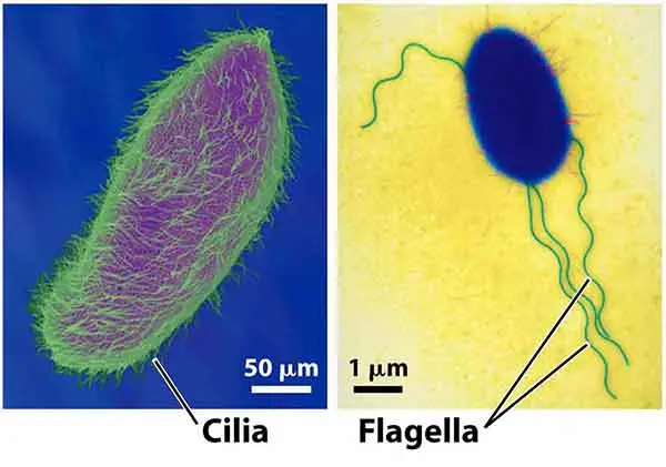
Cilia and flagella are hair-like appendages present on the surface of many eukaryotic cells. These are cell-surface projections surrounded by the plasma membrane. Both structures contain microtubules and associated proteins in their internal part.
Cilia are generally short and present in large numbers on the cell surface. Flagella are usually longer and only one or few flagella are present in a cell. Both have almost similar basic internal organization in eukaryotic cells.
The main discussion of cilia and flagella generally refers to eukaryotic cilia and flagella. These structures are different from the flagella present in bacteria. Bacterial flagella have a different structure and mechanism of movement.
Cilia and flagella are mainly involved in movement. Flagella help in locomotion of cells such as sperm cells and many unicellular eukaryotes. Cilia can move the cell or can move fluid and other materials over the cell surface. Some cilia also perform sensory functions and take part in cell signalling.
The internal microtubule-containing core of cilia and flagella is called the axoneme. It forms the main projecting part of the structure. At the base of each cilium or flagellum, a basal body is present. The basal body is a centriole-like structure from which the axoneme develops and is anchored inside the cell.
What Are Cilia and Flagella?
Cilia and flagella are hair-like structures projecting out from the surface of eukaryotic cells. Cilium is the singular form of cilia. A single flagella is called a flagellum.
They are the specialized structures related with the cytoskeleton. Microtubules make their major internal framework, along with several associated proteins. The arrangement is highly organized and forms an axial core inside the structure, called the axoneme.
In eukaryotes, the entire projecting part is covered by the plasma membrane. This membrane is continuous with the cell membrane itself. Inside lies the microtubular framework.
Cilia and flagella are widely present in eukaryotic organisms. They occur in many unicellular organisms, and also in specialized cells of multicellular organisms. For example, cilia are present over the epithelial cells of respiratory passages, while the sperm cell bears a long flagellum.
Traditionally, both structures were mainly differentiated by their length, number and type of movement. Cilia are usually short. They occur in large number over the cell surface and commonly beat by an effective stroke followed by a recovery stroke. Flagella, on the other hand, are longer, generally one or few in number. Their movement is more commonly wave-like.
But this external difference does not indicate that both have completely different basic structures. In eukaryotic cells, cilia and flagella have fundamentally related internal organization, with an axoneme forming their main structural core. For this structural similarity, the terms are also considered closely related in modern cell biology.
General Characteristics of Cilia and Flagella
The general characteristics of cilia and flagella are as follows-
- Cilia and flagella are thin hair-like projections extending from the cell surface. They are mainly found in eukaryotic cells.
- Both are formed of a microtubule-based internal skeleton. The main internal core is called the axoneme, made up of microtubules together with several associated proteins.
- In eukaryotes, the projecting part is surrounded by the plasma membrane. This membrane is continuous with that of the cell membrane itself.
- Each cilium or flagellum develops from a basal body present at its base. Basal bodies are structurally related to centrioles and contain nine sets of microtubule triplets.
- Cilia are usually short and present in large numbers. Flagella are longer, and commonly one or few occur on a cell. This is the traditional difference between these two structures.
- Their pattern of movement also differs. Cilia generally show repeated effective and recovery strokes, whereas flagella mostly produce wave-like or undulating movement.
- A typical motile cilium or flagellum contains nine peripheral microtubule doublets around two central microtubules. This arrangement is referred to as 9+2 arrangement. However, all cilia do not follow this pattern. Many non-motile primary cilia have a 9+0 arrangement, and some motile cilia also lack the central pair.
- Dynein motor proteins are associated with the axoneme of motile forms. ATP is utilized by these motors. Sliding between adjacent microtubule doublets is produced, which is converted into bending of the axoneme.
- Cilia and flagella are involved in locomotion and movement of materials. Flagella propel sperm cells and several unicellular eukaryotes. Cilia may move a cell itself or move mucus, fluid and other substances over a cell surface.
- Not all cilia are used for movement. Primary cilia are generally non-motile and mostly one is present on a cell. They have important sensory and cell-signalling functions.
- Cilia occur on many animal cells and numerous unicellular eukaryotes. Motile cilia are common in the respiratory tract, oviduct and ependymal cells. A long flagellum is present in mammalian sperm.
- The proteins required for formation and maintenance of these structures are transported along the cilium by intraflagellar transport (IFT). This transport takes place along the axonemal microtubules.
- Although cilia and flagella were traditionally given different names on the basis of their length, number and movement, their basic organization is closely related in eukaryotes. Both have the same general axoneme-basal body organization and are considered fundamentally similar microtubule-based structures.
Where Are Cilia and Flagella Found?
Cilia and flagella are found in different groups of eukaryotic organisms. Their occurrence differs according to cell type and function. Cilia are common on several epithelial cells and protists, while flagella are found in sperm cells and many unicellular eukaryotes.
Cilia in Humans and Animals
- Respiratory epithelium – Motile cilia are present on epithelial cells lining much of the respiratory tract. They help in movement of mucus and trapped particles.
- Oviduct – Ciliated cells are present in the oviduct or uterine tube. Their beating contributes to movement of the oocyte and tubal fluid towards the uterus.
- Ventricular ependyma – Ependymal cells lining the brain ventricles contain motile cilia. These cilia help in local movement and circulation of cerebrospinal fluid (CSF).
- Primary cilia – A single primary cilium is present on most mammalian cell types, especially in non-dividing or differentiated cells. These are generally non-motile and mainly perform sensory and signalling functions.
Cilia in Protists
Paramecium is one of the common examples of a ciliated protist. Its cell surface is covered by thousands of motile cilia arranged in rows. These cilia are mainly used for swimming.
Cilia are also associated with feeding in Paramecium. Cilia around the oral region produce water currents and help in directing food particles towards the oral groove. Thus, the same organism contains cilia involved in both locomotion and feeding.
Flagella in Eukaryotic Cells
- Sperm cells – Mammalian sperm contains a single long flagellum. Its beating produces the force required for sperm movement.
- Chlamydomonas reinhardtii – It is a unicellular green alga having two anterior motile flagella. These flagella are used for swimming and Chlamydomonas is commonly used as a model for studying eukaryotic cilia and flagella.
- Euglena gracilis – It is another flagellated unicellular eukaryote. A prominent emergent flagellum produces a characteristic three-dimensional beating pattern and is used during swimming.
Structure of Cilia and Flagella
The basic structure of cilia and flagella is formed by a membrane, axoneme and basal part. Both have almost similar internal organization in eukaryotic cells.
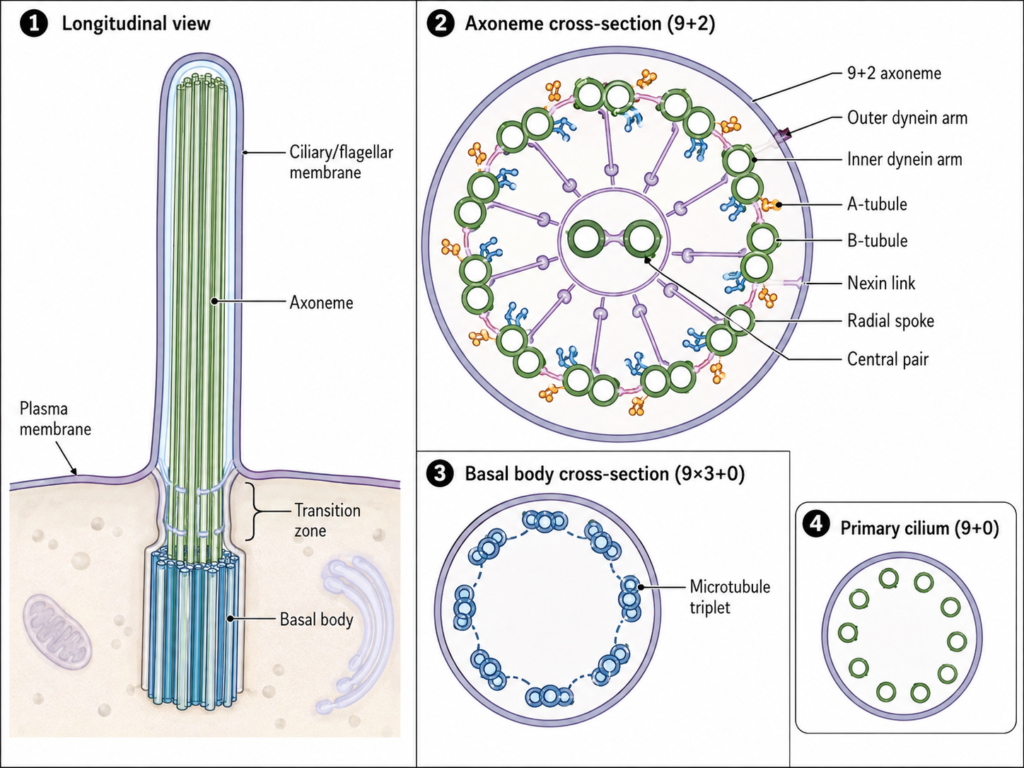
- Ciliary or Flagellar Membrane – The outer surface is covered by a membrane which is continuous with the plasma membrane of the cell. It surrounds the projecting portion. Different membrane proteins and receptors are also present in this membrane.
- Axoneme – The main structural core of cilia and flagella is called the axoneme. It extends along almost the complete length of the projection and is mainly formed of microtubules with several associated proteins.
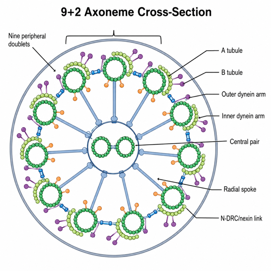
- 9+2 Microtubule Arrangement – A typical motile axoneme consists of nine peripheral microtubule doublets surrounding two single microtubules in the center. This arrangement is referred to as 9+2 arrangement. It is commonly found in motile cilia and eukaryotic flagella. Some cilia, particularly primary cilia, do not contain the central pair and generally show 9+0 arrangement.
- Peripheral Microtubule Doublets – Each of the nine outer doublets contains two microtubules, called A-tubule and B-tubule. A-tubule is a complete microtubule. The B-tubule is incomplete and remains attached with the A-tubule.
- Central Pair – Two single microtubules are present in the central region of a typical 9+2 axoneme. These form the central pair complex. The central pair is surrounded by associated proteins and has a role in regulation of ciliary and flagellar beating.
- Dynein Arms – Axonemal dynein is attached mainly with the A-tubule of peripheral doublets. Inner and outer dynein arms are present in many motile axonemes. These are ATP-dependent motor proteins. Their activity produces relative sliding between adjacent microtubule doublets.
- Radial Spokes – Radial spokes extend inward from the outer microtubule doublets towards the central pair region. They are protein complexes. These structures participate in control and coordination of axonemal movement.
- Nexin and Other Interdoublet Links – Adjacent outer microtubule doublets are joined by protein linkages. Nexin-dynein regulatory complex is one important linkage system. These connections restrict free sliding of the microtubules, due to which the sliding force is changed into bending of the axoneme.
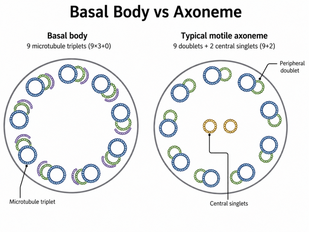
- Basal Body – At the base of every cilium or flagellum is a basal body. It is a centriole-like cylindrical structure and acts as the anchoring region. A typical basal body contains nine peripheral sets of microtubule triplets and no central pair, therefore having a 9×3+0 organization.
- Transition Zone – The region between the basal body and axoneme is called the transition zone. Here, the microtubular organization changes from basal-body triplets towards the doublets of the axoneme. It also forms an important boundary between the ciliary compartment and rest of the cell.
The microtubules of the axoneme are continuous with those organized from the basal region. Two microtubules of each basal-body triplet continue into the outer doublet of the axoneme, while the third microtubule ends around the transition region.
In some flagella, extra structures can also occur around the axoneme. Mammalian sperm flagellum, for example, contains outer dense fibres and other accessory structures in addition to the normal axoneme. These are not the basic structural components of every cilium and flagellum.
Axoneme vs Basal Body
| Characteristics | Axoneme | Basal Body |
|---|---|---|
| Definition | The internal microtubule core of a cilium or flagellum. | A centriole-like structure present at the base of a cilium or flagellum. |
| Location | Present inside the projecting part of cilia and flagella. | Located at the base, just below the cell surface. |
| Main structure | Made up of microtubule doublets with associated proteins. | Made up of microtubule triplets. |
| Typical arrangement | Motile forms commonly show a 9+2 arrangement. | Usually has 9 triplets and no central pair. |
| Microtubules | Contains A- and B-tubules in the outer doublets. | Each triplet contains A-, B- and C-tubules. |
| Central pair | Present in typical 9+2 axonemes. | Absent. |
| Dynein arms | Present in many motile axonemes. | Axonemal dynein arms are not a main basal-body feature. |
| Main function | Produces and supports ciliary or flagellar movement. | Anchors the structure and helps in formation of the axoneme. |
| Role in movement | Directly involved in bending and beating. | Mainly provides attachment and organization at the base. |
| Relation with centriole | Not a centriole-like structure itself. | Structurally closely related to a centriole. |
| Membrane relation | Lies inside the membrane-covered ciliary or flagellar shaft. | Present at the basal region where the projection originates. |
| Continuity | Extends outward from the basal region. | Gives rise to and organizes the axonemal microtubules. |
Types of Cilia
Cilia are of different types based mainly on their motility, number and arrangement of microtubules in the axoneme. They are generally divided into motile cilia, primary cilia and a specialized type called nodal cilia.
1. Motile Cilia
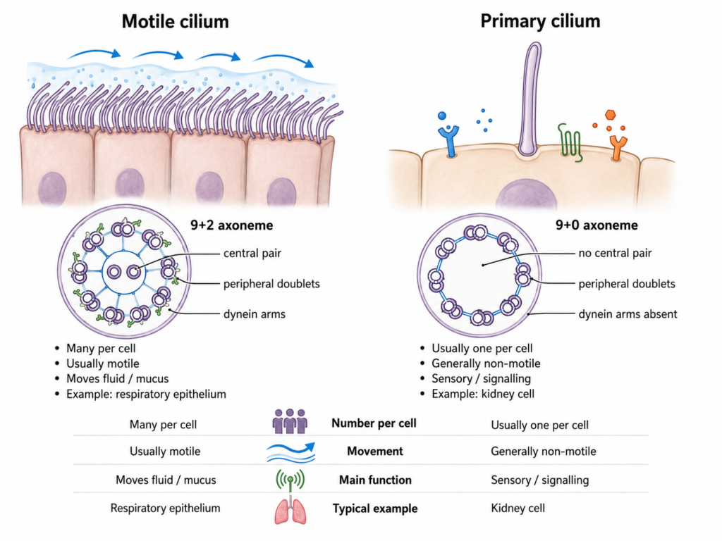
Motile cilia are cilia capable of regular beating movement. These are generally present in large numbers over the surface of a cell. Most of them contain the typical 9+2 axoneme, with nine peripheral microtubule doublets and two central microtubules. Dynein arms are also present and produce ciliary movement by ATP-dependent activity.
They mainly function in moving fluid, mucus or other substances along a cell surface. In the respiratory tract, the beating of these cilia moves mucus towards the pharynx. Motile cilia are also present in the fallopian tubes and on ependymal cells lining the brain ventricles.
2. Primary Cilia
Primary cilia are generally non-motile cilia and occur singly on the cell surface. One cilium is usually present on a cell. Their axoneme commonly has a 9+0 arrangement, in which nine peripheral doublets are present but the central pair is absent. Most primary cilia also lack the axonemal dynein machinery required for regular beating.
These cilia mainly perform sensory and signalling functions rather than movement. They receive different chemical and physical signals from outside the cell. Primary cilia are found on most vertebrate cell types and have important roles during development and maintenance of tissues.
Primary cilia are involved in different signalling pathways. Hedgehog signalling is one of the best-known examples. Defects in their structure or functioning are associated with a group of disorders called ciliopathies.
3. Nodal Cilia
Nodal cilia are a specialized type of motile cilia present during early vertebrate embryonic development. Unlike most motile cilia, they generally have a 9+0 microtubule arrangement. Dynein motors are present, therefore these cilia can move even without the central pair.
Their movement is mainly rotational rather than the typical back-and-forth beating of many 9+2 cilia. This rotation produces a directional fluid flow at the embryonic node. The flow has an important role in establishment of the normal left-right body asymmetry of internal organs.
Thus, the 9+2 arrangement cannot always be considered motile and 9+0 as non-motile. Nodal cilia are an important exception, being 9+0 and motile. Some specialized sensory cilia can also show structural variations from these usual patterns.
Motile vs Primary Cilia
| Characteristics | Motile Cilia | Primary Cilia |
|---|---|---|
| Movement | Usually show regular beating movement. | Generally non-motile. |
| Number per cell | Commonly present in large numbers. | Usually one cilium is present on a cell. |
| Axoneme | Most commonly have a 9+2 arrangement. | Usually have a 9+0 arrangement. |
| Central pair | Usually present. | Generally absent. |
| Dynein arms | Present in typical motile cilia. | Usually lack the axonemal dynein machinery needed for beating. |
| Main function | Mainly move fluid, mucus or other materials. | Mainly perform sensory and signalling functions. |
| Occurrence | Respiratory epithelium, oviduct and ependymal cells. | Present on many vertebrate cell types. |
| Beating pattern | Show coordinated beating with effective and recovery strokes. | Do not normally show this type of beating. |
| Signalling role | Can also have signalling functions. | Strongly involved in cell signalling pathways. |
| Example | Respiratory cilia. | Primary cilium of kidney or fibroblast cells. |
| Important exception | Some motile cilia, such as nodal cilia, can have a 9+0 arrangement. | Some specialized cilia can differ from the usual primary cilium pattern. |
How Do Cilia and Flagella Move?
The movement of cilia and flagella is produced by the bending of their axoneme. It is an ATP-dependent process. Axonemal dynein present between the peripheral microtubule doublets plays the major role in this movement.
Dynein-Driven Microtubule Sliding
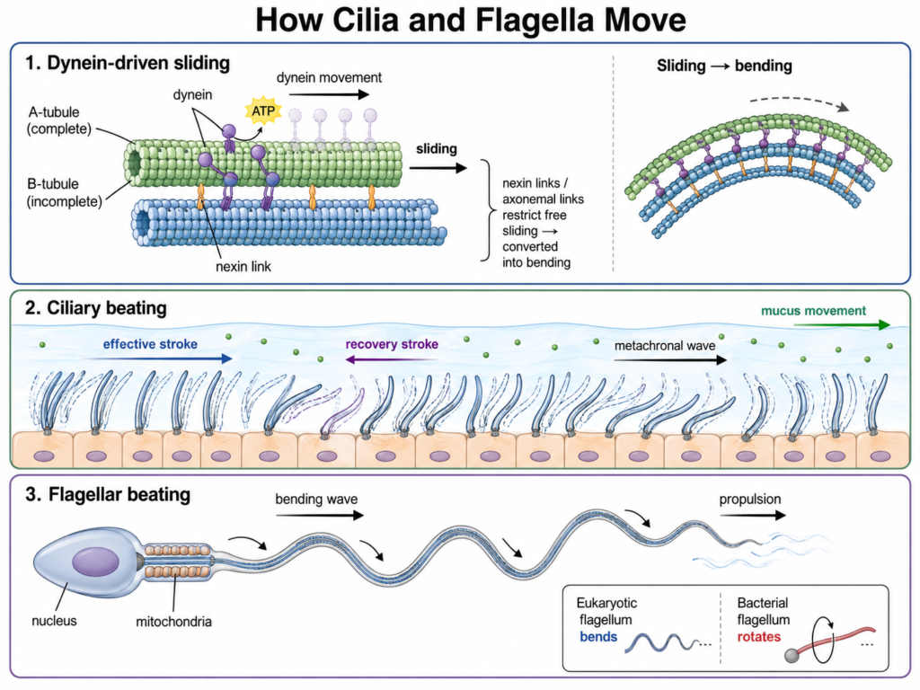
During this process, ATP binds to dynein and is hydrolyzed. The energy obtained is used by dynein for movement along the neighbouring microtubule doublet. Dynein is attached to the A-tubule, while its motor region interacts with the B-tubule of an adjacent doublet.
The action of dynein generates force between two neighbouring doublets. Due to this, the microtubule doublets tend to slide past each other. This is referred to as microtubule sliding.
But the doublets cannot slide freely for a long distance. They are connected by different axonemal structures and are also mechanically restricted within the axoneme. The sliding movement is therefore changed into bending. If these restrictions are removed, the doublets can show much greater sliding instead of normal bending.
Dynein motors are not activated equally throughout the axoneme. Their activity changes between different regions and different sides of the axoneme. Repeated activation of these motors produces successive bends. In this way, regular ciliary beats or flagellar bending waves are produced.
Ciliary Beating
Cilia commonly move by a back-and-forth beating pattern. A usual ciliary beat consists of two main parts, the effective stroke and the recovery stroke.
During the effective or power stroke, the cilium moves in a manner that pushes the surrounding fluid in one main direction. It is followed by a recovery stroke. In this step, the cilium bends and returns towards its original position for the next beat.
When many cilia are present together, their beating is coordinated. They generally do not all beat at exactly the same moment. Instead, a slight difference in timing occurs between neighbouring cilia and a travelling pattern is produced across the ciliated surface. This is called a metachronal wave.
Such coordinated beating is important for movement of fluid and other materials. In the respiratory tract, for example, ciliary beating helps in movement of the mucus layer containing trapped particles towards the pharynx.
Flagellar Beating
Eukaryotic flagella generally move by the formation of bending waves. The bend is generated in the axoneme by the same basic dynein-dependent sliding mechanism.
These bends pass along the length of the flagellum. A repeated wave-like or undulating movement is therefore formed. The exact shape and direction of the wave can vary between different flagellated cells.
Flagellar beating is used for propulsion of many eukaryotic cells. The flagellum of a sperm cell, for example, generates bending waves that help to move the sperm through the surrounding fluid.
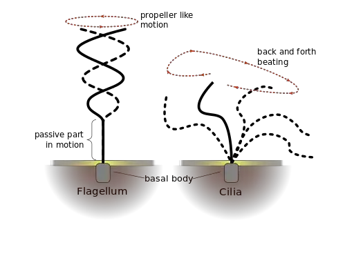
Eukaryotic flagella do not normally move by the rotary mechanism found in bacterial flagella. Bacterial flagella are structurally different and work by rotation, whereas movement of the eukaryotic flagellum is mainly produced by bending of a microtubule-containing axoneme.
Functions of Cilia and Flagella
Functions of Cilia
- Movement of mucus – Cilia move mucus over the respiratory epithelium.
- Fluid movement – They help in movement of fluid over the cell surface.
- Oocyte transport – Cilia in the uterine tube help in movement of the oocyte.
- Cerebrospinal fluid movement – Ependymal cilia help in movement and mixing of cerebrospinal fluid.
- Sensory function – Primary cilia receive different chemical and mechanical signals.
- Cell signalling – They take part in signalling pathways such as Hedgehog signalling.
- Left-right development – Nodal cilia help in establishment of left-right body asymmetry during embryonic development.
- Locomotion – In some unicellular organisms, cilia help in movement of the whole cell.
Functions of Flagella
- Cell locomotion – Flagella help in movement of many unicellular eukaryotes.
- Sperm movement – The sperm flagellum produces propulsion of the sperm cell.
- Swimming in liquid medium – Flagellar beating helps cells move through water or other fluids.
- Directional movement – Bending waves of flagella produce forward or turning movement.
- Reproductive role – Flagellar motility is important for normal sperm function and fertilization.
- Sensory role – Some eukaryotic flagella also contain sensory and signalling components.
Difference Between Cilia and Flagella
Cilia and flagella are closely related microtubule-based cell-surface structures. Cilia are generally shorter and numerous, while flagella are usually longer and one or few in number. Their main difference is seen in length, number, beating pattern and usual function.
| Characteristics | Cilia | Flagella |
|---|---|---|
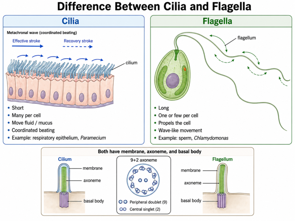
| Length | Usually shorter. | Usually longer. |
| Number | Present in large numbers on a cell. | Generally one or few are present. |
| Distribution | Often cover a large part of the cell surface. | Usually arise from a limited region of the cell. |
| Movement | Show repeated back-and-forth beating. | Mostly show wave-like or undulating movement. |
| Type of stroke | Commonly have effective and recovery strokes. | Bending waves pass along the flagellum. |
| Main function | Mainly move fluid or materials over the cell surface. | Mainly help in locomotion of the cell. |
| Locomotion | Can move some unicellular organisms. | Commonly propel motile cells such as sperm. |
| Coordination | Numerous cilia may beat together in coordinated pattern. | Usually works as one or a few long structures. |
| Metachronal waves | Commonly formed when many cilia beat together. | Generally not described as metachronal beating. |
| Examples | Respiratory epithelial cilia, cilia of Paramecium. | Sperm flagellum, flagellum of Chlamydomonas. |
| Basic structure | Contains a microtubule-based axoneme. | Also contains a similar microtubule-based axoneme. |
| Basal attachment | Arises from a basal body. | Also arises from a basal body. |
Similarities Between Cilia and Flagella
The major similarities between cilia and flagella are as follows-
- Both are hair-like projections present on the surface of eukaryotic cells.
- Cilia and flagella are covered by the plasma membrane.
- Both contain a microtubule-based internal core called the axoneme.
- The axoneme of typical motile forms commonly shows a 9+2 microtubule arrangement.
- Both arise from a basal body present at their base.
- Axonemal dynein is involved in movement of motile cilia and flagella.
- Their movement requires ATP as an energy source.
- In both structures, movement is produced by sliding of adjacent microtubule doublets followed by bending of the axoneme.
- Both can take part in movement. They may move the whole cell or help in movement of materials around the cell.
- Cilia and flagella have closely related basic structure in eukaryotes, even though they differ in length, number and usual beating pattern.
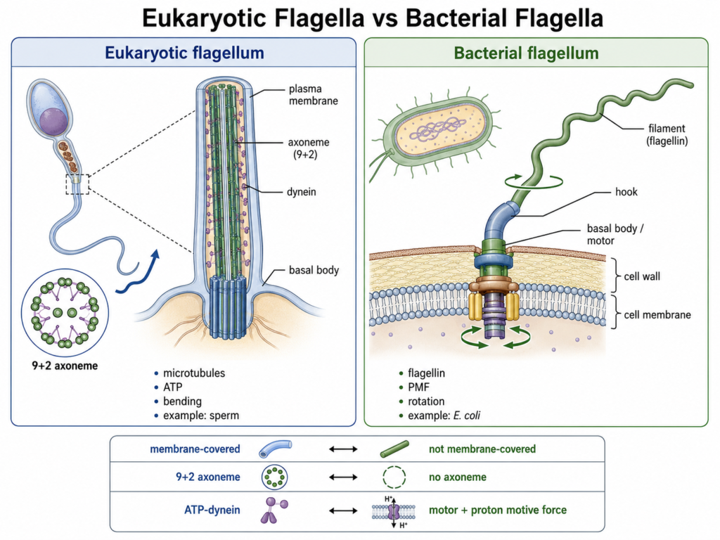
Eukaryotic Flagella vs Bacterial Flagella
| Characteristics | Eukaryotic Flagella | Bacterial Flagella |
|---|---|---|
| Basic structure | Made up of microtubules and associated proteins. | Mainly made up of the protein flagellin. |
| Internal organization | Contains an axoneme, commonly with a 9+2 microtubule arrangement in motile forms. | Does not contain an axoneme or 9+2 microtubule arrangement. |
| Membrane covering | Covered by the plasma membrane. | Not covered by the plasma membrane. |
| Basal region | Arises from a basal body related to centrioles. | Attached to a basal body and hook complex present in the cell envelope. |
| Movement | Movement takes place by bending or wave-like beating. | Movement occurs mainly by rotation of the flagellar filament. |
| Motor system | Movement is produced by axonemal dynein acting on microtubules. | Rotation is produced by a membrane-associated flagellar motor. |
| Energy source | ATP is used directly by dynein motors. | Usually driven by proton motive force, and in some bacteria by sodium ion gradient. |
| Type of motion | Produces bending waves along the flagellum. | Rotates like a helical propeller. |
| Size | Generally thicker and structurally more complex. | Thinner and structurally simpler than eukaryotic flagella. |
| Occurrence | Found in cells such as sperm and many unicellular eukaryotes. | Found in many motile bacterial species. |
| Examples | Sperm flagellum, Chlamydomonas flagella. | Flagella of Escherichia coli and Salmonella. |
| Main function | Mainly used for locomotion of eukaryotic cells. | Mainly used for bacterial motility and movement towards or away from environmental signals. |
What Happens When Ciliary or Flagellar Function Is Disrupted?
Defects in cilia and flagella can affect movement, fluid transport and sensory functions of different cells. The effects depend on which type of cilia or flagella is affected.
- Impaired mucociliary clearance – Defective respiratory cilia cannot move mucus properly. Mucus and trapped particles therefore remain in the airways.
- Chronic respiratory infections – Poor mucus clearance increases the chance of repeated respiratory infections. Long-term airway disease can also develop.
- Reduced fertility – Defects of motile cilia in the reproductive tract can reduce fertility in both males and females.
- Abnormal sperm motility – Structural or functional defects of the sperm flagellum can result in poorly motile or immotile sperm. Normal sperm movement is therefore affected.
- Laterality defects – Abnormal function of embryonic nodal cilia can disturb normal left-right body patterning. This may result in conditions such as situs inversus or other laterality abnormalities.
- Ciliopathies – Disorders produced by defects in cilia, their associated proteins or ciliary functions are generally called ciliopathies. These disorders may involve motile cilia, primary cilia or both. Different organ systems can therefore be affected.
Primary Ciliary Dyskinesia
Primary Ciliary Dyskinesia (PCD) is a genetic disorder mainly affecting the structure or function of motile cilia. Abnormal ciliary beating results in poor clearance of mucus from the respiratory tract.
Repeated respiratory problems are common. Chronic wet cough, sinus disease and recurrent respiratory infections can occur because mucociliary clearance is reduced.
Fertility may also be affected. Male fertility can be reduced due to abnormal sperm flagellar function in some forms of PCD, while impaired motile cilia of the female reproductive tract can also cause subfertility. These effects are not identical in every affected individual.
Some people with PCD also have abnormal arrangement of internal organs. This results from defects of nodal cilia during early embryonic development. Laterality abnormalities occur only in a proportion of affected individuals and depend partly on the underlying genetic defect.
Frequently Asked Questions (FAQs)
1. What are cilia and flagella?
Cilia and flagella are hair-like projections present on the surface of many eukaryotic cells. Both contain a microtubule-based internal core called the axoneme.
2. What is the main function of cilia and flagella?
Cilia mainly help in movement of fluid, mucus or other materials over the cell surface. Flagella are commonly used for locomotion of the whole cell.
3. What is the difference between cilia and flagella?
Cilia are usually shorter and present in large numbers. Flagella are generally longer and only one or few are present on a cell.
4. What is the 9+2 arrangement?
The 9+2 arrangement consists of nine peripheral microtubule doublets surrounding two central microtubules. It is commonly found in motile eukaryotic cilia and flagella.
5. What is an axoneme?
The axoneme is the main internal microtubule-containing core of a cilium or flagellum. It extends through most of the projecting part.
6. What is the basal body of a cilium or flagellum?
The basal body is a centriole-like structure present at the base of cilia and flagella. It anchors the structure and helps in organization of the axoneme.
7. How do cilia and flagella move?
Their movement is produced by bending of the axoneme. Dynein causes sliding between adjacent microtubule doublets, and this sliding is converted into bending.
8. Which motor protein causes ciliary and flagellar movement?
Axonemal dynein is the main motor protein involved. It uses ATP to generate force between neighbouring microtubule doublets.
9. Are cilia and flagella made of microtubules?
Yes. Eukaryotic cilia and flagella contain microtubules along with several associated proteins.
10. Are cilia and flagella membrane-bound?
Eukaryotic cilia and flagella are covered by a membrane continuous with the plasma membrane of the cell.
11. Where are cilia found in the human body?
Cilia are present in the respiratory tract, uterine tubes and ventricular ependyma. Primary cilia are also present on many other cell types.
12. Is a sperm tail a flagellum?
Yes. The tail of a mammalian sperm cell is a specialized flagellum that produces the movement required for sperm propulsion.
13. What is the difference between 9+2 and 9+0 cilia?
A 9+2 cilium has nine outer microtubule doublets and two central microtubules. A 9+0 cilium lacks the central pair and commonly occurs in primary cilia, although some motile cilia also have 9+0 organization.
14. Are cilia present in prokaryotic cells?
True eukaryotic-type cilia are not present in prokaryotic cells. Bacteria may have flagella, but these are structurally very different from eukaryotic cilia and flagella.
15. Are bacterial and eukaryotic flagella the same?
No. Eukaryotic flagella contain microtubules and move mainly by bending. Bacterial flagella are mainly composed of flagellin and move by rotation.
16. What happens when cilia do not work properly?
Defective cilia can cause poor mucus clearance, repeated respiratory infections, fertility problems and abnormalities in left-right body development. Disorders caused by ciliary defects are generally called ciliopathies.
References
- Alberts, B., Johnson, A., Lewis, J., Raff, M., Roberts, K., & Walter, P. (2002). Molecular biology of the cell (4th ed.). Garland Science. https://www.ncbi.nlm.nih.gov/books/NBK21054/
- Bernabé-Rubio, M., & Alonso, M. A. (2017). Routes and machinery of primary cilium biogenesis. Cellular and Molecular Life Sciences, 74(22), 4077–4095. https://doi.org/10.1007/s00018-017-2570-5
- Bouhouche, K., Valentine, M. S., Le Borgne, P., Lemullois, M., Yano, J., Lodh, S., Nabi, A., Tassin, A. M., & Van Houten, J. L. (2022). Paramecium, a model to study ciliary beating and ciliogenesis: Insights from cutting-edge approaches. Frontiers in Cell and Developmental Biology, 10, 847908. https://doi.org/10.3389/fcell.2022.847908
- Brette, R. (2021). Integrative neuroscience of Paramecium, a “swimming neuron.” eNeuro, 8(3), ENEURO.0018-21.2021. https://doi.org/10.1523/ENEURO.0018-21.2021
- Bustamante-Marin, X. M., & Ostrowski, L. E. (2017). Cilia and mucociliary clearance. Cold Spring Harbor Perspectives in Biology, 9(4), a028241. https://doi.org/10.1101/cshperspect.a028241
- Chang, C.-F., Schock, E. N., Attia, A. C., Stottmann, R. W., & Brugmann, S. A. (2015). The ciliary baton: Orchestrating neural crest cell development. Current Topics in Developmental Biology, 111, 97–134. https://doi.org/10.1016/bs.ctdb.2014.11.004
- Cicconofri, G., Noselli, G., & DeSimone, A. (2021). The biomechanical role of extra-axonemal structures in shaping the flagellar beat of Euglena gracilis. eLife, 10, e58610. https://doi.org/10.7554/eLife.58610
- Cooper, G. M. (2000). The cell: A molecular approach (2nd ed.). Sinauer Associates. https://www.ncbi.nlm.nih.gov/books/NBK9833/
- Cui, Z., Wang, Y., & den Toonder, J. M. J. (2024). Metachronal motion of biological and artificial cilia. Biomimetics, 9(4), 198. https://doi.org/10.3390/biomimetics9040198
- Despotes, K. A., Zariwala, M. A., Davis, S. D., & Ferkol, T. W. (2024). Primary ciliary dyskinesia: A clinical review. Cells, 13(11), 974. https://doi.org/10.3390/cells13110974
- Fliegauf, M., Benzing, T., & Omran, H. (2007). When cilia go bad: Cilia defects and ciliopathies. Nature Reviews Molecular Cell Biology, 8, 880–893. https://doi.org/10.1038/nrm2278
- Goetz, S. C., & Anderson, K. V. (2010). The primary cilium: A signalling centre during vertebrate development. Nature Reviews Genetics, 11(5), 331–344. https://doi.org/10.1038/nrg2774
- Hilgendorf, K. I., Myers, B. R., & Reiter, J. F. (2024). Emerging mechanistic understanding of cilia function in cellular signalling. Nature Reviews Molecular Cell Biology, 25(7), 555–573. https://doi.org/10.1038/s41580-023-00698-5
- Ishikawa, T. (2017). Axoneme structure from motile cilia. Cold Spring Harbor Perspectives in Biology, 9(1), a028076. https://doi.org/10.1101/cshperspect.a028076
- Ji, W., Tang, Z., Chen, Y., Wang, C., Tan, C., Liao, J., Tong, L., & Xiao, G. (2022). Ependymal cilia: Physiology and role in hydrocephalus. Frontiers in Molecular Neuroscience, 15, 927479. https://doi.org/10.3389/fnmol.2022.927479
- King, S. M., & Sale, W. S. (2018). Fifty years of microtubule sliding in cilia. Molecular Biology of the Cell, 29(6), 698–701. https://doi.org/10.1091/mbc.E17-07-0483
- Kubo, A., Yuba-Kubo, A., Tsukita, S., & Tsukita, S. (2008). Sentan: A novel specific component of the apical structure of vertebrate motile cilia. Molecular Biology of the Cell, 19(12), 5338–5346. https://doi.org/10.1091/mbc.E08-07-0691
- Long, H., Wang, Q., & Huang, K. (2015). Ciliary/flagellar protein ubiquitination. Cells, 4(3), 474–482. https://doi.org/10.3390/cells4030474
- Malicki, J. J., & Johnson, C. A. (2017). The cilium: Cellular antenna and central processing unit. Trends in Cell Biology, 27(2), 126–140. https://doi.org/10.1016/j.tcb.2016.08.002
- Nachury, M. V., & Mick, D. U. (2019). Establishing and regulating the composition of cilia for signal transduction. Nature Reviews Molecular Cell Biology, 20(7), 389–405. https://doi.org/10.1038/s41580-019-0116-4
- Pablo, J. L., DeCaen, P. G., & Clapham, D. E. (2017). Progress in ciliary ion channel physiology. Journal of General Physiology, 149(1), 37–47. https://doi.org/10.1085/jgp.201611696
- Reiter, J. F., & Leroux, M. R. (2017). Genes and molecular pathways underpinning ciliopathies. Nature Reviews Molecular Cell Biology, 18(9), 533–547. https://doi.org/10.1038/nrm.2017.60
- Roberts, A. J., Kon, T., Knight, P. J., Sutoh, K., & Burgess, S. A. (2013). Functions and mechanics of dynein motor proteins. Nature Reviews Molecular Cell Biology, 14(11), 713–726. https://doi.org/10.1038/nrm3667
- Shapiro, A. J., Davis, S. D., Polineni, D., Manion, M., Rosenfeld, M., Dell, S. D., Chilvers, M. A., Ferkol, T. W., Zariwala, M. A., Sagel, S. D., Josephson, M., Morgan, L., Yilmaz, O., Olivier, K. N., Milla, C., Pittman, J. E., Daniels, M. L. A., Jones, M. H., Janahi, I. A., . . . Lavergne, V. (2018). Diagnosis of primary ciliary dyskinesia: An official American Thoracic Society clinical practice guideline. American Journal of Respiratory and Critical Care Medicine, 197(12), e24–e39. https://doi.org/10.1164/rccm.201805-0819ST
- Sironen, A., Shoemark, A., Patel, M., Loebinger, M. R., & Mitchison, H. M. (2020). Sperm defects in primary ciliary dyskinesia and related causes of male infertility. Cellular and Molecular Life Sciences, 77(11), 2029–2048. https://doi.org/10.1007/s00018-019-03389-7
- Spassky, N., & Meunier, A. (2017). The development and functions of multiciliated epithelia. Nature Reviews Molecular Cell Biology, 18(7), 423–436. https://doi.org/10.1038/nrm.2017.21
- Veland, I. R., Awan, A., Pedersen, L. B., Yoder, B. K., & Christensen, S. T. (2009). Primary cilia and signaling pathways in mammalian development, health and disease. Nephron Physiology, 111(3), p39–p53. https://doi.org/10.1159/000208212
- Wloga, D., Joachimiak, E., Osinka, A., Ahmadi, S., & Majhi, S. (2024). Motile cilia in female and male reproductive tracts and fertility. Cells, 13(23), 1974. https://doi.org/10.3390/cells13231974