

Reactive Oxygen Species (ROS) are a group of reactive oxygen-derived chemical species that are normally formed in biological systems.
They are not only harmful cellular by-products. At regulated amounts, ROS take part in redox signaling and cellular defense, including antimicrobial responses, whereas excessive or poorly controlled ROS can result in oxidative stress and molecular damage.
ROS and free radicals are not exactly the same. Some ROS are free radicals, such as superoxide anion radical (O₂•⁻) and hydroxyl radical (•OH), while hydrogen peroxide (H₂O₂) is a non-radical oxidant. Cells continuously generate and remove ROS with different antioxidant defense systems. This balance is referred to as redox homeostasis, and disturbance of this control towards excessive oxidant activity can interfere with normal redox signaling and increase oxidative damage.
The biological action of ROS varies with the particular species, its amount, cellular location and duration, together with the antioxidant capacity present in that cell or cellular compartment.
Causes of reactive oxygen species (ROS)
Reactive oxygen species (ROS) can arise through controlled enzymatic production and as products of normal cellular metabolism.
ROS production is not limited to a single cellular organelle. Mitochondria and reduced nicotinamide adenine dinucleotide phosphate (NADPH) oxidases (NOXs) are prominent sources, along with peroxisomes, endoplasmic reticulum (ER) and several other enzyme systems. The amount produced by each source varies with cell type and cellular condition.
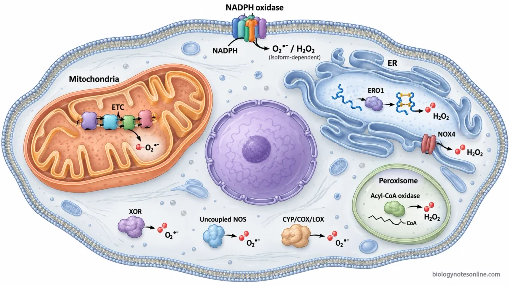
- Mitochondria- During oxidative phosphorylation, electrons are transferred through the mitochondrial electron transport chain (ETC). Some electrons can pass directly to molecular oxygen, producing superoxide instead of completing normal reduction of oxygen to water. Complex I is an important site, while the ETC and several other mitochondrial redox enzymes can take part in mitochondrial ROS production.
- NOXs- These enzymes are specialized for the controlled production of ROS. NOXs transfer electrons from NADPH to molecular oxygen and form superoxide or hydrogen peroxide (H₂O₂), depending on the enzyme system. In phagocytic cells, NOX2 produces a large amount of ROS during the respiratory burst, whereas other NOXs produce localized ROS for different cellular processes.
- Peroxisomes– Many oxidative metabolic reactions take place in peroxisomes. Fatty-acid β-oxidation and different flavin-dependent oxidases generate H₂O₂ during their catalytic reactions. Acyl-CoA oxidases are one of the important peroxisomal ROS-producing enzyme systems.
- ER- Oxidative protein folding is one source of ROS in the ER. Endoplasmic reticulum oxidoreductin 1 (ERO1) transfers electrons during disulfide-bond formation and molecular oxygen is reduced, producing H₂O₂. ER-localized NOXs and cytochrome P450 (CYP) enzyme reactions can also contribute to ROS formation.
- Xanthine oxidoreductase (XOR)- During purine metabolism, xanthine oxidase (XO) oxidizes hypoxanthine to xanthine and then xanthine to uric acid. Electrons can be transferred to oxygen in these reactions, producing superoxide and H₂O₂. ROS formation through this system can increase under conditions such as hypoxia and ischemia.
- Uncoupled nitric oxide synthase (NOS)- Normally, NOS uses electrons for the formation of nitric oxide (NO). When the enzyme becomes “uncoupled”, electrons are transferred to oxygen and superoxide is produced instead. Deficiency of tetrahydrobiopterin (BH₄), low L-arginine availability and some oxidative modifications can promote NOS uncoupling.
- Lipid-metabolizing enzyme systems- Cyclooxygenase (COX), lipoxygenase (LOX) and CYP enzymes can contribute to cellular ROS production during oxidation of fatty acids, arachidonic acid and other substrates. Their contribution differs considerably between tissues and physiological conditions.
External Factors That Increase ROS Generation
Different environmental and chemical factors can increase reactive oxygen species (ROS) generation in the cells. Some factors directly produce reactive species, while others increase cellular ROS-producing reactions or interfere with the antioxidant defence. Some of the important external factors are as follows-
- Ultraviolet (UV) radiation- UV radiation, mainly ultraviolet A (UVA) and ultraviolet B (UVB), increases ROS formation in exposed tissues such as skin. Cellular chromophores absorb the radiation and become excited. The energy or electrons can then be transferred to molecular oxygen, forming singlet oxygen, superoxide and other ROS.
- Ionizing radiation (IR)- X-rays, gamma rays and other IR can produce ROS by radiolysis of cellular water. During this process, hydroxyl radicals and hydrogen peroxide (H₂O₂) are formed rapidly. IR can also disturb mitochondrial metabolism. Further ROS may be generated after the initial radiation exposure.
- Air pollutants- Ozone, traffic exhaust and fine particulate matter (PM2.5) increase cellular ROS production. Some pollutants contain redox-active chemicals and metals, while others stimulate the ROS-producing reactions after entering or coming in contact with tissues.
- Cigarette smoke (CS)- CS contains free radicals, peroxides and several other pro-oxidant chemicals. These can react directly in biological fluids. CS also activates cellular systems involved in ROS production, and transition metals present in the smoke take part in redox reactions to form additional radicals.
- Heavy metals- Exposure to iron, copper, chromium, arsenic and several other metals can increase ROS generation. Redox-active metals take part in electron-transfer reactions. Some of them also catalyze hydroxyl radical formation through Fenton or Fenton-like reactions. Metals such as cadmium and lead mainly increase oxidative stress by interfering with the cellular antioxidant systems.
- Pesticides and industrial chemicals- Pesticides, insecticides, industrial solvents and some other environmental chemicals can act as cellular pro-oxidants. Depending on the chemical, ROS is produced through redox cycling, metabolic activation or stimulation of the endogenous ROS-producing enzymes.
- Drugs and other xenobiotics- Certain pharmaceutical drugs and foreign chemicals increase ROS during their metabolism. Some compounds undergo redox reactions or form reactive metabolites, producing superoxide, H₂O₂ and other reactive species inside the cell.
Major Types of Reactive Oxygen Species
Reactive oxygen species (ROS) include both free-radical and non-radical oxygen-derived species. The four major ROS commonly considered in biological systems are superoxide, hydrogen peroxide, hydroxyl radical and singlet oxygen.
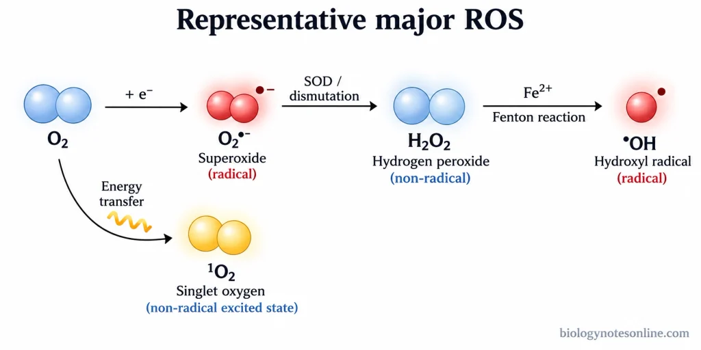
- Superoxide anion radical (O₂•⁻)- It is formed by the one-electron reduction of molecular oxygen (O₂). Superoxide is a free radical and is comparatively less reactive than hydroxyl radical. It can be converted into hydrogen peroxide by spontaneous dismutation or by superoxide dismutase (SOD).
- Hydrogen peroxide (H₂O₂)- H₂O₂ is a non-radical ROS. It is formed from superoxide and is also produced directly by several oxidase enzymes. H₂O₂ is comparatively stable and can participate in cellular redox signalling. In the presence of ferrous or cuprous ions, it can form highly reactive hydroxyl radical through Fenton chemistry.
- Hydroxyl radical (•OH)- It is one of the highly reactive oxygen radicals. Hydroxyl radical is commonly formed from H₂O₂ in metal-catalyzed reactions and reacts rapidly with nearby cellular molecules.
- Singlet oxygen (¹O₂)- This is an electronically excited and non-radical form of molecular oxygen. It can be formed by transfer of energy from an excited photosensitizer to O₂. Singlet oxygen is highly reactive with different biological molecules.
Physiological Functions of ROS
At controlled concentration, reactive oxygen species (ROS) perform different normal functions in the cell. Hydrogen peroxide (H₂O₂) and superoxide (O₂•⁻) are important in these regulated cellular processes. Their production changes according to the cellular condition and requirement.
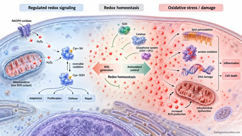
Some of the physiological functions of ROS are as follows-
- Redox signaling- ROS act as intracellular signaling molecules. H₂O₂ is one of the major ROS involved in this process and can reversibly oxidize redox-sensitive proteins, especially cysteine residues. Protein phosphatases, kinases and transcription factors are regulated by such oxidation. The activity of different signaling pathways can then change with cellular redox condition.
- Superoxide in cellular signaling- Superoxide is not only a damaging free radical. Nicotinamide adenine dinucleotide phosphate (NADPH) oxidases (NOXs) and mitochondria produce O₂•⁻ in a regulated manner, where it takes part in local redox signaling. It is short-lived. Superoxide dismutase (SOD) converts a large amount of it into H₂O₂, which can act on other cellular targets.
- Cell proliferation and survival- Growth factors and cytokines can stimulate controlled ROS production, and the ROS regulate proteins involved in cell growth, proliferation and survival. Phosphatidylinositol 3-kinase (PI3K), mitogen-activated protein kinase (MAPK) pathways and protein tyrosine phosphatases are some redox-sensitive signaling systems.
- Cell differentiation– Controlled ROS levels take part in differentiation of different cell types by modifying signaling pathways and transcriptional programs that determine cellular fate. Cellular redox condition and metabolic state also influence stem-cell proliferation and differentiation.
- Regulation of cellular metabolism- ROS interact with metabolic enzymes and signaling pathways. At physiological level, H₂O₂ and O₂•⁻ can alter enzyme activity according to nutrient, energy and redox condition of the cell. Glycolysis, mitochondrial metabolism and pentose phosphate pathway are regulated in part by such ROS-dependent mechanisms.
- Adaptation during cellular stress- A moderate rise in ROS can act as a signal for cellular adaptation rather than cell injury. Redox-sensitive transcription factors activate genes associated with antioxidant defence and cellular protection. During hypoxia, ROS are also involved. Mitochondrial and NOX-derived ROS participate in signaling pathways that respond to reduced oxygen availability.
- Host defense- In neutrophils and other phagocytic cells, NOX2 becomes activated after recognition and engulfment of microorganisms. O₂•⁻ is then formed rapidly. This is referred to as the “respiratory burst”. Superoxide gives rise to H₂O₂ and other antimicrobial oxidants inside the phagocytic compartment. These ROS are used for killing bacteria, fungi and other invading microorganisms.
- Cell migration and tissue repair- During normal wound healing, ROS take part in cellular signaling, and H₂O₂ helps in recruitment and movement of cells towards the damaged region. ROS also regulate proliferation of keratinocytes, fibroblasts and endothelial cells during tissue repair.
- Angiogenesis- Low and regulated ROS concentrations participate in the formation of new blood vessels. Superoxide and H₂O₂ influence endothelial-cell signaling, migration and proliferation, while ROS-dependent pathways also interact with vascular endothelial growth factor (VEGF) signaling during angiogenesis and tissue repair.
Oxidative Stress and Cellular Damage
Oxidative stress occurs when the generation of oxidants, including reactive oxygen species (ROS), becomes higher than the antioxidant and redox defence of the cell. The normal redox balance is disturbed. At high level, ROS can oxidize cellular molecules and also disturb the controlled redox signaling.
Some of the major effects of excessive ROS are as follows-
- Lipid peroxidation- Polyunsaturated fatty acids present in the cell membranes can be attacked by free radicals. Lipid oxidation then continues as a chain reaction. Lipid hydroperoxides and reactive aldehydes are formed. During this process, membrane fluidity and integrity can also be changed.
- Protein oxidation- Proteins and enzymes are oxidized by excessive ROS, which can modify amino-acid residues and the protein structure. Enzyme activity may be reduced or altered. Some oxidized proteins are removed by the cellular protein-degradation systems.
- Deoxyribonucleic acid (DNA) damage- ROS can oxidize DNA bases and also damage its sugar-phosphate backbone. Different DNA lesions are formed, including single-strand damage. If these lesions are not correctly repaired, DNA replication and transcription can be affected.Mitochondrial DNA (mtDNA) is also exposed to oxidative damage, especially because it remains close to the mitochondrial ROS-producing sites.
- Mitochondrial damage- Mitochondria itself can become a target when ROS is high. The mtDNA, membrane components and proteins involved in mitochondrial respiration may be oxidatively damaged. Mitochondrial function becomes disturbed. Severe oxidative injury is also associated with reduced energy production and activation of cell-death pathways. Damaged mitochondria can produce further ROS.
- Disruption of redox signaling- Normal ROS signaling depends on controlled and localized oxidation. With excessive ROS, redox-sensitive proteins can undergo abnormal oxidation and the normal regulation of cellular signaling is changed.
- Inflammation- ROS and inflammation are closely connected. Excess ROS can modify nuclear factor kappa B (NF-κB) signaling, and the response differs with the cell type and cellular condition. Mitochondrial ROS can also take part in activation of the NLR family pyrin domain containing 3 (NLRP3) inflammasome in different inflammatory conditions.During this process, maturation of inflammatory cytokines such as interleukin-1 beta (IL-1β) and interleukin-18 (IL-18) can be promoted. Activated inflammatory cells also produce ROS. More oxidant load can then occur in the surrounding tissue.
- Cell death- When oxidative damage becomes severe and the cellular repair systems cannot maintain the damaged components, apoptotic or necrotic cell death can occur.
- Tissue damage- Persistent or repeated oxidative stress causes accumulation of damage in cellular lipids, proteins and DNA. Such oxidative injury is found in different inflammatory, metabolic, cardiovascular and neurodegenerative disorders, although oxidative stress is not the single cause of these conditions.
Antioxidant Defense Systems
Antioxidant defense systems do not normally remove all reactive oxygen species (ROS) from the cells. The amount of ROS is kept within a controlled range. Reactive species are converted, peroxides are removed and some reactions that produce highly reactive radicals are restricted, while physiological redox signaling can still take place.
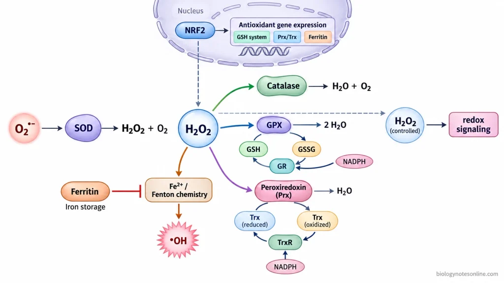
- Superoxide dismutase (SOD) converts superoxide anion radical (O₂•⁻) into hydrogen peroxide (H₂O₂) and molecular oxygen (O₂). The product H₂O₂ is still a ROS and has to be handled by other antioxidant systems.2O₂•⁻ + 2H⁺ → H₂O₂ + O₂
- Catalase (CAT) decomposes H₂O₂ into water and O₂, and in mammalian cells much of this enzyme is associated with peroxisomes.2H₂O₂ → 2H₂O + O₂
- Glutathione peroxidases (GPXs) reduce H₂O₂ and can also reduce lipid hydroperoxides. Reduced glutathione (GSH) provides the reducing equivalents during this reaction. GSH becomes oxidized to glutathione disulfide (GSSG). It has to be regenerated. Glutathione reductase (GR) converts GSSG back to GSH with electrons supplied by reduced nicotinamide adenine dinucleotide phosphate (NADPH).
- Peroxiredoxins (Prxs) react rapidly with H₂O₂ and other peroxides. The oxidized Prxs are reduced again through the thioredoxin system, where thioredoxin (Trx), thioredoxin reductase (TrxR) and NADPH take part in the recycling process. Prxs are also involved in H₂O₂ sensing and redox signaling, so their function is not simply complete removal of H₂O₂.
- GSH also acts as an important cellular redox buffer and helps in maintaining protein thiols in an appropriate redox state.
- Ferritin stores iron in a compact form and decreases the amount of free ferrous iron (Fe²⁺) available for Fenton chemistry. Formation of highly reactive hydroxyl radicals is restricted by this mechanism.
- When oxidant load increases, the antioxidant defence can also be increased at the gene-expression level. Nuclear factor erythroid 2-related factor 2 (NRF2) controls the expression of many antioxidant and detoxification genes. The cellular antioxidant capacity can be adjusted through this response.
- The antioxidant systems are distributed in different cellular compartments rather than acting at a single place, with different SOD forms and peroxide-removing systems controlling ROS close to their sites of production and signaling.
Disease Associations with ROS
Altered reactive oxygen species (ROS) production and oxidative stress are associated with different human diseases. ROS is generally one part of the disease mechanism, rather than being the single cause. The effect varies with the amount, site and duration of ROS production.
- Cardiovascular diseases- Increased ROS occurs in different cardiovascular diseases (CVDs), including hypertension, atherosclerosis, myocardial infarction and heart failure. In the blood vessels, excess ROS can decrease the availability of nitric oxide (NO). Endothelial dysfunction can occur. Vascular inflammation and abnormal remodeling are also associated with this oxidative condition. Nicotinamide adenine dinucleotide phosphate (NADPH) oxidases (NOXs), mitochondria and other oxidase systems take part in the increased ROS production.
- Neurodegenerative diseases- Oxidative stress is commonly observed in Alzheimer’s disease (AD), Parkinson’s disease (PD), Huntington’s disease (HD) and amyotrophic lateral sclerosis (ALS). Proteins, membrane lipids and nucleic acids can undergo oxidative damage, while mitochondrial dysfunction and neuroinflammation are also linked with the redox disturbance in these diseases.
- Cancer- Cancer cells commonly have an altered redox balance and higher basal ROS than many normal cells. Moderate increase in ROS can support proliferative and survival signaling. Oxidative damage to deoxyribonucleic acid (DNA) can also contribute to genomic instability. The effect of ROS is not always tumor-promoting. At excessively high level, ROS can induce different forms of cancer-cell death.
- Type 2 diabetes- In type 2 diabetes (T2D), chronic hyperglycemia and metabolic stress can increase ROS production. Pancreatic β-cells are especially susceptible to oxidative stress because their antioxidant defence is comparatively limited. β-cell dysfunction, impaired insulin secretion and diabetic tissue complications are associated with excess ROS.
- Chronic inflammatory diseases- Activated inflammatory cells produce ROS, while increased ROS can modify inflammatory signaling and cytokine production. ROS and inflammation can occur together in a self-amplifying process in several chronic inflammatory conditions.
- Chronic kidney disease- Increased ROS and disturbed redox signaling are associated with chronic kidney disease (CKD). Oxidative damage, inflammation and renal fibrosis can take place during its progression. Mitochondria and NOXs are among the major renal ROS-producing systems.
Reactive Oxygen Species (ROS) at a Glance
| Topic | Quick exam points |
|---|---|
| Reactive oxygen species (ROS) | Oxygen-derived reactive molecules formed during cellular metabolism and by controlled enzyme systems. ROS include both free-radical and non-radical species. |
| Major intracellular sources | Mitochondria and nicotinamide adenine dinucleotide phosphate (NADPH) oxidases (NOXs) are prominent sources. Peroxisomes, endoplasmic reticulum (ER), xanthine oxidoreductase (XOR), uncoupled nitric oxide synthase (NOS) and other oxidase systems also produce ROS. |
| External factors increasing ROS | Ultraviolet (UV) radiation, ionizing radiation (IR), air pollutants, cigarette smoke, heavy metals, pesticides, industrial chemicals and some xenobiotics can increase ROS generation. |
| Superoxide anion radical (O₂•⁻) | Formed by one-electron reduction of molecular oxygen (O₂). It is a free radical. Superoxide dismutase (SOD) converts O₂•⁻ into hydrogen peroxide (H₂O₂). |
| Hydrogen peroxide (H₂O₂) | A non-radical ROS and important redox-signaling molecule. It is comparatively stable. In the presence of suitable transition metals, H₂O₂ can give rise to highly reactive hydroxyl radical. |
| Hydroxyl radical (•OH) | Highly reactive free radical. It reacts rapidly with nearby lipids, proteins and nucleic acids. |
| Singlet oxygen (¹O₂) | An electronically excited form of O₂. It is non-radical but highly reactive with biological molecules. |
| Physiological functions | Controlled ROS participate in redox signaling, cell proliferation, differentiation, metabolism, cellular adaptation, host defense, cell migration, tissue repair and angiogenesis. H₂O₂ and O₂•⁻ have regulated signaling roles. |
| Host defense | NOX2 produces O₂•⁻ during the “respiratory burst” of phagocytic cells. ROS-derived oxidants take part in killing engulfed microorganisms. |
| Oxidative stress | Occurs when oxidant generation becomes greater than antioxidant and redox defence. Normal cellular redox balance is disturbed. |
| Lipid damage | Excess ROS can cause lipid peroxidation, producing lipid hydroperoxides and reactive aldehydes. Membrane properties can be altered. |
| Protein damage | ROS can oxidize amino-acid residues and modify protein structure. Enzyme activity may be reduced or changed. |
| Deoxyribonucleic acid (DNA) damage | Oxidized bases and strand lesions can be formed. Mitochondrial DNA (mtDNA) is also affected by oxidative damage. |
| Mitochondrial damage | Excess ROS can damage mtDNA, membrane components and respiratory proteins. Mitochondrial function and cellular energy production may become disturbed. |
| ROS and inflammation | Excess ROS can modify inflammatory signaling. Activated inflammatory cells also generate ROS, adding further oxidant load in tissues. |
| Antioxidant defence | Antioxidant systems regulate ROS rather than eliminating them completely. ROS concentrations are maintained within a range compatible with normal redox signaling. |
| SOD | Converts O₂•⁻ into H₂O₂ and O₂. |
| Catalase (CAT) | Converts H₂O₂ into water (H₂O) and O₂. It is strongly associated with peroxisomal H₂O₂ removal. |
| Glutathione system | Glutathione peroxidases (GPXs) reduce H₂O₂ and lipid hydroperoxides using reduced glutathione (GSH). Glutathione reductase (GR) regenerates GSH using reduced NADPH. |
| Peroxiredoxin system | Peroxiredoxins (Prxs) remove peroxides and also participate in H₂O₂-dependent signaling. Thioredoxin (Trx) and thioredoxin reductase (TrxR) help regenerate reduced Prxs. |
| Disease associations | Abnormal ROS and oxidative stress are associated with CVDs, neurodegenerative diseases, cancer, type 2 diabetes (T2D), chronic inflammatory diseases and chronic kidney disease (CKD). ROS is generally one component of these disease mechanisms, not the single cause. |
References
- Abreu, I. A., & Cabelli, D. E. (2010). Superoxide dismutases—A review of the metal-associated mechanistic variations. Biochimica et Biophysica Acta (BBA) – Proteins and Proteomics, 1804(2), 263–274. https://doi.org/10.1016/j.bbapap.2009.11.005
- Averill-Bates, D. A. (2023). The antioxidant glutathione. In G. Litwack (Ed.), Vitamins and hormones (Vol. 121, pp. 109–141). Academic Press. https://doi.org/10.1016/bs.vh.2022.09.002
- Barnham, K. J., Masters, C. L., & Bush, A. I. (2004). Neurodegenerative diseases and oxidative stress. Nature Reviews Drug Discovery, 3(3), 205–214. https://doi.org/10.1038/nrd1330
- Bedard, K., & Krause, K.-H. (2007). The NOX family of ROS-generating NADPH oxidases: Physiology and pathophysiology. Physiological Reviews, 87(1), 245–313. https://doi.org/10.1152/physrev.00044.2005
- Bernardi, P., Di Lisa, F., Fogolari, F., & Lippe, G. (2015). From ATP to PTP and back: A dual function for the mitochondrial ATP synthase. Circulation Research, 116(11), 1850–1862. https://doi.org/10.1161/CIRCRESAHA.115.306557
- Camargo, L. L., Rios, F. J., Montezano, A. C., & Touyz, R. M. (2025). Reactive oxygen species in hypertension. Nature Reviews Cardiology, 22(1), 20–37. https://doi.org/10.1038/s41569-024-01062-6
- Chandimali, N., Bak, S. G., Park, E. H., Lim, H.-J., Won, Y.-S., Kim, E.-K., Park, S.-I., & Lee, S. J. (2025). Free radicals and their impact on health and antioxidant defenses: A review. Cell Death Discovery, 11, Article 19. https://doi.org/10.1038/s41420-024-02278-8
- DeCoursey, T. E., & Ligeti, E. (2005). Regulation and termination of NADPH oxidase activity. Cellular and Molecular Life Sciences, 62(19–20), 2173–2193. https://doi.org/10.1007/s00018-005-5177-1
- Dludla, P. V., Mabhida, S. E., Ziqubu, K., Nkambule, B. B., Mazibuko-Mbeje, S. E., Hanser, S., Basson, A. K., Pheiffer, C., & Kengne, A. P. (2023). Pancreatic β-cell dysfunction in type 2 diabetes: Implications of inflammation and oxidative stress. World Journal of Diabetes, 14(3), 130–146. https://doi.org/10.4239/wjd.v14.i3.130
- Dunnill, C., Patton, T., Brennan, J., Barrett, J., Dryden, M., Cooke, J., Leaper, D., & Georgopoulos, N. T. (2017). Reactive oxygen species (ROS) and wound healing: The functional role of ROS and emerging ROS-modulating technologies for augmentation of the healing process. International Wound Journal, 14(1), 89–96. https://doi.org/10.1111/iwj.12557
- Freinbichler, W., Colivicchi, M. A., Stefanini, C., Bianchi, L., Ballini, C., Misini, B., Weinberger, P., Linert, W., Varešlija, D., Tipton, K. F., & Della Corte, L. (2011). Highly reactive oxygen species: Detection, formation, and possible functions. Cellular and Molecular Life Sciences, 68(12), 2067–2079. https://doi.org/10.1007/s00018-011-0682-x
- Gebicka, L., & Krych-Madej, J. (2019). The role of catalases in the prevention/promotion of oxidative stress. Journal of Inorganic Biochemistry, 197, 110699. https://doi.org/10.1016/j.jinorgbio.2019.110699
- Halliwell, B. (2024). Understanding mechanisms of antioxidant action in health and disease. Nature Reviews Molecular Cell Biology, 25(1), 13–33. https://doi.org/10.1038/s41580-023-00645-4
- Irazabal, M. V., & Torres, V. E. (2020). Reactive oxygen species and redox signaling in chronic kidney disease. Cells, 9(6), 1342. https://doi.org/10.3390/cells9061342
- Juan, C. A., Pérez de la Lastra, J. M., Plou, F. J., & Pérez-Lebeña, E. (2021). The chemistry of reactive oxygen species (ROS) revisited: Outlining their role in biological macromolecules (DNA, lipids and proteins) and induced pathologies. International Journal of Molecular Sciences, 22(9), 4642. https://doi.org/10.3390/ijms22094642
- Lingappan, K. (2018). NF-κB in oxidative stress. Current Opinion in Toxicology, 7, 81–86. https://doi.org/10.1016/j.cotox.2017.11.002
- Lismont, C., Revenco, I., & Fransen, M. (2019). Peroxisomal hydrogen peroxide metabolism and signaling in health and disease. International Journal of Molecular Sciences, 20(15), 3673. https://doi.org/10.3390/ijms20153673
- Manful, C. F., Fordjour, E., Subramaniam, D., Sey, A. A., Abbey, L., & Thomas, R. (2025). Antioxidants and reactive oxygen species: Shaping human health and disease outcomes. International Journal of Molecular Sciences, 26(15), 7520. https://doi.org/10.3390/ijms26157520
- Marchi, S., Giorgi, C., Suski, J. M., Agnoletto, C., Bononi, A., Bonora, M., De Marchi, E., Missiroli, S., Patergnani, S., Poletti, F., Rimessi, A., Duszynski, J., Wieckowski, M. R., & Pinton, P. (2012). Mitochondria-ROS crosstalk in the control of cell death and aging. Journal of Signal Transduction, 2012, 329635. https://doi.org/10.1155/2012/329635
- Murphy, M. P. (2009). How mitochondria produce reactive oxygen species. Biochemical Journal, 417(1), 1–13. https://doi.org/10.1042/BJ20081386
- Murphy, M. P., Bayir, H., Belousov, V., Chang, C. J., Davies, K. J. A., Davies, M. J., Dick, T. P., Finkel, T., Forman, H. J., Janssen-Heininger, Y., Gems, D., Kagan, V. E., Kalyanaraman, B., Larsson, N.-G., Milne, G. L., Nyström, T., Poulsen, H. E., Radi, R., Van Remmen, H., … Halliwell, B. (2022). Guidelines for measuring reactive oxygen species and oxidative damage in cells and in vivo. Nature Metabolism, 4(6), 651–662. https://doi.org/10.1038/s42255-022-00591-z
- Ngo, V., & Duennwald, M. L. (2022). Nrf2 and oxidative stress: A general overview of mechanisms and implications in human disease. Antioxidants, 11(12), 2345. https://doi.org/10.3390/antiox11122345
- Piacenza, L., Zeida, A., Trujillo, M., & Radi, R. (2022). The superoxide radical switch in the biology of nitric oxide and peroxynitrite. Physiological Reviews, 102(4), 1881–1906. https://doi.org/10.1152/physrev.00005.2022
- Ray, P. D., Huang, B.-W., & Tsuji, Y. (2012). Reactive oxygen species (ROS) homeostasis and redox regulation in cellular signaling. Cellular Signalling, 24(5), 981–990. https://doi.org/10.1016/j.cellsig.2012.01.008
- Reczek, C. R., & Chandel, N. S. (2015). ROS-dependent signal transduction. Current Opinion in Cell Biology, 33, 8–13. https://doi.org/10.1016/j.ceb.2014.09.010
- Seo, Y.-S., Park, J.-M., Kim, J.-H., & Lee, M.-Y. (2023). Cigarette smoke-induced reactive oxygen species formation: A concise review. Antioxidants, 12(9), 1732. https://doi.org/10.3390/antiox12091732
- Sharifi-Rad, M., Anil Kumar, N. V., Zucca, P., Varoni, E. M., Dini, L., Panzarini, E., Rajkovic, J., Tsouh Fokou, P. V., Azzini, E., Peluso, I., Mishra, A. P., Nigam, M., El Rayess, Y., Beyrouthy, M. E., Polito, L., Iriti, M., Martins, N., Martorell, M., Docea, A. O., … Sharifi-Rad, J. (2020). Lifestyle, oxidative stress, and antioxidants: Back and forth in the pathophysiology of chronic diseases. Frontiers in Physiology, 11, 694. https://doi.org/10.3389/fphys.2020.00694
- Sies, H. (2017). Hydrogen peroxide as a central redox signaling molecule in physiological oxidative stress: Oxidative eustress. Redox Biology, 11, 613–619. https://doi.org/10.1016/j.redox.2016.12.035
- Sies, H. (2020). Oxidative stress: Concept and some practical aspects. Antioxidants, 9(9), 852. https://doi.org/10.3390/antiox9090852
- Sies, H., & Jones, D. P. (2020). Reactive oxygen species (ROS) as pleiotropic physiological signalling agents. Nature Reviews Molecular Cell Biology, 21(7), 363–383. https://doi.org/10.1038/s41580-020-0230-3
- Stancill, J. S., & Corbett, J. A. (2023). Hydrogen peroxide detoxification through the peroxiredoxin/thioredoxin antioxidant system: A look at the pancreatic β-cell oxidant defense. In G. Litwack (Ed.), Vitamins and hormones (Vol. 121, pp. 45–66). Academic Press. https://doi.org/10.1016/bs.vh.2022.11.001
- Tejero, J., Shiva, S., & Gladwin, M. T. (2019). Sources of vascular nitric oxide and reactive oxygen species and their regulation. Physiological Reviews, 99(1), 311–379. https://doi.org/10.1152/physrev.00036.2017
- Ushio-Fukai, M., Ash, D., Nagarkoti, S., Belin de Chantemèle, E. J., Fulton, D. J. R., & Fukai, T. (2021). Interplay between reactive oxygen/reactive nitrogen species and metabolism in vascular biology and disease. Antioxidants & Redox Signaling, 34(16), 1319–1354. https://doi.org/10.1089/ars.2020.8161
- Valacchi, G., Sticozzi, C., Pecorelli, A., Cervellati, F., Cervellati, C., & Maioli, E. (2012). Cutaneous responses to environmental stressors. Annals of the New York Academy of Sciences, 1271(1), 75–81. https://doi.org/10.1111/j.1749-6632.2012.06724.x
- Varga, G., Gattorno, M., Foell, D., & Rubartelli, A. (2015). Redox distress and genetic defects conspire in systemic autoinflammatory diseases. Nature Reviews Rheumatology, 11(11), 670–680. https://doi.org/10.1038/nrrheum.2015.105
- Wang, Y., Branicky, R., Noë, A., & Hekimi, S. (2018). Superoxide dismutases: Dual roles in controlling ROS damage and regulating ROS signaling. Journal of Cell Biology, 217(6), 1915–1928. https://doi.org/10.1083/jcb.201708007
- Wu, K., El Zowalaty, A. E., Sayin, V. I., & Papagiannakopoulos, T. (2024). The pleiotropic functions of reactive oxygen species in cancer. Nature Cancer, 5(3), 384–399. https://doi.org/10.1038/s43018-024-00738-9
- Xian, D., Song, J., Yang, L., Xiong, X., Lai, R., & Zhong, J. (2019). Emerging roles of redox-mediated angiogenesis and oxidative stress in dermatoses. Oxidative Medicine and Cellular Longevity, 2019, 2304018. https://doi.org/10.1155/2019/2304018
- Yoboue, E. D., Sitia, R., & Simmen, T. (2018). Redox crosstalk at endoplasmic reticulum (ER) membrane contact sites (MCS) uses toxic waste to deliver messages. Cell Death & Disease, 9, Article 331. https://doi.org/10.1038/s41419-017-0033-4
- Yu, Y., Cui, Y., Niedernhofer, L. J., & Wang, Y. (2016). Occurrence, biological consequences, and human health relevance of oxidative stress-induced DNA damage. Chemical Research in Toxicology, 29(12), 2008–2039. https://doi.org/10.1021/acs.chemrestox.6b00265
- Zhang, D. X., & Gutterman, D. D. (2007). Mitochondrial reactive oxygen species-mediated signaling in endothelial cells. American Journal of Physiology-Heart and Circulatory Physiology, 292(5), H2023–H2031. https://doi.org/10.1152/ajpheart.01283.2006
- Zhang, J., Chen, X., Hong, J., Tang, A., Liu, Y., Xie, N., Nie, G., Yan, X., & Liang, M. (2021). Biochemistry of mammalian ferritins in the regulation of cellular iron homeostasis and oxidative responses. Science China Life Sciences, 64(3), 352–362. https://doi.org/10.1007/s11427-020-1795-4