Contents
What is Hyaline cartilage?
- Hyaline cartilage is a type of connective tissue that is characterized by its smooth, translucent, and glassy appearance. It is the most common type of cartilage found in the human body. Hyaline cartilage is composed of specialized cells called chondrocytes, which are embedded in a firm yet flexible extracellular matrix. This matrix consists of collagen fibers (primarily type II collagen) and a ground substance rich in proteoglycans, specifically chondroitin sulfate and hyaluronic acid.
- Hyaline cartilage serves various functions in the body. It provides structural support and maintains the shape of certain body parts, such as the nose, ears, and trachea. It also acts as a cushion and reduces friction between bones at joints, as seen in articular cartilage. Hyaline cartilage is present in the growth plates of developing bones, aiding in their growth and development. Although hyaline cartilage has some degree of flexibility, it is relatively weaker compared to other types of cartilage, such as elastic cartilage or fibrocartilage.
- Due to its limited blood supply and low cellularity, hyaline cartilage has a limited capacity for self-repair. Damage to hyaline cartilage can be challenging to heal and may require medical interventions. In some cases, when hyaline cartilage wears away, it can lead to joint problems and conditions like osteoarthritis, which is characterized by the degeneration of cartilage in the joints.
- Understanding the structure and function of hyaline cartilage is crucial for studying various musculoskeletal conditions, developing treatment strategies, and exploring regenerative medicine approaches to address cartilage injuries and diseases.
Characteristics of Hyaline cartilage
- Matrix Composition: The extracellular matrix of hyaline cartilage is composed of a gel-like substance called ground substance, which is rich in proteoglycans, glycoproteins, and water. The ground substance gives the cartilage its firmness and helps maintain its shape.
- Collagen Fibers: Hyaline cartilage contains a network of collagen fibers, primarily type II collagen. These fibers provide tensile strength to the tissue, allowing it to resist stretching and provide structural support.
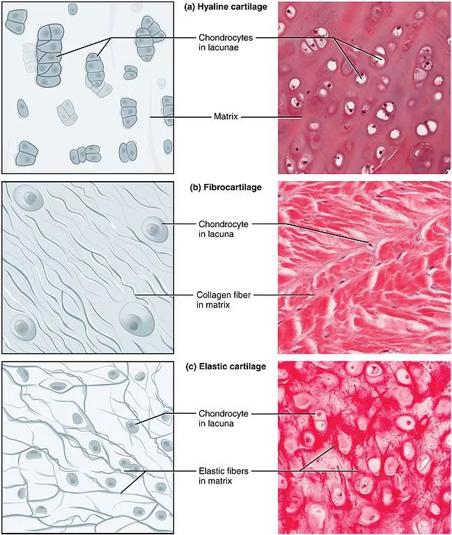
- Chondrocytes: Chondrocytes are the specialized cells found within hyaline cartilage. They are responsible for producing and maintaining the extracellular matrix components, such as collagen and proteoglycans. Chondrocytes are usually located in small spaces called lacunae within the cartilage matrix.
- Smooth and Translucent Appearance: Hyaline cartilage has a smooth and glossy appearance, giving it a translucent quality when viewed under a microscope. This is due to the homogenous distribution of the matrix components, especially the proteoglycans, which mask the collagen fibers.
- Avascular and Aneural: Hyaline cartilage lacks blood vessels and nerves. Nutrients and oxygen are obtained through diffusion from surrounding tissues, primarily from the synovial fluid in the case of articular cartilage found in joints.
- Low Cellularity: Hyaline cartilage has a relatively low density of chondrocytes compared to other types of cartilage. The chondrocytes are typically arranged in small groups or isolated within the matrix, allowing for a more uniform distribution of the extracellular matrix components.
- Resilience and Flexibility: Hyaline cartilage exhibits a balance of stiffness and flexibility, making it resilient and able to withstand compressive forces. This property is important in providing support to various structures while allowing for smooth joint movement and shock absorption.
- Distribution in the Body: Hyaline cartilage is widespread in the body. It is found in the embryonic skeleton, where it serves as a template for bone formation. In adults, it is present in structures such as the nasal septum, tracheal rings, costal cartilages connecting the ribs to the sternum, articular cartilage at joint surfaces, and bronchial tubes.
The characteristics of hyaline cartilage make it well-suited for its various functions, including providing structural support, reducing friction between joint surfaces, facilitating smooth movement, and aiding in the growth and development of skeletal elements.
What is Elastic cartilage?
Elastic cartilage is a type of connective tissue that possesses elastic fibers in addition to the collagen fibers and proteoglycans found in other cartilage types. These elastic fibers, primarily composed of elastin protein, give the cartilage its unique elastic properties. Elastic cartilage is more flexible and resilient compared to hyaline cartilage and fibrocartilage.
The distinguishing characteristic of elastic cartilage is its ability to recoil after being bent or deformed, allowing it to return to its original shape. This elasticity is crucial for structures that require both support and flexibility. Elastic cartilage is found in specific locations in the body where these properties are essential.
The most notable locations of elastic cartilage include:
- External ear (Pinna): The external part of the ear, responsible for collecting sound waves, is composed of elastic cartilage. This allows the ear to maintain its shape while also providing flexibility.
- Epiglottis: The epiglottis is a flap-like structure located at the base of the tongue, covering the entrance to the windpipe during swallowing. Elastic cartilage forms the core of the epiglottis, providing support and elasticity for its movement.
- Laryngeal Cartilages: Elastic cartilage is present in various cartilages of the larynx (voice box), including the epiglottis, cuneiform cartilages, corniculate cartilages, and arytenoid cartilages. These cartilages contribute to the structure, protection, and function of the larynx.
- Auditory Tube/Eustachian Tube: The auditory tube connects the middle ear to the back of the throat, helping to equalize air pressure on both sides of the eardrum. Elastic cartilage forms a part of the auditory tube, assisting in its flexibility.
The presence of abundant elastic fibers in elastic cartilage enables these structures to withstand repeated bending and stretching without losing their shape or functionality. This unique property makes elastic cartilage well-suited for anatomical regions that require both structural support and the ability to recoil or deform.
Characteristics of Elastic cartilage
Elastic cartilage is a specialized type of connective tissue with distinct characteristics that set it apart from other types of cartilage. Here are some key characteristics of elastic cartilage:
- Elastic Fibers: Elastic cartilage is characterized by the presence of abundant elastic fibers within its extracellular matrix. These elastic fibers, composed mainly of the protein elastin, give the cartilage its unique properties of flexibility and elasticity. They allow the cartilage to return to its original shape after being deformed or stretched.
- Matrix Composition: The extracellular matrix of elastic cartilage contains a combination of elastic fibers, collagen fibers (primarily type II collagen), proteoglycans, and glycoproteins. The elastic fibers provide the distinctive elastic quality, while the collagen fibers contribute to the structural integrity of the tissue.
- Chondrocytes: Similar to other types of cartilage, elastic cartilage contains chondrocytes embedded within lacunae in the matrix. These chondrocytes produce and maintain the extracellular matrix components, including elastin and collagen, necessary for the elasticity and strength of the cartilage.
- Yellowish Appearance: Due to the high concentration of elastic fibers, elastic cartilage has a yellowish color, which distinguishes it from hyaline cartilage and fibrocartilage. This yellow tint is particularly noticeable in areas such as the external ear (pinna) where elastic cartilage is abundant.
- Greater Flexibility: Elastic cartilage is highly flexible and resilient, allowing it to bend and recoil repeatedly without permanent deformation. This characteristic makes it well-suited for structures that require elasticity, such as the external ear, epiglottis, laryngeal cartilages, and auditory tubes.
- Structural Support: While elastic cartilage is flexible, it still provides structural support to the surrounding tissues and organs. It helps maintain the shape and stability of certain structures, such as the external ear, by providing a framework for soft tissues to attach to.
- Limited Distribution: Elastic cartilage is less widespread compared to other cartilage types. It is primarily found in specific anatomical locations that require elasticity, including the external ear, epiglottis, laryngeal cartilages (such as the cuneiform, corniculate, and arytenoid cartilages), and auditory tubes (eustachian tubes).
The unique characteristics of elastic cartilage make it suitable for providing flexible support and maintaining the shape of specific structures in the body. Its exceptional elasticity allows for movement and resilience while maintaining tissue integrity.
What is Fibrocartilage?
Fibrocartilage is a specialized type of cartilage that combines characteristics of both dense fibrous connective tissue and hyaline cartilage. It contains a dense network of collagen fibers, primarily type I collagen, which gives it its strength and resilience. Fibrocartilage is the toughest and strongest type of cartilage in the body.
The distinguishing features of fibrocartilage include:
- Collagenous Fibers: Fibrocartilage is rich in collagen fibers, which are arranged in thick bundles. These collagen fibers provide strength and stability to the tissue, allowing it to withstand tension, compression, and shear forces.
- Chondrocytes: Similar to other cartilage types, fibrocartilage contains chondrocytes, which are the specialized cells responsible for producing and maintaining the extracellular matrix. Chondrocytes in fibrocartilage are typically arranged in rows or clusters between the collagen fibers.
- Transition Zones: Fibrocartilage is often found at transition zones where there is a gradual shift or interface between different types of tissues. These transition zones help to distribute forces and provide a smooth transition between tissues with varying mechanical properties. Examples of such transition zones include the intervertebral discs in the spine and the attachments of tendons and ligaments to bone.
- Limited Perichondrium: Unlike hyaline cartilage, fibrocartilage does not have a well-developed perichondrium, which is a layer of connective tissue that surrounds and supports cartilage. Instead, fibrocartilage is typically connected to adjacent tissues, such as bone or tendon, without a distinct boundary.
The primary function of fibrocartilage is to provide structural support and absorb shock in areas subjected to high pressure and mechanical stress. It is commonly found in locations where there is a need for both flexibility and strength, such as:
- Intervertebral Discs: Fibrocartilage forms the discs between the vertebrae in the spine, acting as shock absorbers and facilitating movement while providing stability.
- Pubic Symphysis: The fibrocartilaginous joint between the two pubic bones in the pelvis helps to withstand the forces during childbirth and supports the stability of the pelvis.
- Joint Menisci: Fibrocartilage forms the C-shaped menisci in the knee joints, which help to distribute load, absorb shock, and improve joint stability.
- Tendon and Ligament Insertions: Fibrocartilage can be found at the sites where tendons and ligaments attach to bones, providing a smooth transition and enhancing the strength of these attachments.
The presence of dense collagen fibers makes fibrocartilage more resistant to wear and tear compared to hyaline cartilage. It is specifically adapted to withstand mechanical stress and provide support in areas where there is a need for greater strength and durability.
Characteristics of Fibrocartilage
Fibrocartilage is a specialized type of cartilage with distinct characteristics that make it unique. Here are some key characteristics of fibrocartilage:
- Matrix Composition: Fibrocartilage has a unique composition of extracellular matrix. It contains a dense network of collagen fibers, primarily type I collagen, which provide strength and durability to the tissue. The collagen fibers are densely packed and arranged in an orderly manner, giving fibrocartilage its characteristic appearance.
- Chondrocytes: Like other cartilage types, fibrocartilage contains chondrocytes, which are specialized cells responsible for producing and maintaining the extracellular matrix. However, compared to hyaline and elastic cartilage, fibrocartilage has fewer chondrocytes and a higher proportion of collagen fibers.
- Transition Tissue: Fibrocartilage is often found at sites where there is a transition between different types of connective tissues, such as between tendons or ligaments and bone. It serves as a transitional tissue that can withstand both tension and compression forces.
- Strength and Resilience: The dense arrangement of collagen fibers in fibrocartilage provides it with exceptional strength and resistance to mechanical stress. It allows fibrocartilage to withstand significant pressure, tension, and shear forces without being easily deformed or damaged.
- Lack of Perichondrium: Unlike hyaline and elastic cartilage, fibrocartilage lacks a perichondrium, which is a protective layer of connective tissue that surrounds cartilage. The absence of perichondrium limits the ability of fibrocartilage to regenerate and repair itself.
- Intervertebral Discs and Joints: Fibrocartilage is commonly found in areas that require both support and shock absorption. It is a major component of intervertebral discs in the spine, providing cushioning and stability to the vertebral column. Fibrocartilage also forms the articular discs in certain joints, such as the temporomandibular joint and sternoclavicular joint.
- Appearance: Fibrocartilage has a unique appearance that combines features of dense connective tissue and cartilage. It appears as a tough and fibrous tissue with a whitish color, resembling a hybrid between tendons or ligaments and hyaline cartilage.
- Limited Healing Capacity: Due to its avascular nature and lack of perichondrium, fibrocartilage has a limited capacity for self-repair. When damaged, the healing process is slower compared to other tissues, and fibrocartilage injuries may lead to the formation of scar tissue instead of complete regeneration.
Fibrocartilage’s characteristics allow it to fulfill its role in providing strength, support, and shock absorption in areas subjected to mechanical stress. Its unique composition and structure make it well-suited for its specific anatomical locations, such as the intervertebral discs and certain joint structures.
Differences Between Hyaline cartilage, Elastic, and Fibrocartilage
| Characteristic | Hyaline Cartilage | Elastic Cartilage | Fibrocartilage |
|---|---|---|---|
| Appearance | Translucent and glossy | Glossy and yellow | White, dense, and opaque |
| Location | Fetal skeleton until maturation | External ear, epiglottis, auditory tube, laryngeal cartilages, intervertebral discs, pubic symphysis joint, articular discs in sternoclavicular and temporomandibular joints, glenoid labrum in the shoulder blade, acetabular labrum in the hip joint | |
| Main Collagen Type | Type II | Type II | Type I |
| Chondrocytes | Small, arranged in groups of 2 – 8 cells | Large, arranged in groups of 2 – 4 cells | Small, between bundles of collagen fibers, arranged in strips |
| Extracellular Matrix | Homogenous and basophilic | High in elastic fibers | High in collagen fibers, eosinophilic |
| Perichondrium | Present | Present | Absent |
Related Posts
- Difference Between Homologous Chromosomes and Sister Chromatids
- Difference between Monocarpic and Polycarpic Plants
- Differences Between Poisonous and Non-poisonous Snakes
- Differences Between Sensitivity, Specificity, False positive, False negative
- Anabolism vs Catabolism – Differences Between Anabolism and Catabolism


