Amoeba (less often spelled ameba, or amoeba, plural am(o)ebas or am(o)ebae) is often referred to as an amoeboid, is kind of unicellular organism that has the capability of changing its form, mostly by retracting and expanding pseudopods. Amoebae do not belong to a single taxonomic class, They are found in all major lineages of the … Read more
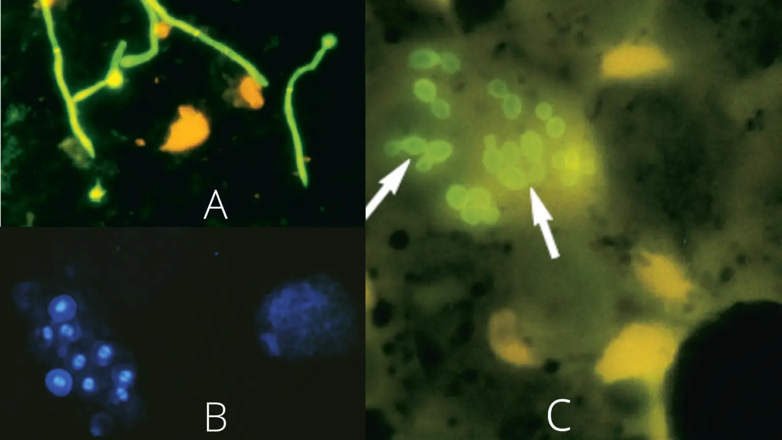
In 1961, Darken reported the uptake of calcofluor white, a fluorescent brightener, by actively growing yeasts and higher fungi. Hageage and Harrington outlined the application of calcofluor white (CFW) to demonstrate hyphae and yeasts in clinical specimens. Monheit et al. utilised this dye for intraoperative identification of fungal infection on frozen sections of lung and … Read more
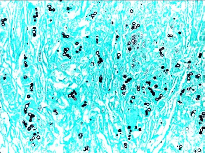
Grocott-Gomori’s Methenamine Silver Staining is a type of special staining technique used in microbiology and histology to visualize fungal organisms, specifically those that form characteristic spores known as arthroconidia. The staining technique was developed in the 1930s by Grocott and Gomori and is based on the use of silver and methenamine. The fungal cells are … Read more
What is silver staining? Silver staining is a powerful and versatile technique used for the detection and identification of proteins in gels. This technique is accomplished by binding silver to the chemical terminal or side chains of amino groups, such as carboxyl and sulfhydryl groups. For decades, silver staining has been used to separate proteins … Read more
What is Auramine- Rhodamine Staining? Principle of Auramine- Rhodamine Staining Auramine and Rhodamine are nonspecific fluorochrome dyes that are attracted to acid-fast microorganisms. In the case of Mycobacteria, the dyes can selectively bind to the mycolic acid present in the cell wall, allowing the stain to penetrate. This compound is resistant to acid-alcohol decolorizer solutions. … Read more
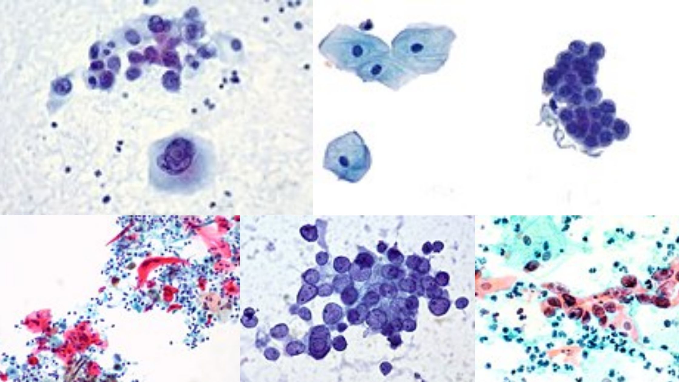
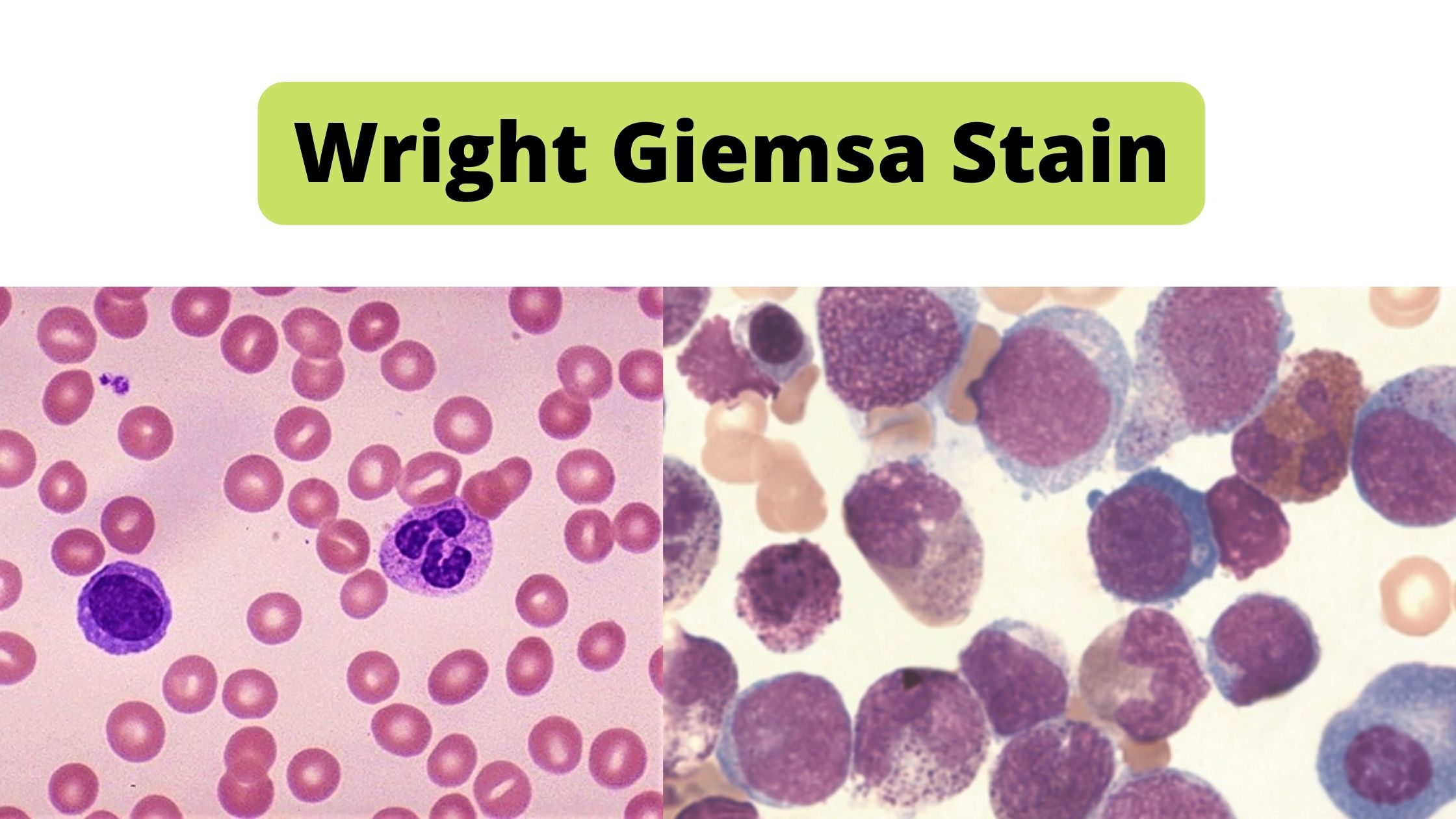
Wright’s stain is a modified Romanowsky stain. In hematology laboratory, it is used for the staining of peripheral blood smear, bone marrow aspirates, and urine samples.
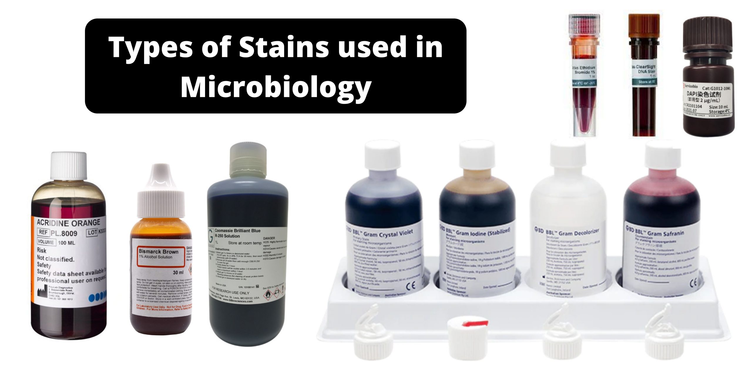
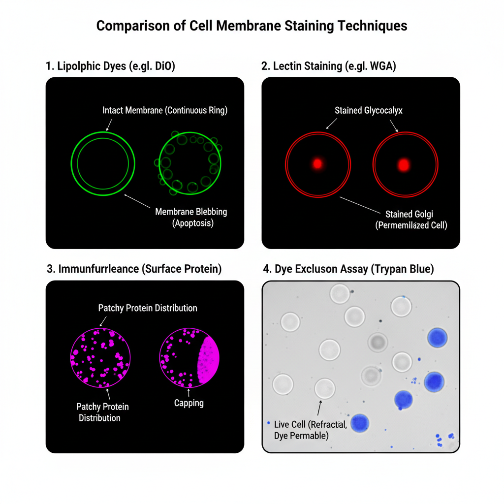
Different stains react or concentrate on different areas of a tissue or cell These properties can be utilized to highlight certain areas or regions. A few of the most well-known biological staining methods can be found below. If not otherwise indicated All of these dyes can be used on tissues and cells that are fixed as well as essential dyes (suitable for use in live organisms) are indicated.
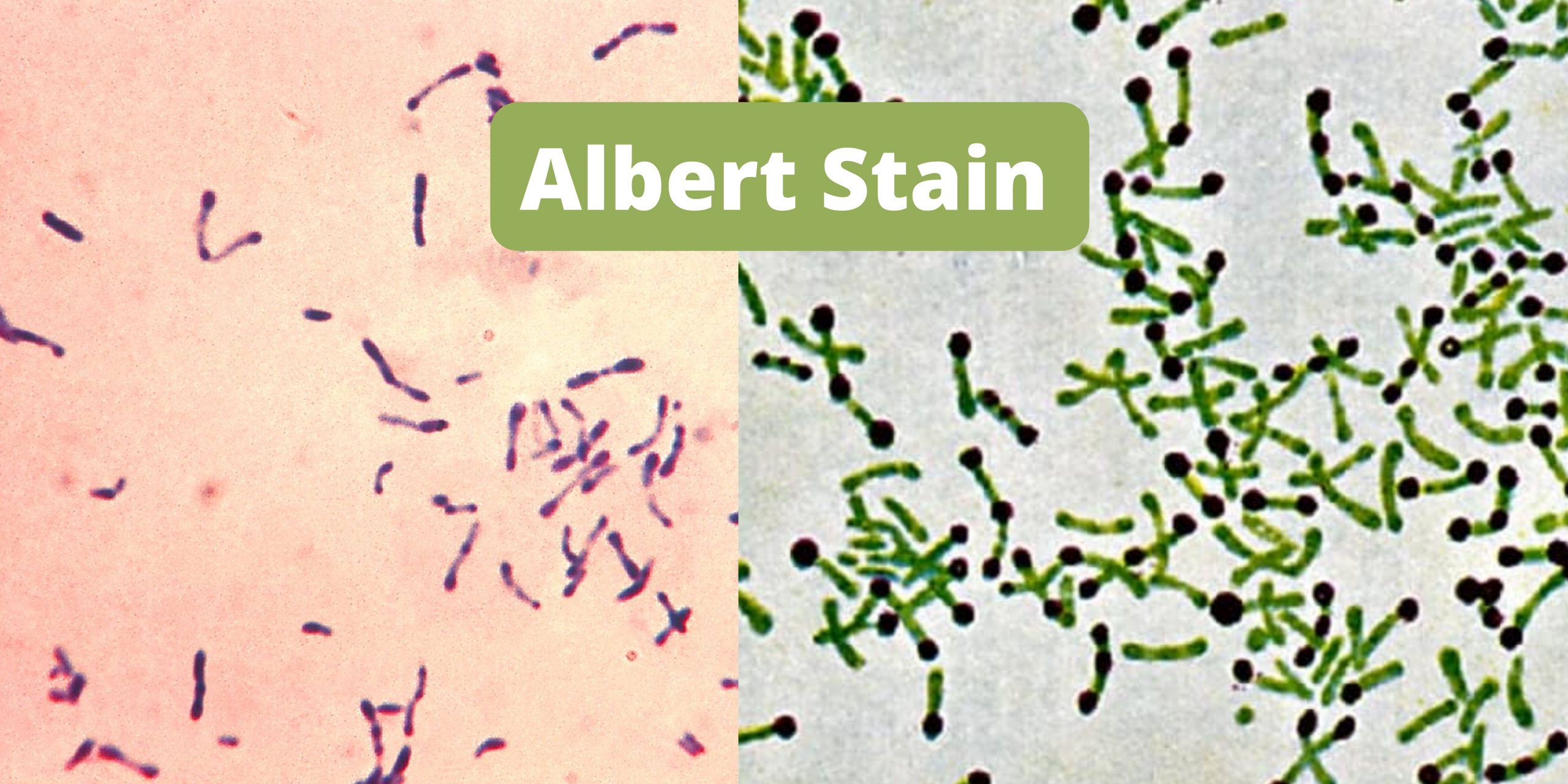
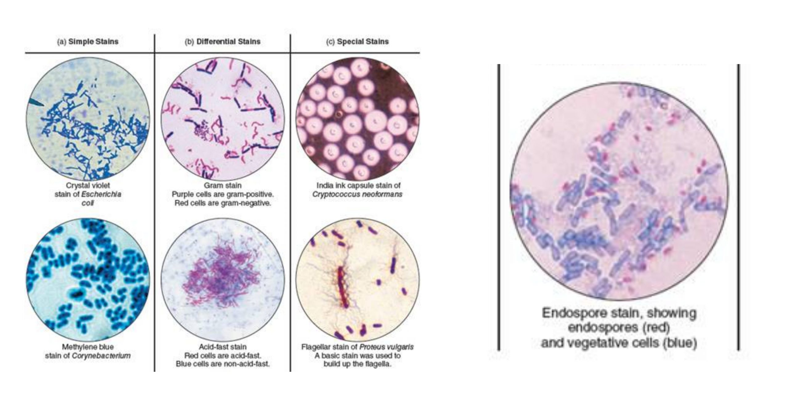
Different stains have been developed over time to distinguish bacteria species, separating them morphologically and the specific characteristics they possess. The most popular stain is Gram staining, acid-fast staining, and endospore staining. Each stain aims at identifying and defining bacteria according to their forms and morphologies.
⚠️
Click on your ad blocker icon in your browser's toolbar
Select "Pause" or "Disable" for this website
Refresh the page if it doesn't automatically reload