

Plant cells are eukaryotic cells that form different tissues and organs of the plant body. They occur in different shapes, sizes and structures depending on their location and function. Some cells remain actively dividing, while others become specialized for photosynthesis, storage, transport, protection and mechanical support. The major types include meristematic cells, parenchyma cells, collenchyma cells, sclerenchyma cells, epidermal cells, xylem cells and phloem cells. These different cell types work together for the normal growth, development and functioning of plants.
What Are Plant Cells?
Plant cells are eukaryotic cells present in plants, which means they have a membrane-bound nucleus. Different cell organelles are present in the cytoplasm and these carry out various functions required for the normal activity of the cell.
A cell wall is present outside the plasma membrane and this is one of the major features of plant cells. It gives shape and mechanical support to the cell. Plant cells also contain plastids. Among them, chloroplasts are present in the green photosynthetic cells and are involved in photosynthesis.
Another characteristic structure is the vacuole, which is generally large in mature plant cells. It is used for storage and also helps in maintaining the turgor of the cell. Plant cells are not similar in all parts of the plant, and their shape, size, organelles and wall structure can vary according to the tissue and function of the cell.
How Are Plant Cells Classified?
Plant cells become differentiated into different types according to their location and function. Some cells continue cell division, while others become specialized for photosynthesis, storage, support and transport. In general, they can be grouped into meristematic cells and different types of mature or differentiated cells.
Meristematic cells are the actively dividing cells of growing regions. The differentiated forms include parenchyma, collenchyma and sclerenchyma cells. Apart from these, plants contain specialized xylem, phloem, guard, root-hair and epidermal cells. Their structure is not similar, and changes according to the work carried out by the particular cell in plant tissues.
Major Types of Plant Cells
The major types of plant cells are as follows-
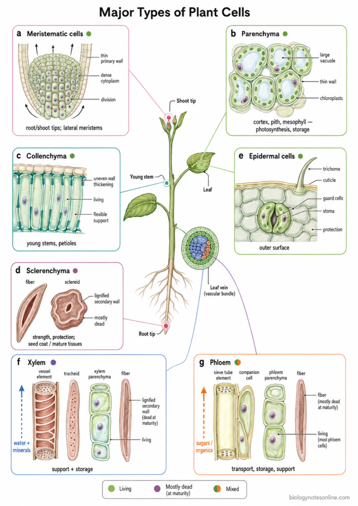
| Cell type | Main structure | Living/dead | Location | Main function | Example |
|---|---|---|---|---|---|
| Meristematic cells | Small cells, thin primary wall and dense cytoplasm. Cells remain capable of division. | Living | Root tips, shoot tips and lateral meristems | Formation of new cells and plant growth. | Root apical meristem |
| Parenchyma cells | Usually thin-walled cells with a large vacuole. Structure varies according to function. | Living | Cortex, pith, leaf mesophyll and vascular tissues | Photosynthesis, storage and different metabolic functions. | Leaf mesophyll |
| Collenchyma cells | Elongated cells having unevenly thickened, pectin-rich primary walls. | Living | Young stems, petioles and other growing organs | Flexible mechanical support. Growth can still continue. | Young stem |
| Sclerenchyma cells | Thick secondary walls, commonly lignified. Includes fibers and sclereids. | Mostly dead at maturity | Mature stems, vascular regions, seed coverings and other hard tissues | Mechanical strength and protection. | Seed coat sclereids |
| Epidermal cells | Closely arranged outer cells. Specialized forms include guard cells and trichomes. | Living | Outer surface of young roots, stems and leaves | Protection, regulation of gas exchange and reduction of water loss. | Leaf epidermis |
| Xylem cells | Contains tracheids, vessel elements, fibers and living parenchyma. Conducting elements have lignified secondary walls. | Both. Tracheids and vessels are dead at maturity, parenchyma is living | Vascular bundles, wood, roots, stems and leaf veins | Transport of water and minerals, mechanical support and storage. | Vessel elements in wood |
| Phloem cells | Contains sieve elements, companion cells, parenchyma and fibers. | Mostly living. Fibers are commonly dead at maturity | Vascular bundles of roots, stems and leaves | Transport of sugars and other organic substances. Storage and support are also performed by some cells. | Sieve-tube elements with companion cells |
1. Parenchyma Cells
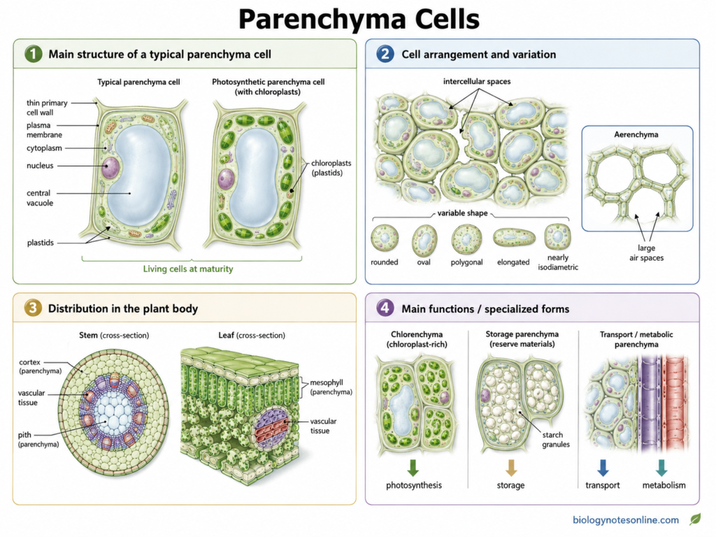
Parenchyma cells are living plant cells that form a major portion of the soft tissues in plants. Their walls are usually thin and made up of primary cell wall. These cells are widely present. Depending upon their location, parenchyma may perform photosynthesis, storage, transport and other metabolic functions.
Characteristics
- Parenchyma cells remain living at maturity.
- These are comparatively less specialized cells. In many tissues they can also regain the ability to divide.
- The cell wall is generally thin. Secondary wall is usually absent.
- Their shape is not always same. Rounded, oval, polygonal, elongated or nearly isodiametric cells can be present.
- Intercellular spaces occur commonly between neighbouring cells. In some forms such as aerenchyma, these spaces become very large.
- Parenchyma is one of the most widely distributed cell types, especially in cortex, pith, mesophyll and vascular tissues.
Structure
- Parenchyma cells usually possess a thin primary cell wall.
- A large central vacuole is commonly present in mature cells.
- Cytoplasm and nucleus remain present because these are living cells.
- Plastids differ according to function. Photosynthetic parenchyma contains chloroplasts, whereas storage cells may contain large amount of reserve substances.
- Cells may be closely arranged or loosely arranged with intercellular spaces. The arrangement depends upon the type and tissue where they are present.
Functions
- Storage is one of the important functions. Starch, sugars, proteins, oils and water can be accumulated in different parenchymatous tissues.
- Chloroplast-containing parenchyma performs photosynthesis.
- In leaves, parenchyma also forms internal spaces that help in movement of gases.
- Some cells take part in short-distance transport of water, nutrients and other substances.
- Ray parenchyma is involved in storage and radial movement within secondary vascular tissues.
- Parenchyma can also participate in wound repair and regeneration because many of these living cells retain considerable developmental capacity.
Types of Parenchyma Cells
The following are some of the important types of parenchyma cells-
- Chlorenchyma – It is parenchyma containing chloroplasts. These cells are present mainly in green parts and carry out photosynthesis. Leaf mesophyll is an important example.
- Aerenchyma – Aerenchyma is specialized parenchyma with large air-filled spaces. It is especially common in aquatic and wetland plants. These spaces help in internal aeration of plant tissues.
- Palisade parenchyma – These are elongated cells generally arranged below the upper epidermis in many dorsiventral leaves. The cells contain numerous chloroplasts and are closely packed. Photosynthesis is its major function.
- Spongy parenchyma – Spongy parenchyma consists of more loosely arranged cells. Large intercellular spaces are present between them, which help in movement of gases inside the leaf. The cells also contain chloroplasts and perform photosynthesis.
- Storage parenchyma – These cells are mainly used for storage of reserve materials. Starch, sugars, proteins and other substances may accumulate in them. Storage roots, tubers, stems and seeds commonly contain this type.
- Ray parenchyma – It forms part of the rays in secondary vascular tissues. The cells are usually arranged in radial direction. They are associated with storage and radial transport through woody stems and roots.
Examples
- Potato tuber contains abundant storage parenchyma in which starch is accumulated.
- The mesophyll of many leaves contains palisade and spongy parenchyma.
- Aquatic and waterlogged plants commonly develop aerenchyma. Large air spaces are characteristic of this tissue.
- Sugarcane stem contains storage parenchyma where large amounts of sucrose can accumulate.
- Wood rays contain ray parenchyma cells extending radially through secondary vascular tissues.
2. Collenchyma Cells
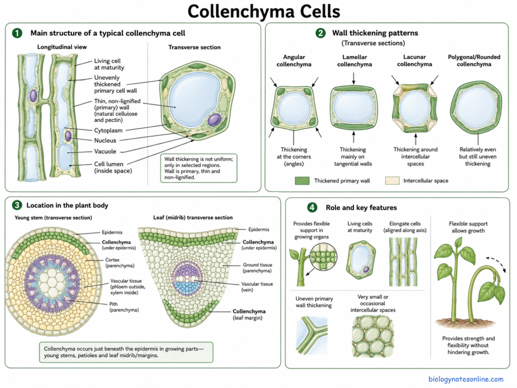
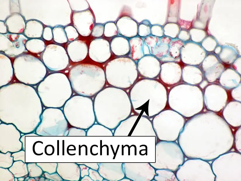
Collenchyma cells are living supporting cells mainly present in the growing parts of plants. Their walls are thickened, but the thickening is generally uneven. Unlike sclerenchyma, these cells remain living at maturity and their walls can continue thickening while the organ is growing. Collenchyma therefore forms an important mechanical tissue in young stems, petioles and leaves.
Characteristics
- Collenchyma consists of living cells and is mainly associated with young and growing plant organs.
- The cell walls become thick during and also after elongation. This allows support without making the growing region completely rigid.
- Wall thickening is not same in every part of the cell. Its pattern forms the basis for different types of collenchyma.
- The tissue is generally non-lignified during normal development. In older organs, however, some collenchyma can become more rigid or undergo sclerification.
- Collenchyma commonly occurs as strands or layers below the epidermis of growing plant parts.
Structure
- Collenchyma cells are commonly elongated. In transverse section, their shape may appear polygonal or rounded.
- The cells possess thick primary cell walls. Cellulose, hemicelluloses and pectic substances form important components of these walls.
- A living protoplast remains present. The cells do not normally lose their nucleus at maturity.
- Wall deposition may be concentrated at the corners, along tangential walls or around intercellular spaces. Different patterns are therefore produced.
- Intercellular spaces are very small in some forms, while noticeable spaces occur in others.
Functions
- The major function of collenchyma is mechanical support of growing plant parts.
- It provides strength to young stems, petioles and leaves. At the same time, elongation of these organs can continue.
- The flexible wall helps tissues withstand bending and other mechanical stresses.
- Collenchyma also prevents soft growing organs from tearing easily. Its supporting role is therefore different from the more rigid sclerenchyma tissue.
Types of Collenchyma
The following are the important types of collenchyma based on the pattern of wall thickening-
- Angular collenchyma – In this type, wall thickening is mainly present at the corners of the cells. Intercellular spaces are generally absent or very small. The angular regions therefore appear more strongly thickened.
- Lamellar collenchyma – It is also referred to as tangential or plate collenchyma. Thickening occurs mainly along the inner and outer tangential walls of the cells.
- Lacunar collenchyma – This type contains well-developed intercellular spaces. Wall thickening is mainly associated with the regions bordering these spaces.
- Annular collenchyma – Here, the walls become more uniformly thickened around the cell. The thickening is therefore not strongly restricted to only the corners or tangential walls.
Examples
- The strings present in celery (Apium graveolens) petiole contain prominent collenchyma tissue and provide mechanical support.
- Collenchyma is commonly present below the epidermis of young dicot stems.
- Leaf petioles of many plants contain collenchyma strands, especially in regions exposed to bending.
- It also occurs along the midrib and larger veins of many leaves, where additional support is required.
3. Sclerenchyma Cells
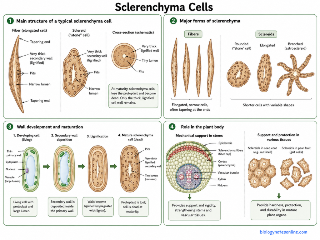
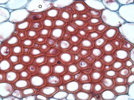
Sclerenchyma cells are specialized supporting cells of plants having thick secondary cell walls. The walls are commonly lignified and become very strong after complete development. Many sclerenchyma cells lose their protoplast and become dead at maturity. Fibers and sclereids are the two major forms of sclerenchyma.
Characteristics
- Sclerenchyma is mainly a mechanical tissue. It gives strength to mature plant parts.
- The cells develop thick secondary walls. Lignin is commonly deposited in these walls.
- Most sclerenchyma cells are dead after complete maturation, although living forms can occur during their development.
- Cell lumen becomes comparatively narrow because of heavy deposition of wall materials.
- Their shape is not similar in all cases. Fibers become long and narrow, whereas sclereids show several shapes and sizes.
- These cells usually occur in plant regions where continued elongation is no longer required.
Structure
- A thick secondary cell wall is the major structural character of sclerenchyma cells.
- Secondary walls contain mainly cellulose and hemicelluloses along with lignin. This wall gives considerable rigidity to the cell.
- Pits occur in the thickened walls.
- Fibers are generally elongated and narrow, often with tapering ends.
- Sclereids are shorter and more variable. Rounded, elongated or branched forms are present depending upon tissue.
- During maturation, the protoplast is commonly lost. The thick wall then remains as the main structural portion of cell.
Functions
- The major function of sclerenchyma is mechanical support.
- Thick lignified walls provide rigidity and strength to stems, leaves and vascular tissues.
- Fibers help the plant tissues to resist pulling and other mechanical forces.
- Sclereids provide hardness. This is particularly important in structures such as hard fruits, seed coverings and shells.
- Sclerenchymatous layers in seeds can also perform a protective function around the internal tissues.
Types of Sclerenchyma Cells
The following are the two major types of sclerenchyma cells-
- Fibers – Fibers are long and slender sclerenchyma cells. Their secondary walls become thick and commonly lignified. They generally occur together as strands or bundles, providing mechanical strength. Fibers are present in vascular tissues and other supporting regions of plant body.
- Sclereids – These are comparatively short sclerenchyma cells with very thick secondary walls. Their shape is highly variable. Sclereids are also referred to as stone cells, especially the short forms present in some fruits. They may occur separately or together in groups.
Examples
- The gritty particles present in pear fruit (Pyrus) are formed by sclereids or stone cells. This gives pear its characteristic gritty texture.
- Seed coats of different plants contain sclerenchymatous cells. In several species these form hard mechanical layers around the seed.
- Flax (Linum usitatissimum) contains long supporting sclerenchyma fibers. These fibers are commercially used in linen and related materials.
- Sclerenchyma fibers also occur around or within vascular tissues, where they provide additional mechanical support to plant organs.
4. Xylem Cells
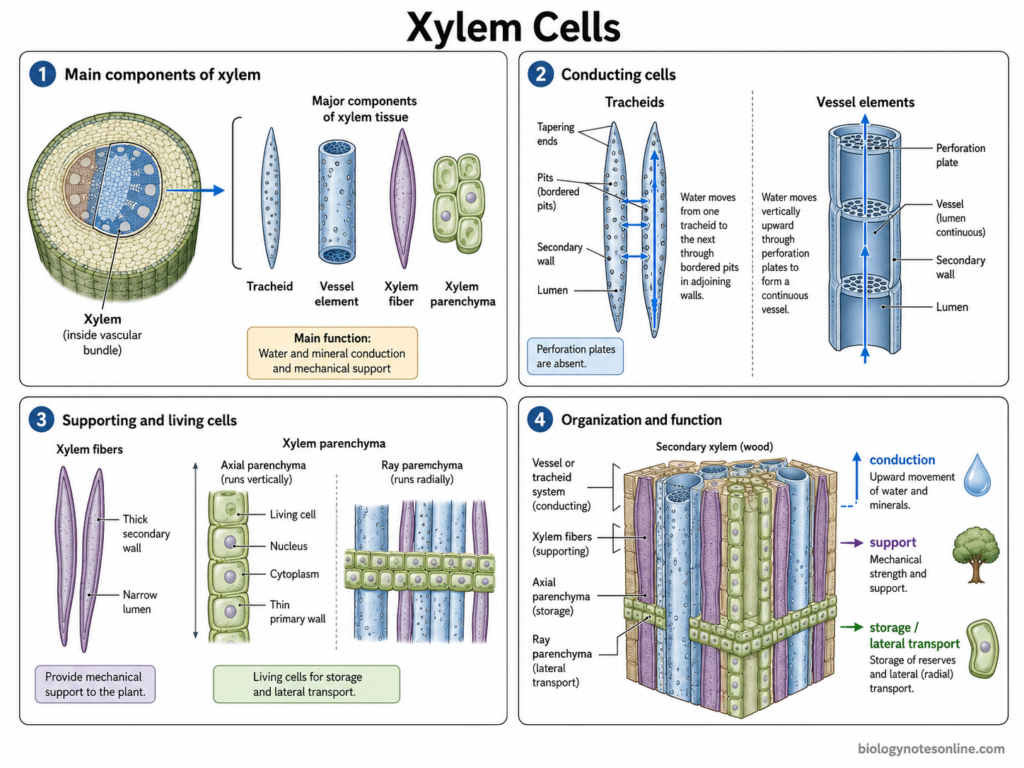
Xylem cells are the cells forming xylem, a complex vascular tissue of plants. It is mainly associated with conduction of water and mineral nutrients. Xylem contains different cell types rather than one uniform cell. Tracheids, vessel elements, xylem fibers and xylem parenchyma are its major components.
Characteristics
- Xylem contains both living and dead cells.
- Tracheids and vessel elements are together referred to as tracheary elements. These are the major water-conducting cells.
- Tracheary elements develop thick and patterned secondary cell walls, commonly lignified. Their protoplast is lost during maturation.
- Xylem fibers also develop thick secondary walls and mainly perform supporting function.
- Xylem parenchyma remains living at maturity. Storage and transport-related functions are carried out by these cells.
- Wood is mainly secondary xylem. Its cellular composition, however, differs considerably among plant groups.
Structure
- Xylem tissue contains elongated conducting cells together with fibers and parenchymatous cells.
- Tracheids are long cells with tapering or pointed ends. Perforation plates are absent. Water moves between adjoining tracheids through pits present in their walls.
- Vessel elements are usually wider and shorter. Several vessel elements are joined end to end, forming a continuous vessel. Their end walls contain perforations.
- Xylem fibers are generally elongated, with thick secondary walls and comparatively narrow lumen.
- Parenchyma forms the living component of xylem. In secondary xylem it may occur as axial parenchyma and ray parenchyma.
Functions
- Water conduction is one of the major functions of xylem. Tracheids and vessels form the principal conducting pathways from roots towards aerial plant parts.
- Mineral nutrients dissolved in xylem sap are also carried through these conducting elements.
- Xylem provides mechanical support. Lignified walls of tracheids and especially fibers give strength to stems and other plant regions.
- Living xylem parenchyma is used for storage of reserve substances. These cells also take part in movement and remobilization of nutrients.
- Ray parenchyma is important in radial transport through woody tissues.
Types of Xylem Cells
The following are the major types of xylem cells-
- Tracheids – These are elongated conducting cells having tapering ends and lignified secondary walls. Pits connect them with neighbouring cells. Tracheids perform both water conduction and mechanical support, and are particularly important in gymnosperm xylem.
- Vessel elements – Vessel elements are shorter and generally wider than tracheids. They join with one another to form long xylem vessels. Perforation plates occur between adjacent elements, providing a pathway for water movement. Vessels are especially characteristic of angiosperms.
- Xylem fibers – These are thick-walled cells mainly involved in mechanical strength. Most become dead after maturation. In many angiosperms, fibers form an important supporting part of secondary xylem.
- Xylem parenchyma – Xylem parenchyma consists of living cells. They occur around conducting elements and also as rays in woody tissues. Storage, nutrient movement and remobilization are some of their important functions.
Examples
- Conifer wood contains large numbers of tracheids. These cells perform water conduction as well as supporting function.
- Angiosperm wood commonly contains vessels made up of vessel elements, along with fibers and parenchyma.
- Wood rays contain living ray parenchyma. Their cells are associated mainly with storage and radial movement of materials.
- Xylem conducting cells are also present in the vascular bundles of roots, stems and leaves, carrying water through different parts of the plant.
5. Phloem Cells
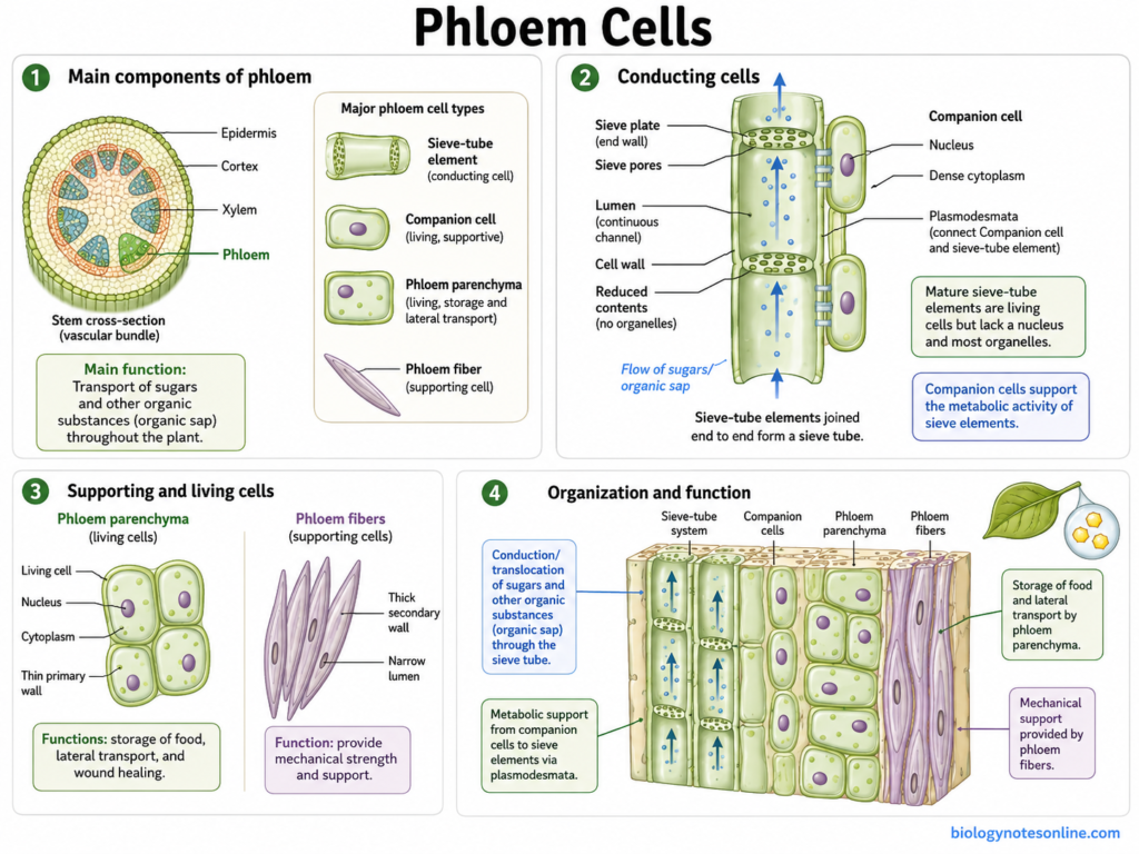
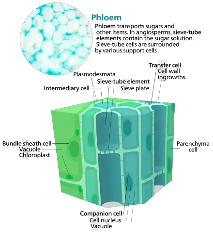
Phloem cells are the cells forming phloem, a complex vascular tissue of plants. It is mainly concerned with transport of sugars and other organic substances. Phloem is not made up of one type of cell. Sieve elements, companion cells, phloem parenchyma and fibers are its important components.
Characteristics
- Phloem contains different types of living and supporting cells.
- Sieve elements form the conducting portion. These cells remain alive at functional maturity, although nucleus and several cellular components are lost during their development.
- In flowering plants, sieve-tube elements remain closely associated with companion cells.
- The tissue mainly transports photosynthates from source regions to different sink regions. Transport can occur over long distances through the plant body.
- Phloem also contains parenchyma and, in many plants, mechanical fibers.
Structure
- The conducting cells of angiosperm phloem are called sieve-tube elements.
- These are elongated cells arranged one after another. Together they form a long sieve tube.
- Their end walls contain perforated regions known as sieve plates. Materials can pass through the pores of these plates.
- A mature sieve-tube element has no nucleus and its internal contents become greatly reduced. Still, the cell remains living.
- Companion cells occur beside sieve-tube elements and are connected with them through numerous plasmodesmata. These cells retain a nucleus and support the metabolic activities of sieve elements.
- Phloem parenchyma and fibers form the other cellular components of the tissue. Their amount is not same in every plant.
Functions
- Transport of sugars is the major function of phloem. Sucrose and other photosynthates are translocated from source tissues towards different sinks.
- Amino acids and some other organic substances are also carried in phloem sap.
- Proteins, RNAs and different signalling molecules can move through the sieve-tube system.
- Companion cells are involved in phloem loading and unloading and provide metabolic support for sieve elements.
- Parenchyma cells perform storage and local transport functions.
- Fibers mainly give mechanical strength to the tissue.
Types of Phloem Cells
The following are the major types of phloem cells-
- Sieve-tube elements – These are the main conducting cells in angiosperms. They join end to end forming sieve tubes, with sieve plates between adjacent cells. Organic solutes are transported through this system.
- Companion cells – Companion cells are living and nucleated cells present along with sieve-tube elements. They are closely connected to them by plasmodesmata. Because mature sieve elements have reduced cellular machinery, companion cells perform several supporting metabolic functions.
- Sieve cells – These are elongated conducting cells found mainly in gymnosperms and seed-free vascular plants. They overlap with neighbouring sieve cells and do not form the highly specialized sieve tubes found in angiosperms.
- Phloem parenchyma – These are living parenchymatous cells associated with the phloem. Storage and short-distance movement of different substances are some of their functions.
- Phloem fibers – These are supporting sclerenchymatous cells present in phloem of many plants. Their walls become thick and provide mechanical strength rather than carrying phloem sap.
Examples
- Sieve-tube elements and companion cells are typical conducting and associated cells of flowering plant phloem.
- Gymnosperm phloem contains sieve cells rather than typical angiosperm sieve tubes.
- Phloem parenchyma is present as living cells associated with conducting tissues.
- Supporting phloem fibers are found in phloem of many stems.
6. Meristematic Cells
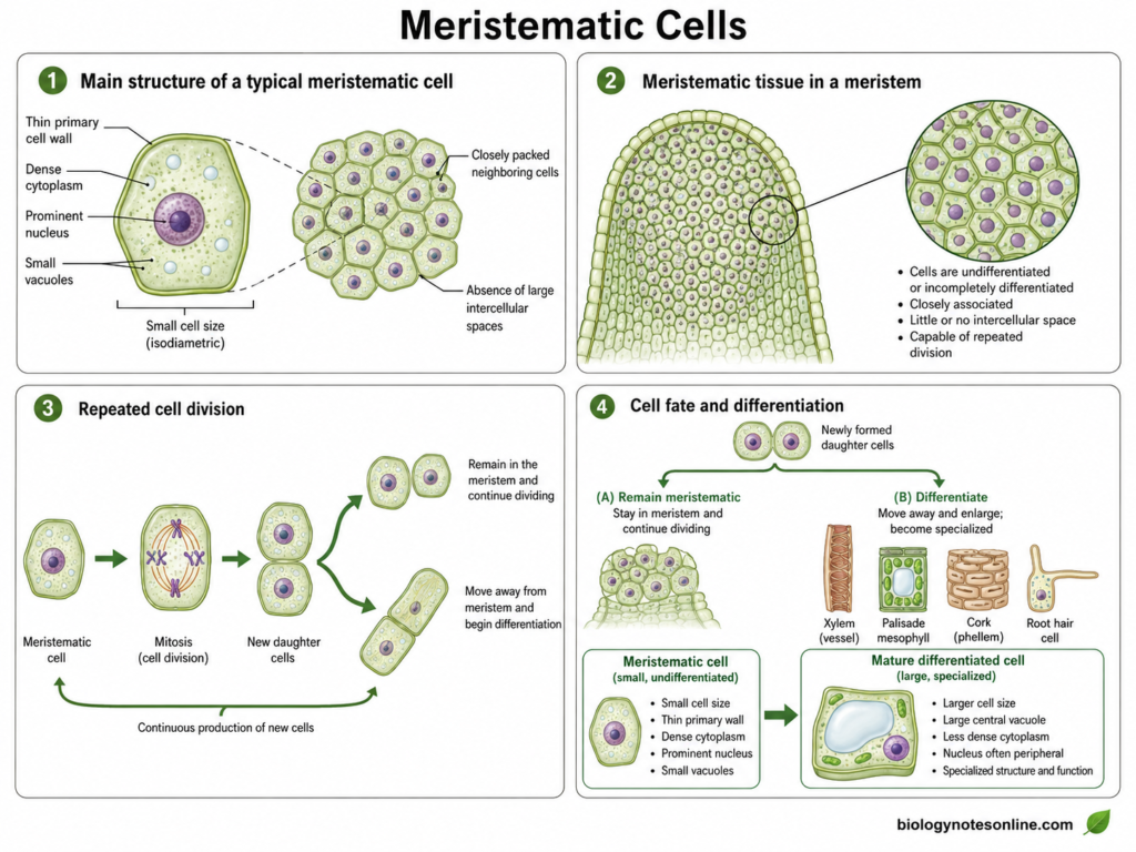
Meristematic cells are undifferentiated or incompletely differentiated plant cells having the capacity for repeated cell division. They are present in specialized growing regions called meristems. New cells are continuously produced from these regions, some remain meristematic while others undergo differentiation and form different tissues and organs.
Characteristics
- Meristematic cells have the ability to divide and produce new cells. However, rate of division is not same throughout a meristem. Some stem-cell regions divide comparatively slowly.
- These cells are generally undifferentiated and have not acquired the specialized structure of mature plant cells.
- Daughter cells formed from meristem can later differentiate into different cell types.
- They are mainly associated with regions where active growth is taking place.
- Meristematic activity may continue for a long period. Due to this, plants can continue producing new roots, shoots and other organs after embryonic development.
Structure
- Meristematic cells are generally small in comparison to many mature cells. Their shape may be rounded, polygonal or nearly isodiametric.
- The cells possess a thin primary cell wall.
- Cytoplasm is dense and the nucleus occupies a noticeable portion of the cell.
- Vacuoles are generally small in young meristematic cells rather than the single large central vacuole commonly found after cell maturation.
- The cells are closely associated with one another. Large intercellular spaces are generally absent.
- Their internal structure changes as the cells leave the meristem and begin differentiation.
Functions
- Formation of new cells is the major function of meristematic tissue.
- Apical meristems are responsible for much of the primary growth of roots and shoots. This increases their length.
- Meristems also give rise to cells which later differentiate into the different tissues of plant body.
- New leaves, stems, roots and other organs are ultimately produced through meristem activity. The shoot apical meristem also initiates leaves and buds.
- Lateral meristems produce secondary tissues and increase the thickness or girth of stems and roots.
- Meristematic activity is also important during regeneration and repair of plant tissues.
Types of Meristems
The following are the major types of meristems based on their position-
- Apical Meristem – Apical meristems are present at the growing tips of roots and shoots. Cells divide in these regions and contribute mainly to primary growth. It results in elongation of the plant body. Shoot and root apical meristems also maintain populations of stem cells from which new tissues are produced.
- Intercalary Meristem – These meristems occur between regions of more mature tissue, and are especially well developed in grasses. In grass stems they commonly occur near the base of internodes, where continued cell division contributes to internode elongation. The presence of intercalary growth also allows many grasses to continue elongation after cutting or grazing.
- Lateral Meristem – Lateral meristems occur parallel to the sides of stems and roots. Their activity causes an increase in diameter rather than mainly length. Vascular cambium and cork cambium are the important lateral meristems associated with secondary growth. Vascular cambium produces secondary xylem and phloem, while cork cambium contributes to formation of the periderm.
Examples
- The growing tip of a root contains the root apical meristem. New cells produced here later enter regions of elongation and differentiation.
- Shoot tips and buds contain shoot apical meristem, from which new shoot tissues and lateral organs are produced.
- Rice, maize and other grasses have intercalary meristems associated with elongating internodes.
- Vascular cambium of woody stems is an example of lateral meristem. It produces secondary vascular tissues during increase in stem girth.
- Cork cambium is another lateral meristem. In woody plants it forms tissues of the periderm as secondary growth proceeds.
7. Epidermal Cells
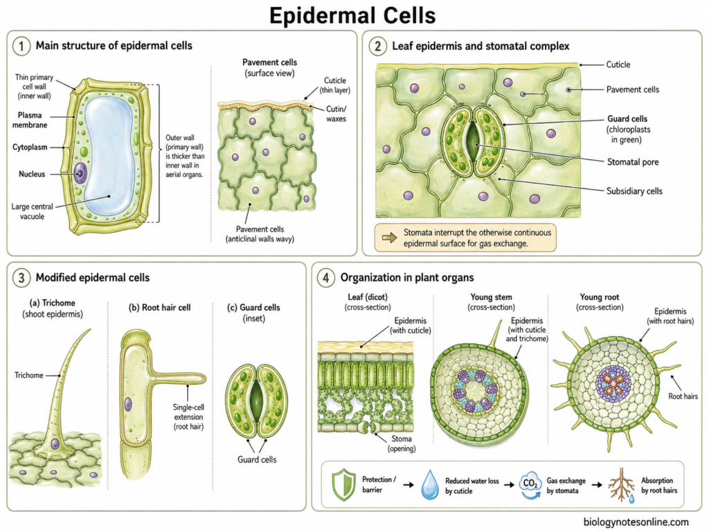
Epidermal cells are the cells forming the epidermis, which covers the primary plant body. It is generally the outermost cell layer of young roots, stems, leaves and other organs. The cells are not all similar. Some remain as ordinary epidermal cells, while others become modified into guard cells, trichomes and root-hair cells.
Characteristics
- Epidermal cells are generally closely arranged with little or no intercellular spaces.
- In most young plant organs, the epidermis consists of a single cell layer. Multiple epidermal layers are also present in some plants.
- The cells are living. Their shape and size, however, differ according to the organ where they occur.
- Aerial epidermal cells commonly have a cuticle over their outer surface. It helps in reducing water loss.
- Different specialized cells may develop within the epidermis. These give the tissue several functions rather than only forming an outer covering.
Structure
- Ordinary epidermal cells generally possess a primary cell wall, plasma membrane, cytoplasm, nucleus and a large vacuole.
- Their outer wall may become thicker than the inner walls, particularly in aerial organs.
- A layer of cutin and waxes forms the cuticle over much of the shoot epidermis.
- Leaf epidermal cells may have straight or irregular, wavy walls when viewed from the surface. Pavement cells form much of this epidermal covering.
- Stomatal complexes interrupt the continuous epidermal surface. Each stomatal pore is surrounded by a pair of guard cells, and subsidiary cells may also be present around them.
- In roots, some epidermal cells form long tubular extensions called root hairs. These are extensions of single cells, not multicellular structures.
Functions
- The epidermis provides protection to the internal tissues of young plant organs.
- Cuticle present over the shoot epidermis decreases uncontrolled loss of water from the surface.
- Guard cells regulate opening and closing of stomata. In this way, gas exchange and water loss through transpiration are controlled.
- Root epidermal cells with root hairs take part in absorption of water and mineral nutrients from soil. Root hairs also greatly increase the contact of root with surrounding soil.
- Trichomes can perform different functions. Protection against herbivores, reduction of water loss and secretion are some of them, depending on the type of trichome.
- Epidermis also acts as a barrier against entry of many harmful organisms and substances.
Types of Epidermal Cells
The following are some of the important types of epidermal cells-
- Pavement cells – These form most of the ordinary surface of the leaf epidermis. Their shape is often irregular and interlocking, although this varies considerably among plants. They form a protective covering and are covered externally by cuticle in aerial regions.
- Guard cells – Two guard cells surround a stomatal pore. Changes in their turgor regulate opening and closing of the pore. Thus, these cells have an important role in gas exchange and transpiration.
- Subsidiary cells – These are specialized epidermal cells present next to guard cells in many plants. They form part of the stomatal complex. Their number and arrangement are not same in every plant group.
- Trichomes – These are epidermal hairs or outgrowths present mainly on aerial plant parts. They may be unicellular or multicellular and glandular or non-glandular. Their functions include protection, secretion and reduction of water loss in different plants.
- Root-hair cells – These are specialized cells of the root epidermis, also called trichoblasts in developmental studies. Each forms a tubular root hair from the cell surface. It helps in uptake of water and mineral nutrients from soil.
Examples
- The epidermis of onion bulb scale leaves contains closely arranged ordinary epidermal cells.
- Leaf epidermis contains pavement cells together with stomata and their guard cells.
- Root hairs are formed from specialized epidermal cells in the maturation region of young roots.
- Hair-like trichomes are present on leaves and stems of many plants. They may form a protective or secretory surface depending upon the plant.
Other Specialized Plant Cells
Apart from the major plant cell types, several other specialized cells are present in particular tissues and organs. Their structure changes according to the function performed by them. Some of the important types are as follows-
Palisade Mesophyll Cells
- Palisade mesophyll cells are elongated photosynthetic cells present in leaves. In many dorsiventral leaves, they occur just below the upper epidermis.
- The cells are closely arranged and contain numerous chloroplasts. Photosynthesis is their major function.
Spongy Mesophyll Cells
- Spongy mesophyll cells are more loosely arranged than palisade cells. Large intercellular air spaces occur between them.
- These cells perform photosynthesis, but the air-space system is also important for movement of carbon dioxide, oxygen and water vapour inside the leaf.
Endodermal Cells
- Endodermal cells form the innermost layer of root cortex and surround the vascular region.
- A characteristic Casparian strip develops in their radial and transverse walls. It forms a barrier to uncontrolled apoplastic movement of water and dissolved substances into the vascular tissues.
Cork Cells
- Cork cells, also called phellem cells, form the outer part of periderm during secondary growth. They are produced outward by the cork cambium or phellogen.
- The cells become closely packed and their walls are suberized. Mature cork cells generally lose their living contents and form a protective covering around older stems and roots.
Pericycle Cells
- Pericycle cells occur just inside the endodermis, around the vascular tissues of roots.
- Some of these cells retain the ability to divide. Lateral roots commonly originate from particular pericycle cells.
- In roots undergoing secondary growth, pericycle cells can also contribute to formation of secondary meristematic tissues.
Secretory Cells and Idioblasts
- Secretory cells are specialized for formation, storage or release of different substances. Oils, resins, mucilage, nectar and other compounds may be produced depending upon the plant and cell type.
- Idioblasts are individual specialized cells which differ noticeably from the surrounding cells in their size, shape or contents. They may occur separately within otherwise uniform tissues.
- Crystal-containing idioblasts are common in many plants. Some contain calcium oxalate crystals, while other idioblasts may contain oils or different stored substances.
Types of Plant Cells in Different Plant Organs
Different plant organs contain different types of cells according to the function performed by them. Roots mainly contain cells for absorption and transport, while stems have large number of supporting and vascular cells. Leaves contain specialized photosynthetic cells. The arrangement is also not same in every plant species.
Cells Found in Roots
The following are some of the important cells found in roots-
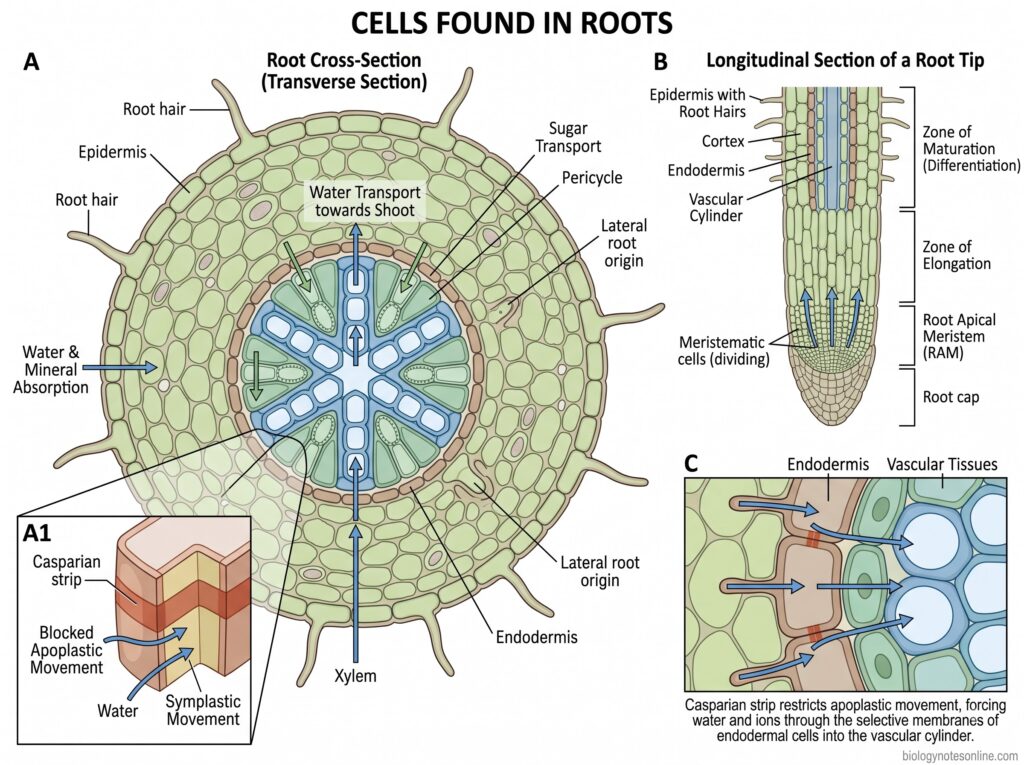
- Epidermal cells – These cells form the outer surface of young roots. Some epidermal cells develop long extensions called root hairs, which increase the surface available for absorption.
- Cortical parenchyma cells – A large portion of root cortex is formed by parenchyma. These cells are living and may store starch and other substances.
- Endodermal cells – Endodermis forms the innermost layer of the cortex. The cells develop Casparian strips, which restrict uncontrolled apoplastic movement of water and dissolved ions into the stele.
- Pericycle cells – These cells are present just inside the endodermis and surround the vascular tissues. Lateral roots commonly originate from particular pericycle cells.
- Xylem cells – Tracheids and vessel elements present in root xylem conduct water and dissolved mineral substances toward the shoot.
- Phloem cells – These occur in the vascular region along with xylem and transport sugars and other organic substances.
- Meristematic cells – Root tip contains actively dividing cells of the root apical meristem. New root tissues are formed from these cells.
Cells Found in Stems
Stem contains supporting, storage and conducting cells. Their arrangement varies greatly between herbaceous, woody, monocot and dicot stems.
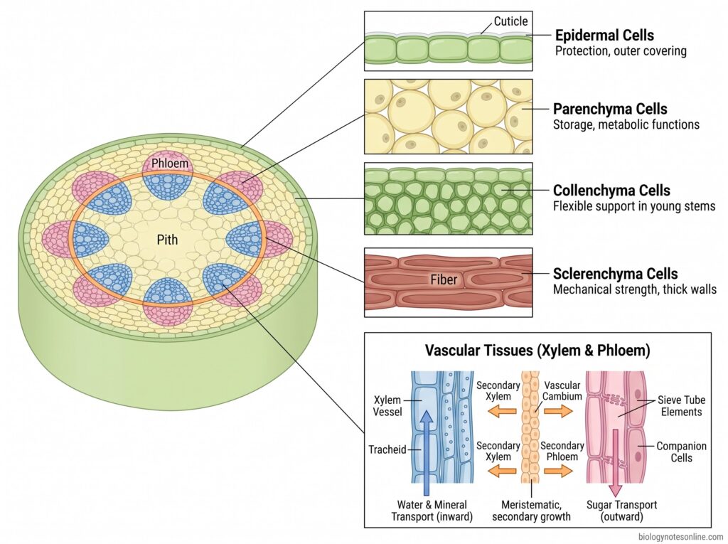
- Epidermal cells form the outer covering of young stems. A cuticle is commonly present over the surface.
- Parenchyma cells occur in cortex, pith and around vascular tissues. These living cells are mainly associated with storage and other metabolic functions.
- Collenchyma cells are common in many young stems, especially below the epidermis. They provide flexible support while the stem is still growing.
- Sclerenchyma cells occur as fibers or other supporting forms. Thick secondary walls provide mechanical strength to mature stem regions.
- Xylem and phloem cells form the vascular tissues of the stem. Xylem occurs toward the inner side and phloem generally toward the outer side in a typical collateral vascular bundle.
- Vascular cambium cells are meristematic cells present between secondary xylem and phloem in stems undergoing secondary growth. New secondary xylem is formed inward and secondary phloem outward.
Cells Found in Leaves
Leaves contain several specialized cells, particularly those used for photosynthesis, gas exchange and transport.
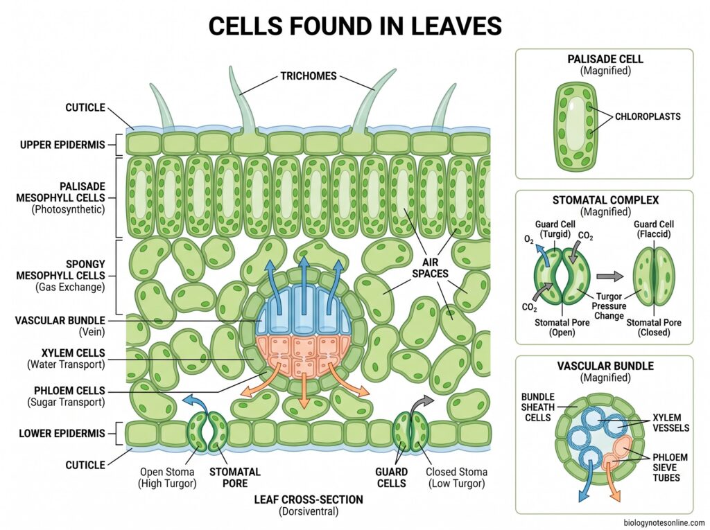
- Epidermal cells form the upper and lower surface of the leaf. These cells mainly provide protection and are usually covered by a cuticle on the exposed surface.
- Guard cells occur around stomatal pores. Their change in turgor controls stomatal opening and closing.
- Palisade mesophyll cells are elongated photosynthetic cells, generally closely arranged below the upper epidermis in many dorsiventral leaves. They contain numerous chloroplasts.
- Spongy mesophyll cells are more loosely arranged. Large air spaces occur between them, which are important for movement of gases within the leaf.
- Xylem cells occur in leaf veins and carry water into different regions of the leaf.
- Phloem cells are also present in veins. These are used for transport of sugars formed during photosynthesis.
- Bundle sheath cells surround the vascular bundles. Their development and function differ among plants, and they are particularly specialized in C4 plants.
- Trichomes may also develop from epidermal cells in many leaves. These are not present in the same form in every plant.
Comparison of Different Types of Plant Cells
The major plant cell types differ in their shape, wall structure, living condition and function. Some of the important differences are given below-
| Cell type | Shape | Wall | Lignin | Living/dead | Chloroplasts | Location | Function |
|---|---|---|---|---|---|---|---|
| Meristematic cells | Usually small, rounded, polygonal or nearly isodiametric | Thin primary cell wall | Generally absent | Living and actively dividing | Usually absent or plastids remain poorly differentiated | Root tips, shoot tips, cambium and other meristematic regions | Formation of new cells. Responsible for primary and secondary growth |
| Parenchyma cells | Rounded, oval, polygonal or elongated | Usually thin primary wall | Generally absent | Living | May be present. Abundant in chlorenchyma | Cortex, pith, mesophyll, fruits, seeds and vascular tissues | Storage, photosynthesis, secretion, transport and wound repair |
| Collenchyma cells | Usually elongated, polygonal in transverse section | Unevenly thickened primary wall, rich in cellulose and pectic substances | Normally absent | Living | Usually absent, but may occur in some green collenchyma cells | Below epidermis of young stems, petioles and leaf regions | Flexible mechanical support to growing organs |
| Sclerenchyma cells | Fibers are long and narrow. Sclereids are short or irregular | Very thick secondary wall | Commonly present | Usually dead at maturity | Absent | Mature stems, vascular tissues, seed coats, shells and hard fruit tissues | Mechanical strength, rigidity and protection |
| Epidermal cells | Variable. Often flattened, polygonal or irregular | Primary wall, with outer wall often thicker | Generally absent | Living | Usually absent from ordinary epidermal cells. Guard cells contain chloroplasts | Outer surface of young roots, stems and leaves | Protection, reduction of water loss and interaction with external environment |
| Xylem cells | Highly variable. Tracheids are elongated, vessel elements are wider and shorter, fibers are narrow | Conducting and supporting cells have thick secondary walls. Parenchyma has thinner walls | Common in tracheids, vessels and fibers | Both. Conducting elements are dead at maturity, xylem parenchyma remains living | Generally absent | Vascular bundles of roots, stems and leaves. Secondary xylem forms wood | Water conduction, mineral transport, mechanical support and storage |
| Phloem cells | Variable. Sieve elements are elongated, companion and parenchyma cells are generally smaller | Mostly primary walls. Fibers develop thick secondary walls | Usually absent from conducting cells. May occur in phloem fibers | Mostly living. Phloem fibers are usually dead at maturity | Generally absent | Vascular bundles and secondary phloem of roots, stems and leaves | Transport of sugars and other organic substances, storage and mechanical support |
| Palisade mesophyll cells | Elongated and column-like | Thin primary wall | Absent | Living | Numerous | Below upper epidermis of many dorsiventral leaves | Major site of photosynthesis |
| Spongy mesophyll cells | Irregular or rounded, loosely arranged | Thin primary wall | Absent | Living | Present, generally fewer than palisade cells | Below palisade mesophyll in many leaves | Photosynthesis and internal gas movement |
| Endodermal cells | Compact, usually barrel-shaped or rectangular in transverse section | Primary wall with specialized Casparian strip | Lignin is present in Casparian strips, often with suberin deposition during development | Living | Generally absent | Innermost layer of root cortex | Controls movement of water and dissolved ions toward vascular tissues |
| Cork cells | Usually compact and flattened | Walls become strongly suberized | May occur depending on tissue and developmental stage, but suberin is the characteristic wall material | Usually dead at maturity | Absent | Periderm of older stems and roots | Protection and reduction of water loss |
| Pericycle cells | Usually parenchymatous and relatively small | Mostly thin primary wall | Generally absent in young functional cells | Living | Generally absent | Just inside the endodermis of roots | Formation of lateral roots and contribution to secondary growth |
| Root-hair cells | Epidermal cell with a long tubular extension | Thin primary wall | Absent | Living | Usually absent | Root epidermis in the zone of differentiation | Absorption of water and mineral nutrients |
| Guard cells | Kidney-shaped in many dicots, dumbbell-shaped in many grasses | Unevenly thickened primary wall | Absent | Living | Present | Epidermis surrounding stomatal pores | Opening and closing of stomata, regulation of gas exchange and transpiration |
| Secretory cells and idioblasts | Highly variable according to type | Variable | May be absent or present depending on specialized cell | Usually living when functionally active | Variable | Leaves, stems, roots, flowers and other tissues | Secretion or storage of oils, resins, mucilage, crystals and other substances |
Parenchyma vs Collenchyma vs Sclerenchyma
The major differences between parenchyma, collenchyma and sclerenchyma are given below-
| Feature | Parenchyma | Collenchyma | Sclerenchyma |
|---|---|---|---|
| Nature of cells | Simple, living and comparatively less specialized cells | Living supporting cells | Strong supporting cells, generally dead at maturity |
| Cell shape | Rounded, oval, polygonal or sometimes elongated | Usually elongated. Polygonal in transverse section | Fibers are long and narrow, while sclereids may be rounded, irregular or branched |
| Cell wall | Usually thin primary cell wall | Unevenly thickened primary wall | Very thick secondary cell wall |
| Wall thickening | Usually not strongly thickened | Thickening is uneven, commonly at corners or particular walls | Heavy and more extensive secondary wall deposition |
| Lignin | Generally absent | Normally absent | Commonly present |
| Pectin | Present in primary wall | Abundant pectic substances are characteristic of the thickened wall | Not a major characteristic of mature secondary wall |
| Living or dead | Living at maturity | Living at maturity | Most cells become dead at maturity |
| Protoplast | Present | Present | Usually lost after maturation |
| Intercellular spaces | Commonly present. May become very large in aerenchyma | Small or absent in some types, larger in lacunar collenchyma | Usually very small or absent |
| Flexibility | Soft and comparatively flexible | Flexible and provides support without stopping growth | Rigid and strong |
| Main location | Cortex, pith, mesophyll, fruits, seeds and vascular tissues | Young stems, petioles and leaf regions, commonly below epidermis | Mature stems, vascular tissues, seed coats, shells and hard plant parts |
| Main function | Storage, photosynthesis, secretion, transport and regeneration | Flexible mechanical support in growing plant organs | Mechanical strength, rigidity and protection |
| Photosynthesis | Performed by chloroplast-containing parenchyma or chlorenchyma | May occur in green collenchyma, but not the major function | Generally absent |
| Storage | One of the important functions | Not a major function | Generally not the main function |
| Mechanical support | Limited compared with the other two | Important in young and growing regions | Major function in mature organs |
| Ability to divide again | Many cells can regain division under suitable conditions | Limited compared with parenchyma | Usually absent after maturity |
| Types | Chlorenchyma, aerenchyma, palisade, spongy, storage and ray parenchyma | Angular, lamellar, lacunar and annular | Fibers and sclereids |
| Examples | Potato storage tissue, leaf mesophyll | Celery strings, young dicot stems | Pear stone cells, seed coats and supporting fibers |
Xylem Cells vs Phloem Cells
The major differences between xylem cells and phloem cells are given below-
| Feature | Xylem Cells | Phloem Cells |
|---|---|---|
| Nature of tissue | Xylem is a complex vascular tissue made up of different cell types | Phloem is also a complex vascular tissue and contains several types of cells |
| Major cell types | Tracheids, vessel elements, xylem fibers and xylem parenchyma | Sieve-tube elements, companion cells, sieve cells, phloem parenchyma and phloem fibers |
| Main conducting cells | Tracheids and vessel elements | Sieve-tube elements in angiosperms and sieve cells in gymnosperms |
| Living or dead | Both are present. Tracheids, vessel elements and most fibers are dead at maturity, while parenchyma remains living | Mostly living. Sieve elements, companion cells and parenchyma are living, while phloem fibers are commonly dead at maturity |
| Cell wall | Conducting cells usually have thick secondary walls | Conducting cells mainly possess primary walls |
| Lignin | Commonly present in tracheids, vessel elements and fibers | Generally absent from conducting cells. It may occur in fibers |
| Perforations | Vessel elements contain perforation plates. Tracheids have pits but no perforation plates | Sieve-tube elements contain sieve plates with pores |
| Nucleus at maturity | Absent in mature tracheids and vessel elements | Mature sieve-tube elements lose their nucleus, while companion cells retain it |
| Associated living cells | Xylem parenchyma remains living | Companion cells and phloem parenchyma remain living |
| Main transported material | Water and dissolved mineral nutrients | Sugars and other organic substances |
| Direction of transport | Mainly from roots towards stems and leaves | Transport occurs from source to sink regions and can move in different directions in different sieve tubes |
| Main function | Water conduction and mineral transport | Translocation of sugars and other organic compounds |
| Mechanical support | Important function. Fibers and lignified conducting cells provide strength | Mainly provided by phloem fibers, when present |
| Storage | Xylem parenchyma stores starch and other substances | Phloem parenchyma stores carbohydrates and other materials |
| Position in vascular bundle | Usually present towards the inner side of a typical collateral vascular bundle | Usually present towards the outer side of xylem in a typical collateral vascular bundle |
| Role in wood formation | Secondary xylem forms most of the wood | Secondary phloem forms part of the inner bark |
| Examples | Vessel elements in angiosperm wood, tracheids in conifers | Sieve-tube elements with companion cells in flowering plants |
References
- Alberts, B., Johnson, A., Lewis, J., Raff, M., Roberts, K., & Walter, P. (2002). Molecular biology of the cell (4th ed.). Garland Science. https://www.ncbi.nlm.nih.gov/books/NBK21054/
- Barros, J., Serk, H., Granlund, I., & Pesquet, E. (2015). The cell biology of lignification in higher plants. Annals of Botany, 115(7), 1053–1074. https://doi.org/10.1093/aob/mcv046
- Baucher, M., El Jaziri, M., & Vandeputte, O. (2007). From primary to secondary growth: Origin and development of the vascular system. Journal of Experimental Botany, 58(13), 3485–3501. https://doi.org/10.1093/jxb/erm185
- Beck, C. B. (2010). An introduction to plant structure and development: Plant anatomy for the twenty-first century (2nd ed.). Cambridge University Press. https://doi.org/10.1017/CBO9780511844683
- Buckley, T. N. (2015). How does leaf anatomy influence water transport outside the xylem? Plant Physiology, 168(4), 1616–1635. https://doi.org/10.1104/pp.15.00731
- Casique, J. V., Cardoso-Gustavson, P., Marinho, C. R., & Teixeira, S. P. (2022). Coryanthes macrantha (Orchidaceae: Stanhopeinae) and its complex secretory structures: Anatomy, histochemistry and floral biology. AoB PLANTS, 14(5), plac039. https://doi.org/10.1093/aobpla/plac039
- Clark, M. A., Douglas, M., & Choi, J. (2018). Biology 2e. OpenStax, Rice University. https://openstax.org/details/books/biology-2e
- De Schepper, V., De Swaef, T., Bauweraerts, I., & Steppe, K. (2013). Phloem transport: A review of mechanisms and controls. Journal of Experimental Botany, 64(16), 4839–4850. https://doi.org/10.1093/jxb/ert302
- Eljebbawi, A., Dolata, A., Strotmann, V. I., & Stahl, Y. (2024). Stem cell quiescence and dormancy in plant meristems. Journal of Experimental Botany, 75(19), 6022–6036. https://doi.org/10.1093/jxb/erae201
- Emonet, A., Hay, A., & Geldner, N. (2022). Development and diversity of lignin patterns. Plant Physiology, 190(1), 31–43. https://doi.org/10.1093/plphys/kiac262
- Glover, B. J. (2000). Differentiation in plant epidermal cells. Journal of Experimental Botany, 51(344), 497–505. https://doi.org/10.1093/jexbot/51.344.497
- Grierson, C., & Schiefelbein, J. (2014). Root hairs. The Arabidopsis Book, 12, e0172. https://doi.org/10.1199/tab.0172
- Horbens, M., Feldner, A., Höfer, M., & Neinhuis, C. (2014). Ontogenetic tissue modification in Malus fruit peduncles: The role of sclereids. Annals of Botany, 113(1), 105–118. https://doi.org/10.1093/aob/mct262
- Jacquemoud, S., & Ustin, S. L. (2019). Leaf optical properties. Cambridge University Press. https://doi.org/10.1017/9781108686457
- Kang, B.-H., Anderson, C. T., Arimura, S.-i., Bayer, E., Bezanilla, M., Botella, M. A., Brandizzi, F., Burch-Smith, T. M., Chapman, K. D., Dünser, K., Gu, Y., Jaillais, Y., Kirchhoff, H., Otegui, M. S., Rosado, A., Tang, Y., Kleine-Vehn, J., Wang, P., & Zolman, B. K. (2022). A glossary of plant cell structures: Current insights and future questions. The Plant Cell, 34(1), 10–52. https://doi.org/10.1093/plcell/koab247
- Kim, J.-Y., Symeonidi, E., Pang, T. Y., Denyer, T., Weidauer, D., Bezrutczyk, M., Miras, M., Zöllner, N., Hartwig, T., Wudick, M. M., Lercher, M., Chen, L.-Q., Timmermans, M. C. P., & Frommer, W. B. (2021). Distinct identities of leaf phloem cells revealed by single cell transcriptomics. The Plant Cell, 33(3), 511–530. https://doi.org/10.1093/plcell/koaa060
- Leroux, O. (2012). Collenchyma: A versatile mechanical tissue with dynamic cell walls. Annals of Botany, 110(6), 1083–1098. https://doi.org/10.1093/aob/mcs186
- Leroux, O., Sørensen, I., & Marcus, S. E. (2018). Comparative in situ analysis reveals the dynamic nature of sclerenchyma cell walls of fern and seed plant species. Annals of Botany, 121(2), 345–359. https://doi.org/10.1093/aob/mcx151
- Meents, M. J., Watanabe, Y., & Samuels, A. L. (2018). The cell biology of secondary cell wall biosynthesis. Annals of Botany, 121(6), 1107–1125. https://doi.org/10.1093/aob/mcy005
- Mehdi, R., Lamm, C. E., Bodampalli Anjanappa, R., Münder, S., Kretzschmar, T., Kaiser, W. M., & Weber, A. P. M. (2019). Symplasmic phloem unloading and radial post-phloem transport via vascular rays in tuberous roots of Manihot esculenta. Journal of Experimental Botany, 70(20), 5559–5573. https://doi.org/10.1093/jxb/erz297
- Otero, S., & Helariutta, Y. (2017). Companion cells: A diamond in the rough. Journal of Experimental Botany, 68(1), 71–78. https://doi.org/10.1093/jxb/erw392
- Pace, M. R., Alcantara, S., Lohmann, L. G., & Angyalossy, V. (2015). Secondary phloem diversity and evolution in Bignonieae (Bignoniaceae). Annals of Botany, 116(3), 333–358. https://doi.org/10.1093/aob/mcv106
- Péret, B., Larrieu, A., & Bennett, M. J. (2009). Lateral root emergence: A difficult birth. Journal of Experimental Botany, 60(13), 3637–3643. https://doi.org/10.1093/jxb/erp232
- Procko, C., Lee, T., Borsuk, A., Bargmann, B. O. R., Crenshaw, C. M., Nery, J. R., Heger, A., Albrecht, V., Ecker, J. R., & Chory, J. (2022). Leaf cell-specific and single-cell transcriptional profiling reveals a role for the palisade layer in UV light protection. The Plant Cell, 34(9), 3261–3279. https://doi.org/10.1093/plcell/koac167
- Rudall, P. J. (2020). Anatomy of flowering plants: An introduction to plant structure and development (4th ed.). Cambridge University Press. https://doi.org/10.1017/9781108782104
- Rusnak, B., Clark, F. K., Vadde, B. V. L., & Roeder, A. H. K. (2024). What is a plant cell type in the age of single-cell biology? It’s complicated. Annual Review of Cell and Developmental Biology, 40, 301–328. https://doi.org/10.1146/annurev-cellbio-111323-102412
- Seo, D. H., Jeong, H., Choi, Y. D., & Jang, G. (2021). Auxin controls the division of root endodermal cells. Plant Physiology, 187(3), 1577–1586. https://doi.org/10.1093/plphys/kiab341
- Słupianek, A., Dolzblasz, A., Sokołowska, K., & Święch, Ł. (2021). Xylem parenchyma—Role and relevance in wood functioning in trees. Plants, 10(6), 1247. https://doi.org/10.3390/plants10061247
- Spicer, R. (2014). Symplasmic networks in secondary vascular tissues: Parenchyma distribution and activity supporting long-distance transport. Journal of Experimental Botany, 65(7), 1829–1848. https://doi.org/10.1093/jxb/ert459
- Stahl, J. M., Nepi, M., Galetto, L., Guimarães, E., & Machado, S. R. (2012). Functional aspects of floral nectar secretion of Ananas ananassoides, an ornithophilous bromeliad from the Brazilian savanna. Annals of Botany, 109(7), 1243–1252. https://doi.org/10.1093/aob/mcs053
- Steeves, T. A., & Sussex, I. M. (1989). Patterns in plant development (2nd ed.). Cambridge University Press. https://doi.org/10.1017/CBO9780511626227
- Torii, K. U. (2021). Stomatal development in the context of epidermal tissues. Annals of Botany, 128(2), 137–148. https://doi.org/10.1093/aob/mcab052
- Truernit, E. (2014). Phloem imaging. Journal of Experimental Botany, 65(7), 1681–1688. https://doi.org/10.1093/jxb/ert445
- Turley, E. K., & Etchells, J. P. (2022). Laying it on thick: A study in secondary growth. Journal of Experimental Botany, 73(3), 665–679. https://doi.org/10.1093/jxb/erab455
- Uchida, N., & Torii, K. U. (2019). Stem cells within the shoot apical meristem: Identity, arrangement and communication. Cellular and Molecular Life Sciences, 76, 1067–1080. https://doi.org/10.1007/s00018-018-2980-z
- Xiao, Y., Tholen, D., & Zhu, X.-G. (2016). The influence of leaf anatomy on the internal light environment and photosynthetic electron transport rate: Exploration with a new leaf ray tracing model. Journal of Experimental Botany, 67(21), 6021–6035. https://doi.org/10.1093/jxb/erw359