

Red blood cells (RBCs) are also known as erythrocytes. They are the most abundant formed cells of blood. A large amount of hemoglobin is present inside these cells, which gives the blood its red colour and binds oxygen. RBCs mainly take part in the transport of respiratory gases. Oxygen is carried from the lungs to the tissues, while part of the carbon dioxide produced in tissues is also transported with the help of red blood cells. These cells are formed by erythropoiesis from erythroid precursor cells, mainly in the bone marrow. During maturation, mammalian erythrocytes lose their nucleus. The mature RBC therefore does not contain a nucleus.
What Are Red Blood Cells?
Red blood cells (RBCs) are specialized cells present in the blood. They are also known as erythrocytes. These are the most common type of cells found in human blood.
The mature human RBC is a small biconcave disc-shaped cell. It does not contain a nucleus. Most of its internal space is occupied by hemoglobin (Hb), which is the major oxygen-carrying protein of the cell.
The major function of red blood cells is the transport of oxygen (O₂) from the lungs to different tissues of the body. Hemoglobin present inside the cells binds oxygen and makes this transport possible. Red blood cells also take part in the transport of carbon dioxide from tissues towards the lungs.
Red blood cells are formed through the process called erythropoiesis. The developing cells are present in the red bone marrow. Reticulocytes are released from the bone marrow into peripheral blood, where they complete their maturation into mature RBCs.
Why Are They Called Erythrocytes?
The term erythrocyte refers to a red blood cell. The word is formed from erythro-, meaning “red”, and -cyte, meaning “cell”. Thus, the term erythrocyte simply means a red cell.
The red colour is mainly due to the large amount of hemoglobin present within these cells. Hemoglobin is an iron-containing protein and gives RBCs, and therefore blood, its characteristic red colour.
Hence, red blood cell and erythrocyte are two names used for the same blood cell. The term erythrocyte is commonly used in hematology and other biological studies.
Where Are Red Blood Cells Found?
Red blood cells are mainly found in the circulating blood. They remain suspended in the liquid portion of blood called plasma along with white blood cells and platelets.
During circulation, RBCs pass through the arteries, capillaries and veins. They also pass through the chambers of the heart and pulmonary circulation. Their flexible structure allows them to move through very narrow blood capillaries.
The developing stages of red blood cells are mainly found in the red bone marrow. Here, erythroid precursor cells develop and finally produce reticulocytes. These immature cells are then released into the bloodstream and develop into mature erythrocytes.
Red Blood Cell at a Glance
| Feature | Description |
|---|---|
| Scientific name | Erythrocyte |
| Abbreviation | RBC (Red Blood Cell) |
| Shape | Biconcave, disc-shaped cell. |
| Diameter | About 7–8 µm, with a mean diameter near 7.5 µm. |
| Nucleus | Absent in mature human red blood cells. |
| Major protein | Hemoglobin (Hb), the oxygen-carrying protein. |
| Formation site | Mainly formed in the red bone marrow in adults through erythropoiesis. |
| Major function | Transport of oxygen (O₂) from lungs to tissues. They also participate in transport of carbon dioxide (CO₂) back towards the lungs. |
| Approximate lifespan | About 120 days in circulation. |
| Destruction/recycling sites | Old and damaged RBCs are removed mainly by macrophages of the spleen. The liver and bone marrow also participate in RBC clearance and recycling. |
Characteristics of Red Blood Cells
- Red blood cells (RBCs) are circular, biconcave disc-shaped cells. The centre of the cell is thinner as compared to its peripheral region.
- The normal RBC has a diameter of about 7.5–8.7 µm and thickness ranges between 1.7–2.2 µm.
- Mature human red blood cells do not contain a nucleus and other internal cell organelles. Mitochondria are also absent.
- A large amount of hemoglobin (Hb) is present inside the erythrocytes. This protein gives red colour to the cells and carries oxygen in the blood.
- RBC membrane is highly flexible and the cells can change their shape while moving through very small blood vessels. Thus, they can pass through capillaries having diameter smaller than the normal RBC.
- The biconcave form provides comparatively large surface area for exchange of respiratory gases. It also reduces the distance through which oxygen has to diffuse within the cell.
- Since mitochondria are absent, erythrocytes cannot carry out oxidative phosphorylation. The energy required by the cell is obtained mainly by anaerobic glycolysis.
- Mature RBCs also lack the normal machinery required for synthesis of new cellular proteins. They therefore have limited ability to repair their cellular components during their life in circulation.
- The major function of the RBC is the transport of oxygen (O₂) from lungs to different tissues. They also take part in transport of carbon dioxide (CO₂) from tissues to lungs.
- Red blood cells are produced from erythroid precursor cells in the bone marrow. The process of their formation is referred to as erythropoiesis.
- A mature human erythrocyte remains in the circulation for about 120 days. After this period, old and damaged erythrocytes are removed mainly by macrophages.
- Red blood cells are present in very large numbers in the circulating blood. Their biconcave shape, absence of nucleus, high hemoglobin content and deformable membrane are some of the main characteristics that make them suitable for transport of respiratory gases.
Size of a Red Blood Cell
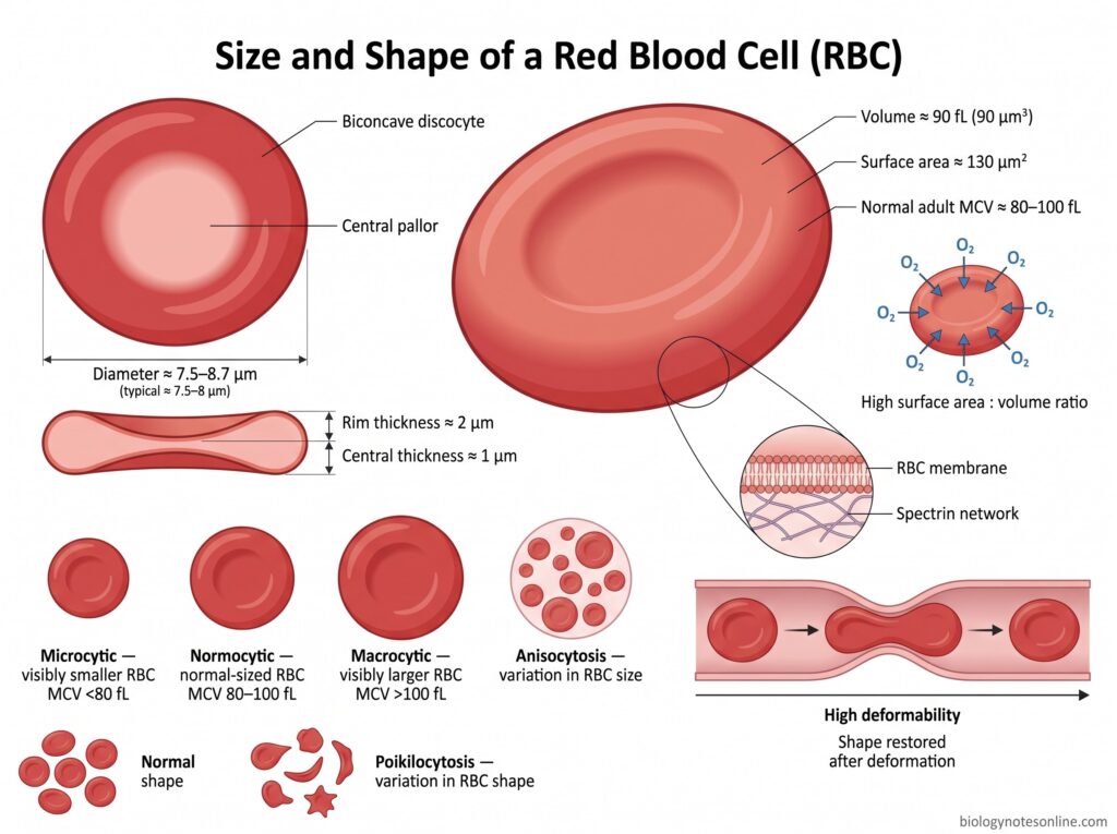
- The normal human red blood cell (RBC) has a diameter of about 7.5–8.7 µm. A value of about 7.5–8 µm is commonly taken for a mature erythrocyte.
- The thickness of RBC is not same throughout the cell. It is about 1.7–2.2 µm in thickness because of its biconcave form.
- At the peripheral or rim region, the cell is nearly 2 µm thick. The central region is much thinner, around 1 µm.
- A typical mature erythrocyte has a volume of approximately 90 µm³ (90 fL). Its surface area is around 130 µm².
- In hematology, the average size or volume of RBCs is expressed as Mean Corpuscular Volume (MCV). Normal adult MCV generally ranges between 80–100 fL.
- RBCs having a lower than normal volume are referred to as microcytic cells, whereas larger RBCs are called macrocytic cells. An MCV below about 80 fL is considered microcytic and above 100 fL is generally macrocytic.
- The size of every RBC in a blood sample may not always remain uniform. Variation in the size of red blood cells is called anisocytosis.
Shape of a Red Blood Cell
- Normal mature human red blood cell (RBC) is a biconcave disc-shaped cell. This normal form is also called a discocyte. Both surfaces are depressed towards the centre.
- The RBC is circular when viewed from the surface. Under a peripheral blood smear, its middle portion appears paler and is referred to as central pallor.
- The central region of the erythrocyte is thin while its outer rim is thicker. Therefore the cell does not have a flat disc-like appearance throughout. It forms the characteristic biconcave shape.
- The biconcave form provides a comparatively high surface area-to-volume ratio. It increases the surface available for diffusion of oxygen across the RBC.
- Red blood cells are highly deformable. They can change their normal disc shape while passing through narrow capillaries and other small spaces of the circulation.
- After the external force is removed, a healthy RBC generally returns to its normal biconcave form. Thus the cell can undergo repeated deformation during circulation without permanently losing its shape.
- The shape is maintained mainly by the RBC membrane and its underlying membrane cytoskeleton. Spectrin forms an important part of this supporting network.
- Changes from the normal erythrocyte shape may occur in different blood disorders. The presence of abnormally shaped RBCs in blood is referred to as poikilocytosis.
Why Do Red Blood Cells Have a Biconcave Shape?
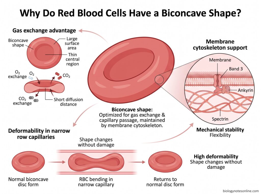
The biconcave shape of red blood cells is important for the exchange of respiratory gases. In this form, the cell has a large surface area in relation to its volume. More membrane surface is therefore available for the movement of oxygen (O₂) and carbon dioxide (CO₂). The thin central region also reduces the distance for diffusion within the cell.
This shape also makes the red blood cell highly deformable. RBCs have to move through very narrow capillaries during circulation. The biconcave form with extra membrane surface allows the cell to bend and change its shape without being damaged. After passing through the narrow vessels, it can again return towards its normal disc form.
The shape is maintained by the red cell membrane and its membrane cytoskeleton. Spectrin forms an important supporting network below the membrane and is connected with proteins such as ankyrin and band 3. These structures provide mechanical stability but at the same time allow flexibility of the cell. Thus, the biconcave form is suitable both for gas transport and movement of RBCs through the microcirculation.
Structure of a Red Blood Cell
Red blood cell has a comparatively simple structure. A mature human RBC is mainly formed of a cell membrane, membrane skeleton and hemoglobin containing cytoplasm. The nucleus and other major cell organelles are absent.
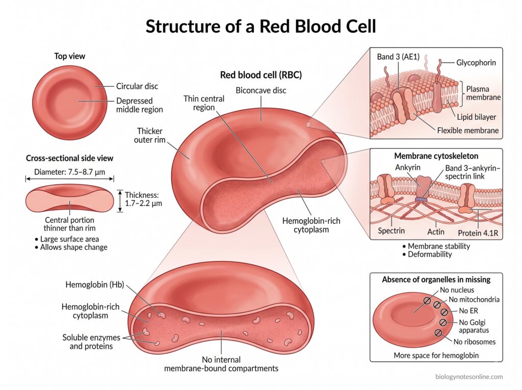
The following are the main structural features of red blood cells-
- Biconcave structure
- Mature RBC is a biconcave disc-shaped cell, also called a discocyte. Both sides of the cell are depressed at the centre.
- It is about 7.5–8.7 µm in diameter and around 1.7–2.2 µm in thickness. The central portion is thinner as compared to the outer rim.
- This arrangement gives the RBC a large surface area and also allows considerable changes in its shape.
- Cell membrane
- The RBC is surrounded by a thin and flexible plasma membrane. It is made up of a lipid bilayer containing different membrane proteins.
- Several proteins are inserted within this membrane. Band 3 (AE1) and glycophorins are some of the major membrane proteins.
- Band 3 is the most abundant integral membrane protein of the human erythrocyte. It also forms important attachment sites between the membrane and its internal skeleton.
- Membrane cytoskeleton
- Just below the lipid bilayer is a thin protein network called the membrane cytoskeleton. It gives mechanical support to the RBC membrane.
- Spectrin is the major structural protein of this network. It is arranged with short actin filaments and other proteins such as ankyrin and protein 4.1R.
- The spectrin skeleton is attached to the cell membrane at different points. One important connection is formed by band 3–ankyrin–spectrin.
- This structure maintains the membrane stability and at the same time allows the RBC to remain highly deformable.
- Cytoplasm and hemoglobin
- The interior part of a mature RBC contains a large amount of hemoglobin (Hb). It is the major oxygen carrying protein of the erythrocyte.
- The cytoplasm also contains soluble enzymes and proteins required for maintaining normal metabolism of the cell. Mature RBC itself has no internal membrane-bound compartments.
- Absence of nucleus and cell organelles
- Mature human red blood cell does not contain a nucleus. The nucleus is expelled during the later stages of erythrocyte development.
- The mature RBC also lacks mitochondria, endoplasmic reticulum, Golgi apparatus and ribosomes. These structures are removed during maturation of reticulocytes.
- Therefore, most of the internal space of the mature cell is available for hemoglobin and other soluble components.
Why Do Mature Red Blood Cells Have No Nucleus?
- Mature human red blood cells (RBCs) do not contain a nucleus. During the final stage of erythrocyte development, the nucleus becomes highly condensed and is removed from the erythroblast. This process is called enucleation.
- Removal of the nucleus provides more internal space for hemoglobin (Hb). Thus, the mature RBC can contain a large amount of hemoglobin required for transport of oxygen.
- The absence of nucleus also helps the RBC to form its characteristic biconcave shape. This form is thin at the centre and has a large surface area for exchange of respiratory gases.
- RBCs have to pass through very narrow blood capillaries. A large rigid nucleus would limit such changes in cell shape. The enucleated cell is more flexible and deformable, which helps in its movement through microcirculation.
- Enucleation is one of the final changes during formation of a mature mammalian erythrocyte. Other cell organelles and ribosomes are also removed during later maturation of the reticulocyte.
- The mature RBC therefore becomes a highly specialised cell, mainly packed with hemoglobin and suitable for transport of oxygen (O₂) and carbon dioxide (CO₂).
- Absence of the nucleus also has a limitation. Mature erythrocytes cannot divide, and with loss of ribosomes and other cellular machinery they have very limited ability to replace damaged cellular proteins.
- Enucleation is mainly a characteristic of mammalian red blood cells. Mature RBCs of many non-mammalian vertebrates, such as birds and fish, normally retain their nucleus.
Why Do RBCs Lack Mitochondria?
- Mature red blood cells (RBCs) do not possess mitochondria. During maturation of reticulocytes, mitochondria are removed from the cell by mitophagy.
- The major function of RBC is to carry oxygen (O₂) from lungs to different tissues. If mitochondria are present, some amount of this oxygen would be used by the RBC itself during cellular respiration.
- Due to absence of mitochondria, mature RBC cannot perform Krebs cycle, electron transport chain and oxidative phosphorylation. Thus, oxygen is not used for mitochondrial energy production inside the cell.
- The energy required by RBC is mainly obtained from glycolysis. During this process, glucose is converted into pyruvate and finally lactate. Only a small amount of ATP is produced but it is sufficient for different activities of mature RBC.
- Absence of mitochondria also provides more internal space inside the cell. This space is mainly occupied by hemoglobin (Hb), which is required for transport of oxygen.
- During formation of mature erythrocytes, mitochondria are not the only structures removed. Ribosomes and several other cell organelles are also lost during maturation of reticulocytes.
- Mitochondria are one of the sources of reactive oxygen species (ROS) in cells. Their absence also reduces mitochondrial production of ROS, which can otherwise cause oxidative damage to cellular components.
- Therefore, mature RBC is mainly dependent on glycolysis for its energy requirement. It does not consume the oxygen carried by hemoglobin and remains specialised for transport of respiratory gases.
Do Red Blood Cells Contain Other Organelles?
- Mature human red blood cells (RBCs) do not contain the common cell organelles. Nucleus and mitochondria are absent, also the endoplasmic reticulum and ribosomes.
- Golgi apparatus and other internal membrane structures are also lost. Thus, the mature erythrocyte is without most of the membrane-bound organelles which are normally present in other cells.
- These organelles, however, are present during the early stages of erythropoiesis.
- After removal of the nucleus, the young red cell is known as a reticulocyte. It still has some RNA, ribosomes and remaining organelle materials, which are gradually removed when the cell becomes mature.
- During this maturation process, mitochondria and several other cellular components are degraded. The mature RBC finally becomes almost free of internal cell organelles.
- Mature red blood cell is not an empty cell. A large amount of hemoglobin (Hb) is present in its cytoplasm together with different soluble enzymes, proteins and metabolites required for the cell. These are not organelles.
- The outer plasma membrane remains in the erythrocyte. Just below this membrane is the membrane cytoskeleton, giving support to the cell and maintaining its characteristic shape.
- In some immature or abnormal RBCs, remaining cellular materials may still be seen. These are called cellular inclusions, and are not the normal organelles of a mature erythrocyte.
How Are Red Blood Cells Adapted to Their Function?
Red blood cells show several structural and functional adaptations for transport of respiratory gases. Some of the important adaptations are-
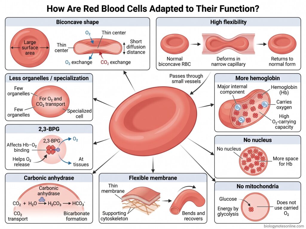
- Biconcave shape – RBC is a biconcave disc-shaped cell, with the central region thinner than the outer region. This form provides large surface area for gaseous exchange. Diffusion distance is also less.
- High flexibility – The red blood cell can easily change its shape. During circulation, it has to pass through very narrow blood capillaries, sometimes smaller than the normal diameter of RBC. This is possible because the cell is highly deformable.
- More hemoglobin – A major part of the RBC is occupied by hemoglobin (Hb). It is the respiratory pigment that combines with oxygen. Thus, a large amount of oxygen can be carried in the blood.
- No nucleus – Mature mammalian erythrocytes have no nucleus. This provides more internal space for hemoglobin inside the cell.
- No mitochondria – Mitochondria are absent in mature RBCs. Therefore, energy is mainly obtained by glycolysis, without using the oxygen carried by hemoglobin.
- Flexible membrane – The membrane of RBC is thin and flexible, with an underlying protein cytoskeleton. It gives support to the cell, but does not make it rigid. During passage through small vessels, the membrane bends and again returns towards its normal form.
- Carbonic anhydrase – RBC contains carbonic anhydrase, an enzyme involved in carbon dioxide transport. It rapidly converts carbon dioxide and water into carbonic acid, which then forms bicarbonate. A large portion of CO₂ is transported in this form.
- 2,3-BPG – 2,3-bisphosphoglycerate (2,3-BPG) is present in erythrocytes. It affects the binding of oxygen with hemoglobin and helps in release of oxygen at the tissue level.
- Less organelles – Mature RBC lacks most of the common cell organelles. The cell is therefore highly specialised, mainly for carrying hemoglobin and transport of oxygen (O₂) and carbon dioxide (CO₂).
How Do Red Blood Cells Transport Oxygen?
Red blood cells (RBCs) carry oxygen from lungs towards different tissues of the body. Most of this oxygen is transported by hemoglobin (Hb). The process occurs in following steps-
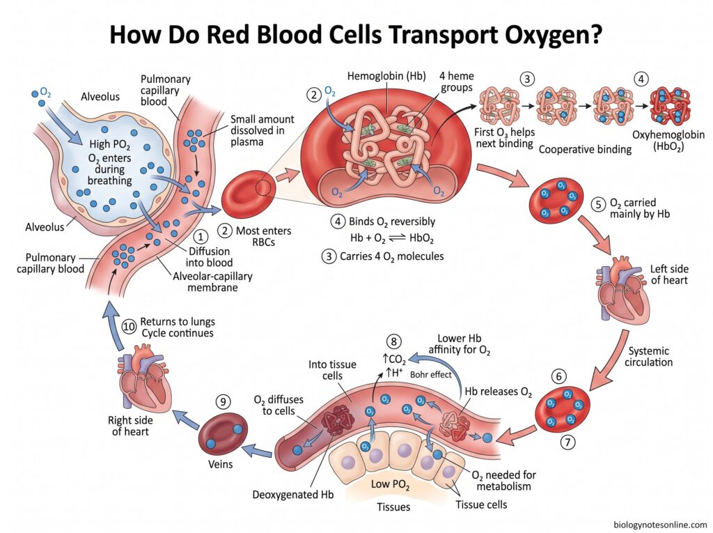
Step 1- Oxygen reaches alveoli
During breathing, oxygen enters into the alveoli of lungs. Here, oxygen partial pressure (PO₂) is high.
Step 2- Oxygen enters blood
Oxygen is diffused from alveoli across the alveolar-capillary membrane into pulmonary blood. A very small portion remains dissolved in plasma, most of it enters the red blood cells.
Step 3- Oxygen binds with hemoglobin
Inside RBC is hemoglobin, the major oxygen carrying protein. Oxygen combines reversibly with the iron-containing heme groups of hemoglobin.
A hemoglobin molecule has four heme groups and can carry four oxygen molecules.
The reaction can be represented as-
Hb + O₂ ⇌ HbO₂
Step 4- Oxyhemoglobin is formed
Hemoglobin combined with oxygen is called oxyhemoglobin (HbO₂).
Binding of first O₂ makes binding of the next oxygen easier. This type of binding is referred to as cooperative binding.
Step 5- Carried towards tissues
The oxygenated RBCs leave lungs, pass through the left side of heart and are then distributed to the body by systemic circulation. During this transport, oxygen remains largely bound with hemoglobin.
Step 6- At the tissue level
Body cells continuously consume oxygen for metabolism. Therefore, PO₂ is lower in the tissues.
Hemoglobin now begins to release its oxygen.
Step 7- Oxygen is unloaded
In actively metabolizing tissues, carbon dioxide and H⁺ are increased. The affinity of hemoglobin for oxygen becomes lower and more O₂ is released.
This effect of increased CO₂ and H⁺ on oxygen release is referred to as the Bohr effect.
Step 8- Oxygen enters cells
The released oxygen moves out from RBC, through plasma and tissue fluid and finally enters the cells according to the difference in oxygen partial pressure.
Hemoglobin becomes deoxygenated.
Step 9- RBC returns to lungs
After oxygen delivery, the deoxygenated RBCs are carried through veins to the right side of heart and then towards lungs. Oxygen again enters these cells, combines with hemoglobin and the same process continues.
Formation of Red Blood Cells — Erythropoiesis
What Is Erythropoiesis?
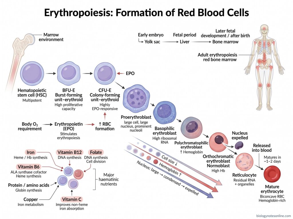
Erythropoiesis is the process by which new red blood cells (RBCs) or erythrocytes are formed. It starts from hematopoietic stem cells and finally forms mature erythrocytes. During this process, the developing cell passes through different stages, becomes smaller and hemoglobin (Hb) content is increased. The nucleus is finally removed.
Old RBCs are continuously removed from circulation and therefore new cells are also formed continuously. The process is mainly controlled according to oxygen requirement of the body. Erythropoietin (EPO) has the major role in this regulation.
Where Does Erythropoiesis Occur?
In adults, erythropoiesis takes place mainly in the red bone marrow. The active marrow is present mainly in vertebrae, ribs, sternum, skull and pelvic bones. Some amount is also present near the ends of certain long bones.
The site is not same throughout human development. During early embryonic period, red blood cell formation starts in the yolk sac. Later the fetal liver becomes the major site. Bone marrow gradually takes over during later fetal development and becomes the main site after birth.
Stages of Erythropoiesis
Erythropoiesis occurs through several stages of cell differentiation and maturation. The following are the stages-
Stage 1- Hematopoietic stem cell
The process begins from hematopoietic stem cell (HSC) in bone marrow. It is a multipotent cell and can give rise to different blood cells. Some cells become committed towards erythrocyte formation.
Stage 2- BFU-E
The committed cells form burst-forming unit-erythroid (BFU-E). It is an early erythroid progenitor and has high capacity for cell division.
Stage 3- CFU-E
BFU-E is further changed into colony-forming unit-erythroid (CFU-E). At this stage, the developing cells become highly responsive to erythropoietin (EPO).
Stage 4- Proerythroblast
CFU-E develops into proerythroblast. It is the first erythroid cell that can be clearly identified by its morphology.
The cell is large and nucleated. Prominent nucleoli are also present.
Stage 5- Basophilic erythroblast
Proerythroblast is converted to basophilic erythroblast. The cytoplasm stains blue because of large amount of ribosomal RNA.
Cell size now starts decreasing.
Stage 6- Polychromatophilic erythroblast
More hemoglobin is formed during this stage. Both ribosomal RNA and hemoglobin are present, therefore the cytoplasm shows a mixed grey-blue colour.
The cell becomes smaller.
Stage 7- Orthochromatic erythroblast
The polychromatophilic erythroblast changes into orthochromatic erythroblast, also referred to as normoblast. Hemoglobin content is now high and the nucleus becomes small and highly condensed.
At the end of this stage, nucleus is expelled from the cell.
Stage 8- Reticulocyte
After removal of nucleus, the cell is called a reticulocyte. Some RNA and remaining cell organelles are still present in it.
Reticulocytes are released from bone marrow into blood. During maturation, these remaining cellular materials are gradually removed.
Stage 9- Mature erythrocyte
The reticulocyte finally develops into a mature erythrocyte. This generally occurs within about 1–2 days after entering peripheral blood.
The mature RBC has no nucleus and most of the cell organelles are absent. It becomes a biconcave cell, containing a large amount of hemoglobin.
What Changes During RBC Maturation?
During maturation of red blood cells (RBCs), the developing erythroid cell undergoes several changes. The cell becomes smaller, hemoglobin increases and finally most of the internal cellular structures are lost. These changes continue until the mature erythrocyte is formed.
The following are the major changes during RBC maturation-
- Cell size – The size of erythroblast gradually decreases with maturation. After each successive cell division, smaller cells are produced.
- Hemoglobin – Hemoglobin (Hb) starts accumulating in increasing amount inside the cytoplasm. Thus, the developing cell becomes more hemoglobinized towards the later stages.
- Cytoplasm – Early erythroblasts have more ribosomal RNA and their cytoplasm is basophilic. With increase in hemoglobin and reduction of RNA, the cytoplasm changes towards the typical colour of a mature red cell.
- Nucleus – The nucleus becomes smaller and chromatin is increasingly condensed. In the orthochromatic erythroblast, it becomes highly condensed or pyknotic.
- Enucleation – Finally the condensed nucleus is expelled from the erythroblast. This process is called enucleation, producing an anucleated reticulocyte.
- Organelles – Reticulocytes still possess some mitochondria, ribosomes and other cellular materials. During further maturation these are degraded or removed. Mitochondria are mainly cleared by autophagic mechanisms.
- RNA content – The amount of residual RNA also decreases. Reticulocytes are named because this remaining ribosomal RNA forms a reticular appearance with special staining, but it disappears as the cell becomes mature.
- Cell membrane – Considerable remodeling of the plasma membrane takes place during reticulocyte maturation. Some membrane and unwanted membrane proteins are removed, while the membrane becomes suitable for the mature erythrocyte.
- Cell shape – The newly formed reticulocyte is not yet a completely mature biconcave RBC. During its final maturation, membrane and cytoskeletal remodeling results in formation of the characteristic biconcave erythrocyte.
- Final cell – At the end, a mature RBC is formed which lacks nucleus and practically all of the usual cell organelles. It is mainly filled with hemoglobin and is specialised for transport of respiratory gases.
Role of Erythropoietin (EPO)
Erythropoietin (EPO) is a glycoprotein hormone involved in regulation of erythropoiesis. In adults, it is produced mainly by the kidneys. The following are some of the important roles of EPO-
- Hypoxia response – The production of EPO is increased when oxygen supply to tissues becomes low. During this condition, the HIF pathway is activated in kidney and more EPO is released into blood.
- Marrow action – EPO reaches the bone marrow through blood. Here, it acts on erythroid progenitor and precursor cells having erythropoietin receptor (EPOR).
- Cell survival – One of the major actions of EPO is survival of developing erythroid cells. It prevents excessive apoptosis, especially in EPO-dependent erythroid progenitors. Thus, more cells remain available for further development.
- Cell proliferation – EPO stimulates proliferation of erythroid progenitor cells. More erythroid cells are therefore formed in bone marrow.
- Differentiation – It also promotes differentiation and maturation of erythroid precursors towards red blood cells. CFU-E and the following erythroblast stages are particularly associated with EPO action.
- RBC production – When EPO level is increased, erythropoiesis is stimulated and more red blood cells (RBCs) are produced. The oxygen-carrying capacity of blood is increased with the increase in red cell mass.
- Feedback control – EPO forms an important part of oxygen-dependent control of RBC production. Low tissue oxygen increases EPO formation. After sufficient RBC production and improvement of oxygen delivery, the stimulus for high EPO production becomes reduced.
Nutrients Required for RBC Formation
Formation of red blood cells (RBCs) needs a continuous supply of certain nutrients. Some are required for hemoglobin formation, while others take part in DNA synthesis and maturation of erythroid cells. The following are the major nutrients required-
- Iron – Iron is the major mineral required for hemoglobin (Hb) synthesis. It becomes a part of the heme group of hemoglobin. During erythropoiesis, large amount of body iron is used by developing erythroblasts. Iron deficiency therefore decreases normal hemoglobin formation.
- Vitamin B12 – Vitamin B12 (cobalamin) is required for normal DNA synthesis in dividing erythroid precursor cells. Its deficiency causes ineffective erythropoiesis and megaloblastic changes.
- Folate – Folate is also necessary for DNA formation and rapid cell division. Developing erythroblasts divide several times before a mature RBC is formed, therefore sufficient folate is required. Deficiency results in abnormal maturation of these cells.
- Vitamin B6 – Vitamin B6 (pyridoxine) takes part in heme synthesis. Its active form, pyridoxal phosphate, acts as a cofactor for ALA synthase, the first and rate-limiting enzyme of this pathway.
- Protein – Amino acids are required for synthesis of the globin chains of hemoglobin and many other proteins of developing red cells. During erythropoiesis, amino acids also support growth and metabolism of erythroid cells.
- Copper – Copper has an important role in normal iron metabolism. It helps in movement and proper utilization of iron, making iron available for hemoglobin formation. Copper deficiency can therefore result in anemia.
- Vitamin C – Vitamin C is not a structural component of RBC, but it improves absorption of non-heme iron from the intestine. In this way it helps in maintaining iron available for normal erythropoiesis.
Among these, iron, folate and vitamin B12 are particularly important haematinic nutrients and their deficiencies are common nutritional causes of defective erythropoiesis and anemia.
Life Cycle of a Red Blood Cell
The red blood cell (RBC) passes through formation, maturation, circulation and finally destruction. In healthy adults, new RBCs are continuously formed in the bone marrow to replace the old cells. A mature erythrocyte remains in blood for about 120 days.
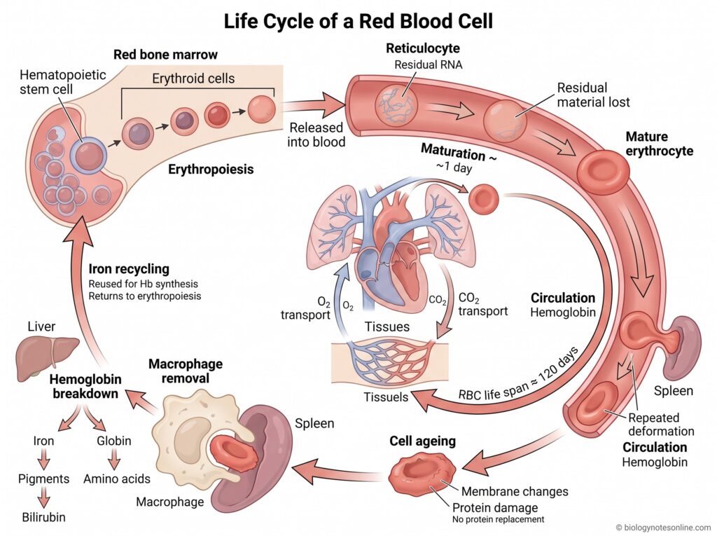
Some of the important stages in the life cycle of red blood cells are-
- Formation – RBC formation takes place in the red bone marrow by the process called erythropoiesis. Hematopoietic stem cells give rise to erythroid cells which pass through several stages before reticulocytes are formed.
- Reticulocyte stage – After removal of nucleus, the developing red cell becomes a reticulocyte. It leaves the bone marrow and enters into blood. Some residual RNA is still present at this stage.
- Maturation – Reticulocyte further matures in the circulation, generally within about a day. The remaining RNA and cellular materials are lost and a mature biconcave erythrocyte is formed.
- Circulation – Mature RBCs circulate continuously through the heart, lungs and different body tissues. Their major work during this period is transport of oxygen (O₂) with hemoglobin. Carbon dioxide transport is also carried out by these cells.
- Life span – A normal human erythrocyte remains in circulation for approximately 120 days. During its life it undergoes repeated deformation, particularly while passing through small capillaries and the spleen.
- Cell ageing – With time, different changes occur in the RBC membrane and cellular proteins. Mature erythrocytes cannot replace these damaged components because nucleus and normal protein-synthesizing machinery are absent. Old cells gradually become more suitable for removal from circulation.
- Removal – Aged or damaged RBCs are removed mainly by macrophages, particularly in the spleen. Liver macrophages also take part. The spleen acts as an important site for filtering old erythrocytes from blood.
- Hemoglobin breakdown – After phagocytosis, hemoglobin (Hb) is broken down into heme and globin components. Globin can be degraded into amino acids. Heme is further processed, producing iron and pigments that finally contribute to bilirubin formation.
- Iron recycling – Iron obtained from old RBCs is not simply lost. It is recycled by macrophages and can return to blood, where it becomes available again for hemoglobin synthesis in developing erythrocytes. A large part of iron required for daily erythropoiesis comes through this recycling process.
How Long Do Red Blood Cells Live?
Red blood cells (RBCs) have an average life span of about 120 days, nearly four months. During this period, they remain in circulation and continuously move through blood vessels, lungs and different body tissues.
Some of the important points about RBC life span are-
- Life span – A normal mature erythrocyte lives for approximately 120 days in circulation. This is an average value, not exactly same for every red blood cell. Measurements of normal RBC survival also show some variation around this period.
- Circulation – During its life, an RBC passes repeatedly through arteries, veins and very small capillaries. It also passes through the spleen many times. For nearly four months, the cell continues its major work of respiratory gas transport.
- Ageing – With time, changes occur in the membrane of RBC. The cell gradually loses some of its normal deformability, membrane proteins are also changed. Old erythrocytes therefore become less flexible than younger cells.
- Removal – After reaching old age, senescent RBCs are recognized and engulfed by macrophages. The spleen is an important site for this removal. Liver and bone marrow macrophages also take part.
- Recycling – Hemoglobin of the removed RBC is broken down. Iron is recovered from heme and can again be used for formation of new red blood cells. Other components are also further degraded or reused.
- Replacement – RBC destruction and formation continue together. The bone marrow therefore continuously produces new erythrocytes to replace the cells being removed from blood, roughly 0.8–1% of circulating RBCs each day under normal conditions.
What Happens to Old Red Blood Cells?
Red blood cells remain in the circulation for about 120 days. After this period, aged or damaged RBCs are gradually removed from blood. Macrophages have the major role in this process.
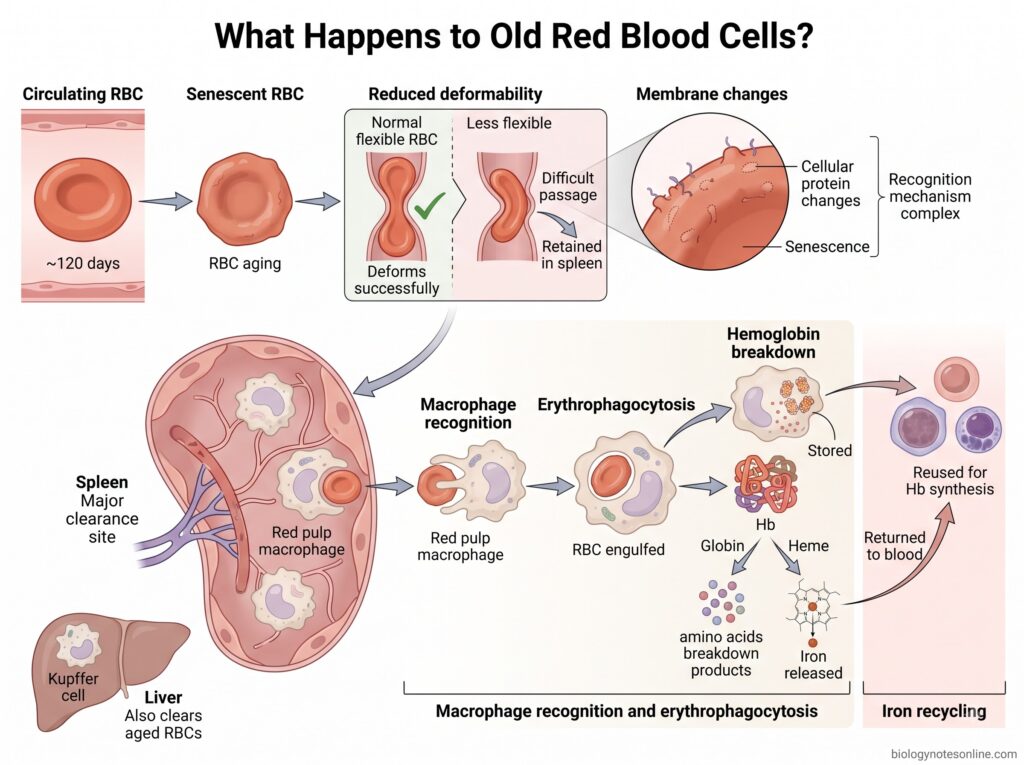
The following changes take place-
- RBC aging – With increasing age, different changes occur in the red blood cell. Membrane and cellular proteins are changed, and the cell gradually becomes old or senescent.
- Less deformability – The aged RBC loses some of its normal flexibility. Its deformability becomes reduced, making passage through the narrow spaces of spleen more difficult. Old cells can therefore become trapped in the splenic circulation.
- Membrane changes – Changes also take place on the RBC membrane during aging. These changes, together with decreased deformability, help in distinguishing old erythrocytes from normal circulating cells. The exact mechanism for recognition of naturally aged RBCs is complex and is not completely understood.
- Macrophage recognition – Senescent RBCs are recognized and engulfed by macrophages. This removal of erythrocytes by macrophages is called erythrophagocytosis.
- Spleen and liver – The spleen is one of the major sites where old RBCs are removed, particularly by red pulp macrophages. Kupffer cells of liver also take part in clearance.
- Hemoglobin breakdown – After the RBC is engulfed, hemoglobin (Hb) is degraded inside the macrophage. The globin part is broken down, while heme is further degraded. Iron is then released from heme.
- Iron recycling – The released iron is not normally wasted. It can be stored in macrophages or returned to blood and again supplied for hemoglobin formation in developing RBCs. A major portion of iron required for normal erythropoiesis comes from recycling of old red blood cells.
Thus, the process can be shown as-
Aging RBC → reduced deformability and membrane changes → macrophage recognition → spleen/liver clearance → hemoglobin degradation → iron recycling
Red Blood Cells Under the Microscope
Red blood cells (RBCs) can be examined under light microscope from a thin peripheral blood smear. The blood film is generally stained with stains such as Wright, Giemsa or Leishman stain for better observation of the blood cells.
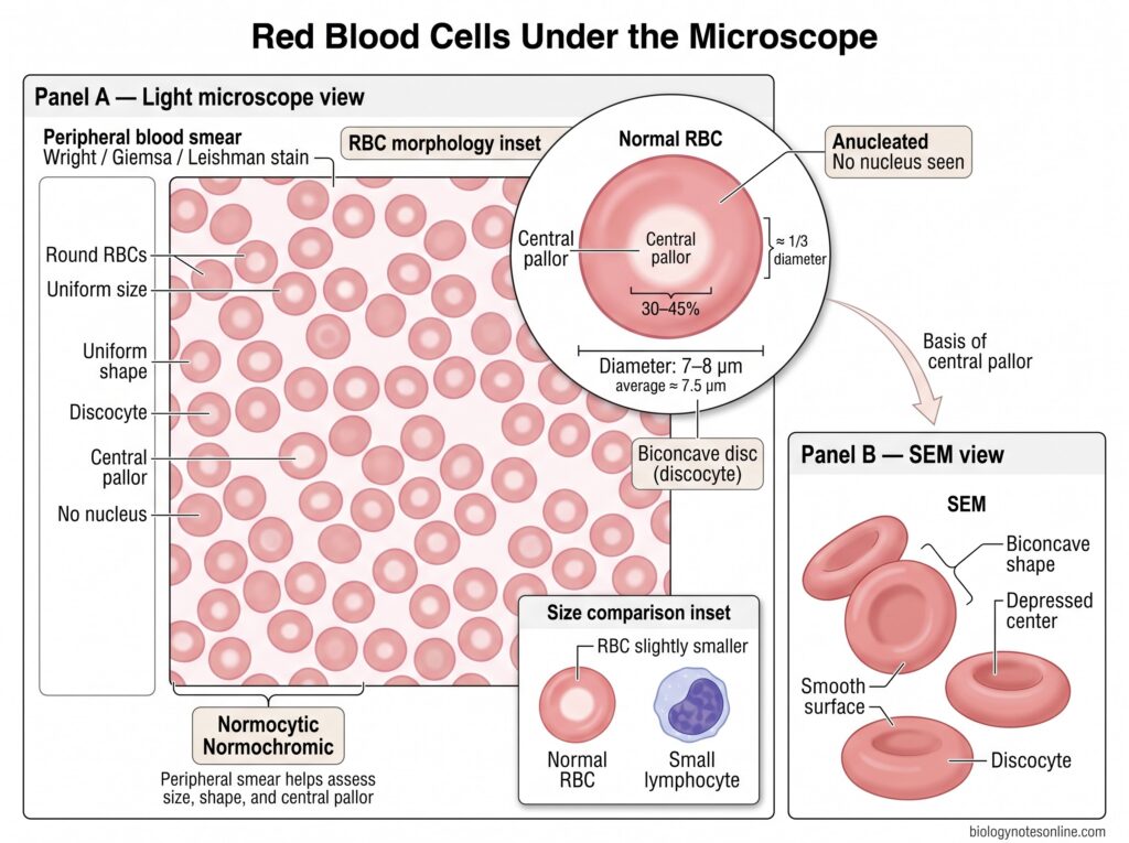
Some of the main microscopic features of normal red blood cells are-
- Appearance – Under the light microscope, normal RBCs appear as round or circular cells. They are fairly uniform in their normal size and shape.
- Shape – The normal erythrocyte is a biconcave disc, also referred to as a discocyte. In the blood smear it is seen mainly as a circular cell with a pale centre.
- Size – A normal RBC measures about 7–8 µm in diameter, with an average diameter around 7.5 µm. It is slightly smaller than a small lymphocyte.
- Central pallor – The middle portion appears pale because of the biconcave structure of RBC. This central pallor normally occupies about one-third of the cell diameter, approximately 30–45%.
- No nucleus – Mature human erythrocytes are anucleated. Thus, no nucleus is seen within a normal mature RBC under the microscope.
- Normal cells – RBCs having normal size and normal hemoglobinization are termed normocytic and normochromic. Changes in size, shape or central pallor can therefore be observed during examination of a peripheral blood smear.
- Under SEM – Under a Scanning Electron Microscope (SEM), the biconcave form becomes clearly visible. The cell shows a depressed central region with a smooth surface, giving the typical discocyte appearance.
Red Blood Cell Count
What Does RBC Count Measure?
Red blood cell (RBC) count is the number of erythrocytes present in a particular volume of blood. It is generally measured as a part of complete blood count (CBC).
The RBC count gives information about the number of circulating red cells, but it is not used alone. Hemoglobin, hematocrit and RBC indices are also considered. The normal reference value is not exactly same in every laboratory, and the range given by the testing laboratory should be followed.
How Is RBC Count Measured?
For RBC count, a small blood sample is collected, usually from a vein. The sample is then examined by an automated hematology analyzer.
Most modern analyzers count the red cells by methods such as electrical impedance. The final value represents the number of RBCs present per unit volume of blood.
What Does a Low RBC Count Mean?
A low RBC count means the number of circulating erythrocytes is below the reference range. It is commonly seen in anemia.
Blood loss, increased destruction of RBCs, decreased production in bone marrow, nutritional deficiency and kidney disease are some of the conditions where RBC count can be reduced. The cause cannot be determined from RBC count only.
What Does a High RBC Count Mean?
A high RBC count means an increased number of erythrocytes in blood. It may occur when red cell production is increased or when plasma volume becomes reduced, as during dehydration.
High altitude, long-term low oxygen conditions, some heart and lung diseases and polycythemia vera may also show an increased RBC count.
For normal ranges, causes and interpretation in detail, see the dedicated RBC Count article.
Disorders Affecting Red Blood Cells
Different disorders affect red blood cells (RBCs) in different ways. The number may decrease or increase, hemoglobin can be abnormal, or the cell membrane itself is affected. Some conditions cause early destruction of RBCs.
Disorders With Reduced RBC Number
- Anemia – Anemia is mainly defined by a reduced hemoglobin concentration. In many types, the circulating RBC number is also decreased. It may develop from decreased production, blood loss or increased destruction of red cells.
- Blood loss – During bleeding, RBCs are lost along with whole blood. Large acute loss can produce acute anemia. Repeated or chronic blood loss is different, it can gradually cause iron deficiency and decreased hemoglobin formation.
- Impaired erythropoiesis – Here the bone marrow does not produce sufficient normal RBCs. Iron, vitamin B12 or folate deficiency can affect erythropoiesis, also kidney disease with inadequate erythropoietin and different bone marrow disorders. New red cells are therefore not produced sufficiently to replace the normal loss.
Disorders With Increased RBC Number
- Erythrocytosis – Erythrocytosis refers to an increased red cell concentration or mass. It may be relative, due to reduction in plasma volume, or absolute when the amount of circulating RBCs is actually increased. Hypoxia and excess erythropoietin are among the causes of secondary erythrocytosis.
- Polycythemia – The term polycythemia is also used for an increased RBC mass. Polycythemia vera (PV) is a primary clonal bone marrow disorder where red cell production becomes increased independently. White blood cells and platelets may also increase in this disease.
Disorders Affecting Hemoglobin
- Sickle cell disease – It is an inherited disorder of hemoglobin caused by production of abnormal hemoglobin S (HbS). On deoxygenation, HbS can polymerize and RBCs become rigid and sickled. Hemolysis occurs, along with repeated obstruction of small blood vessels.
- Thalassemia – These are inherited disorders where production of α-globin or β-globin chains is reduced. The globin chains become unbalanced and normal hemoglobin formation is affected. In β-thalassemia, ineffective erythropoiesis and shortened survival of circulating red cells are important features.
Disorders Affecting RBC Membranes
- Hereditary spherocytosis – It is an inherited disorder affecting proteins of the RBC membrane and membrane skeleton. Spectrin, ankyrin, band 3 or protein 4.2 may be involved. The cells lose their normal biconcave form and spherocytes are produced, which are more readily trapped and destroyed in spleen.
Disorders Causing RBC Destruction
- Hemolytic anemia – In hemolytic anemia, RBCs are destroyed earlier than their normal life span. This destruction is referred to as hemolysis and may occur inside blood vessels or mainly after uptake by macrophages in spleen and liver. When destruction becomes more than the bone marrow can replace, anemia develops.
Infections Affecting Red Blood Cells
- Malaria – Malaria is caused by Plasmodium parasites. During the blood stage, merozoites invade RBCs and develop inside them. Infected erythrocytes are eventually ruptured or removed, while uninfected RBCs may also be cleared. Reduced erythropoiesis can occur as well, therefore malarial anemia has more than one mechanism.
Functions of Red Blood Cells
The major function of red blood cells (RBCs) is the transport of respiratory gases. Besides this, erythrocytes also take part in acid-base balance and normal blood flow. Some of the important functions are-
- Oxygen transport – RBCs transport oxygen (O₂) from lungs to different body tissues. Oxygen combines reversibly with hemoglobin (Hb) present inside the cell. This is the major function of erythrocytes.
- CO₂ transport – Red blood cells also take part in carrying carbon dioxide (CO₂) from tissues towards the lungs. Inside RBC, carbonic anhydrase converts CO₂ and water rapidly into carbonic acid, which forms bicarbonate and H⁺. A smaller amount of CO₂ is also carried by hemoglobin as carbaminohemoglobin.
- pH balance – Hemoglobin acts as an important buffer inside the red blood cell. It can bind the hydrogen ions formed during CO₂ transport, thus taking part in maintenance of normal acid-base condition of blood.
- Oxygen release – RBC does not only carry oxygen. It also helps in its release where the tissues require it. Increased CO₂ and H⁺ in tissues decrease the affinity of hemoglobin for oxygen, referred to as the Bohr effect.
- Blood flow – Erythrocytes have an important role in blood viscosity and its flow through the circulation. RBCs are highly deformable, allowing their movement even through very small blood vessels.
- Vascular regulation – RBCs can also participate in regulation of vascular function. Interaction of erythrocytes with nitric oxide (NO) and other signalling mechanisms has a role in adjusting local blood flow according to oxygen requirement of tissues.
- Immune role – Red blood cells have some functions in innate immune responses also. They can bind different circulating molecules, including chemokines, nucleic acids and some pathogens. This is an additional function, apart from their main role in gas transport.
Normal Red Blood Cells vs Abnormal Red Blood Cells
| Feature | Normal Red Blood Cells | Abnormal Red Blood Cells |
|---|---|---|
| Shape | Normal RBC is biconcave disc-shaped. | Shape may become irregular, spherical, sickle-shaped, elliptical or fragmented. |
| Size | Usually about 7–8 µm in diameter. | Cells may be smaller (microcytes) or larger (macrocytes). |
| Colour | Normally normochromic, with usual hemoglobin content. | Cells may become pale (hypochromic) or show altered staining. |
| Central pallor | A pale central region is normally present. | Central pallor may be increased, reduced or absent. |
| Nucleus | Mature human RBC has no nucleus. | Nucleated red cells may appear in blood during some abnormal conditions or marked marrow stress. |
| Hemoglobin | Contains normal amount and type of hemoglobin (Hb). | Hemoglobin amount or structure may be abnormal, as in iron deficiency, thalassemia or sickle cell disease. |
| Deformability | Highly flexible and can pass through narrow capillaries. | Deformability may become reduced, making movement through small vessels difficult. |
| Membrane | Membrane and cytoskeleton maintain normal biconcave form. | Membrane defects may produce cells such as spherocytes or elliptocytes. |
| Life span | About 120 days in circulation. | Life span may become shortened, especially in hemolytic disorders. |
| Examples | Discocytes, normocytic and normochromic RBCs. | Spherocytes, sickle cells, target cells, schistocytes, elliptocytes, acanthocytes and others. |
Red Blood Cells vs White Blood Cells
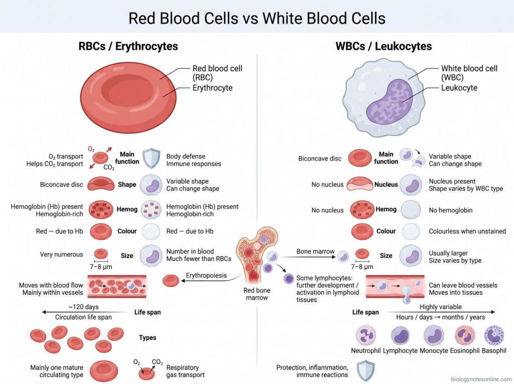
| Feature | Red Blood Cells (RBCs) | White Blood Cells (WBCs) |
|---|---|---|
| Other name | Erythrocytes | Leukocytes |
| Main function | Transport of oxygen (O₂) and also help in carbon dioxide transport. | Mainly involved in body defense and immune responses. |
| Shape | Usually biconcave disc-shaped. | Shape is variable. Many WBCs can also change their shape during movement. |
| Nucleus | Mature human RBCs have no nucleus. | Nucleus is present. Its shape differs among different WBC types. |
| Hemoglobin | Contains a large amount of hemoglobin (Hb). | Hemoglobin is absent. |
| Colour | Red in colour because of hemoglobin. | Colourless in unstained condition. |
| Size | Generally about 7–8 µm in diameter. | Usually larger than RBCs, but size depends upon the WBC type. |
| Number in blood | Present in very large number. | Present in much lower number as compared to RBCs. |
| Formation | Mainly formed in red bone marrow by erythropoiesis. | Formed mainly from hematopoietic cells in bone marrow. Some lymphocytes further develop or become activated in lymphoid tissues. |
| Movement | Remain mainly within blood vessels and move with blood flow. | Some WBCs can leave blood vessels and move into tissues. |
| Life span | Mature RBCs remain in circulation for about 120 days. | Life span varies greatly. Some survive for hours or days, while others may remain for months or years. |
| Types | Mature circulating RBCs are mainly one general cell type. | Major types include neutrophils, lymphocytes, monocytes, eosinophils and basophils. |
| Major role | Mainly respiratory gas transport. | Mainly protection against infection, inflammation and immune reactions. |
Mammalian vs Non-Mammalian Red Blood Cells
| Feature | Mammalian Red Blood Cells | Non-Mammalian Red Blood Cells |
|---|---|---|
| Nucleus | Mature RBCs generally have no nucleus. | Mature RBCs usually retain a nucleus. |
| Shape | Mostly biconcave disc-shaped. | Commonly oval or elliptical-shaped. |
| Size | Generally smaller. Human RBCs are about 7–8 µm in diameter. | Usually larger than mammalian RBCs, but size differs among species. |
| Cell organelles | Most common cell organelles are absent in mature RBCs. | Several internal cell structures are retained along with the nucleus. |
| Hemoglobin | Contains a large amount of hemoglobin (Hb) in the cytoplasm. | Hemoglobin is also present and carries oxygen. |
| Flexibility | Highly flexible and deformable. | Generally less deformable because of the presence of nucleus and different cell structure. |
| Main function | Mainly transport of oxygen (O₂) and participation in carbon dioxide transport. | Also mainly involved in transport of respiratory gases. |
| Surface area | Biconcave structure provides a high surface area in relation to cell volume. | Oval nucleated structure has a different surface area-to-volume arrangement. |
| Cell division | Mature RBCs cannot divide. | Mature circulating RBCs are nucleated, although their ability to divide depends on the vertebrate group and stage. |
| Examples | Humans, dogs, cats, cattle and most other mammals. | Birds, reptiles, amphibians and fish. |
Importance of Red Blood Cells
Red blood cells (RBCs) are important blood cells mainly involved in transport of respiratory gases. They carry oxygen to tissues, and also help in removal of carbon dioxide. Some of the important roles of red blood cells are-
- Oxygen transport – The major importance of RBC is transport of oxygen (O₂). Oxygen combines with hemoglobin (Hb) inside the cell and is carried from lungs to different body tissues.
- Tissue oxygenation – Body cells require oxygen for aerobic metabolism and production of energy. RBCs continuously supply this oxygen through blood circulation. If sufficient RBCs or hemoglobin are not present, tissue oxygen supply becomes reduced.
- Carbon dioxide transport – RBCs also participate in transport of carbon dioxide (CO₂) from tissues towards lungs. Inside the erythrocyte is carbonic anhydrase, which rapidly converts CO₂ and water into carbonic acid and then bicarbonate. A major amount of CO₂ is transported in this form.
- pH maintenance – Hemoglobin present inside RBC acts as an important buffer. It binds hydrogen ions formed during carbon dioxide transport. In this way, erythrocytes also take part in maintenance of acid-base balance of blood.
- Capillary circulation – Red blood cells are highly flexible. During circulation they have to move through very narrow capillaries, and the cells can change their shape for passing through these vessels. Thus oxygen can be supplied even to small tissue spaces.
- Oxygen unloading – Hemoglobin binds oxygen strongly in lungs but its binding is not same in all conditions. In actively metabolizing tissues, increased CO₂ and H⁺ help in release of oxygen from hemoglobin. More oxygen is therefore available where cellular activity is high.
- Blood flow – RBCs form a major cellular component of blood and influence its viscosity and flow. Their normal deformability is important, especially in microcirculation where blood vessels are very small.
- Vascular function – Erythrocytes also interact with different vasoactive substances including nitric oxide (NO) related pathways. These interactions have a role in adjustment of local blood flow according to oxygen requirement of tissues.
References
- Alberts, B., Johnson, A., Lewis, J., Raff, M., Roberts, K., & Walter, P. (2002). Molecular biology of the cell (4th ed.). Garland Science. https://www.ncbi.nlm.nih.gov/books/NBK21054/
- Anderson, H. L., Brodsky, I. E., & Mangalmurti, N. S. (2018). The evolving erythrocyte: Red blood cells as modulators of innate immunity. The Journal of Immunology, 201(5), 1343–1351. https://doi.org/10.4049/jimmunol.1800565
- Barbalato, L., & Pillarisetty, L. S. (2022). Histology, red blood cell. In StatPearls. StatPearls Publishing. https://www.ncbi.nlm.nih.gov/books/NBK539702/
- Dean, L. (2005). Blood groups and red cell antigens. National Center for Biotechnology Information. https://www.ncbi.nlm.nih.gov/books/NBK2261/
- Dean, L. (2005). Blood and the cells it contains. In Blood groups and red cell antigens. National Center for Biotechnology Information. https://www.ncbi.nlm.nih.gov/books/NBK2263/
- Diez-Silva, M., Dao, M., Han, J., Lim, C. T., & Suresh, S. (2010). Shape and biomechanical characteristics of human red blood cells in health and disease. MRS Bulletin, 35(5), 382–388. https://doi.org/10.1557/mrs2010.571
- Doyle, J., & Cooper, J. S. (2023). Physiology, carbon dioxide transport. In StatPearls. StatPearls Publishing. https://www.ncbi.nlm.nih.gov/books/NBK532988/
- Dzierzak, E., & Philipsen, S. (2013). Erythropoiesis: Development and differentiation. Cold Spring Harbor Perspectives in Medicine, 3(4), a011601. https://doi.org/10.1101/cshperspect.a011601
- Ford, J. (2013). Red blood cell morphology. International Journal of Laboratory Hematology, 35(3), 351–357. https://doi.org/10.1111/ijlh.12082
- Gottlieb, Y., Topaz, O., Cohen, L. A., Yakov, L. D., Haber, T., Morgenstern, A., Weiss, A., Berman, K. C., Fibach, E., & Meyron-Holtz, E. G. (2012). Physiologically aged red blood cells undergo erythrophagocytosis in vivo but not in vitro. Haematologica, 97(7), 994–1002. https://pmc.ncbi.nlm.nih.gov/articles/PMC3396668/
- Ji, P., Murata-Hori, M., & Lodish, H. F. (2011). Formation of mammalian erythrocytes: Chromatin condensation and enucleation. Trends in Cell Biology, 21(7), 409–415. https://pmc.ncbi.nlm.nih.gov/articles/PMC3134284/
- Keerthivasan, G., Wickrema, A., & Crispino, J. D. (2011). Erythroblast enucleation. Stem Cells International, 2011, 139851. https://pmc.ncbi.nlm.nih.gov/articles/PMC3189604/
- Klei, T. R. L., Meinderts, S. M., van den Berg, T. K., & van Bruggen, R. (2017). From the cradle to the grave: The role of macrophages in erythropoiesis and erythrophagocytosis. Frontiers in Immunology, 8, 73. https://doi.org/10.3389/fimmu.2017.00073
- Li, H., Lu, L., Li, X., Buffet, P. A., Dao, M., Karniadakis, G. E., & Suresh, S. (2021). Mechanics of diseased red blood cells in human spleen and consequences for hereditary blood disorders. Proceedings of the National Academy of Sciences of the United States of America, 115(38), 9574–9579. https://pmc.ncbi.nlm.nih.gov/
- Li, H., Lu, L., Li, X., Buffet, P. A., Dao, M., Karniadakis, G. E., & Suresh, S. (2021). How the spleen reshapes and retains young and old red blood cells: A computational investigation. PLoS Computational Biology, 17(11), e1009516. https://pmc.ncbi.nlm.nih.gov/articles/PMC8584971/
- Menon, V., Ghaffari, S., & Erythroid Enucleation Group. (2021). Erythroid enucleation: A gateway into a “bloody” world. Experimental Hematology, 95, 13–22. https://pmc.ncbi.nlm.nih.gov/articles/PMC8147720/
- Mohandas, N., & Gallagher, P. G. (2008). Red cell membrane: Past, present, and future. Blood, 112(10), 3939–3948. https://doi.org/10.1182/blood-2008-07-161166
- Moras, M., Lefevre, S. D., & Ostuni, M. A. (2017). From erythroblasts to mature red blood cells: Organelle clearance in mammals. Frontiers in Physiology, 8, 1076. https://doi.org/10.3389/fphys.2017.01076
- Namvar, A., Blanch, A. J., Dixon, M. W. A., Carmo, O. M. S., Liu, B., Tiash, S., Looker, O., Andrew, D., Chan, L. J., Tham, W.-H., & Tilley, L. (2021). Surface area-to-volume ratio, not cellular viscoelasticity, is the major determinant of red blood cell traversal through small channels. Cellular Microbiology, 23(1), e13270. https://doi.org/10.1111/cmi.13270
- Ney, P. A. (2011). Normal and disordered reticulocyte maturation. Current Opinion in Hematology, 18(3), 152–157. https://pmc.ncbi.nlm.nih.gov/articles/PMC3157046/
- Onishi, M., Yamano, K., Sato, M., Matsuda, N., & Okamoto, K. (2021). Molecular mechanisms and physiological functions of mitophagy. The EMBO Journal, 40(3), e104705. https://pmc.ncbi.nlm.nih.gov/articles/PMC7849173/
- Ovchynnikova, E., Aglialoro, F., von Lindern, M., & van den Akker, E. (2018). The shape shifting story of reticulocyte maturation. Frontiers in Physiology, 9, 829. https://doi.org/10.3389/fphys.2018.00829
- Patel, S., Jose, A., & Mohiuddin, S. S. (2023). Physiology, oxygen transport and carbon dioxide dissociation curve. In StatPearls. StatPearls Publishing. https://www.ncbi.nlm.nih.gov/books/NBK539815/
- Pretini, V., Koenen, M. H., Kaestner, L., Fens, M. H. A. M., Schiffelers, R. M., Bartels, M., & Van Wijk, R. (2019). Red blood cells: Chasing interactions. Frontiers in Physiology, 10, 945. https://doi.org/10.3389/fphys.2019.00945
- Rai, D., & Wilson, A. M. (2023). Histology, reticulocytes. In StatPearls. StatPearls Publishing. https://www.ncbi.nlm.nih.gov/books/NBK542172/
- Rhodes, C. E., Denault, D., & Varacallo, M. A. (2022). Physiology, oxygen transport. In StatPearls. StatPearls Publishing. https://www.ncbi.nlm.nih.gov/books/NBK538336/
- Slusarczyk, P., Mleczko-Sanecka, K., & Hentze, M. W. (2021). The multiple facets of iron recycling. Genes, 12(9), 1364. https://pmc.ncbi.nlm.nih.gov/articles/PMC8469827/
- Thiagarajan, P., Parker, C. J., & Prchal, J. T. (2021). How do red blood cells die? Frontiers in Physiology, 12, 655393. https://doi.org/10.3389/fphys.2021.655393
- Baldwin, C., Pandey, J., & Olarewaju, O. (2023). Hemolytic anemia. In StatPearls. StatPearls Publishing. https://www.ncbi.nlm.nih.gov/books/NBK558904/
- Billett, H. H. (1990). Hemoglobin and hematocrit. In H. K. Walker, W. D. Hall, & J. W. Hurst (Eds.), Clinical methods: The history, physical, and laboratory examinations (3rd ed.). Butterworths. https://www.ncbi.nlm.nih.gov/books/NBK259/
- Clinical Methods Editorial Group. (1990). Peripheral blood smear. In H. K. Walker, W. D. Hall, & J. W. Hurst (Eds.), Clinical methods: The history, physical, and laboratory examinations (3rd ed.). Butterworths. https://www.ncbi.nlm.nih.gov/books/NBK263/
- Maner, B. S., & Moosavi, L. (2022). Mean corpuscular volume. In StatPearls. StatPearls Publishing. https://www.ncbi.nlm.nih.gov/books/
- Sanchez, J. R., & Lynch, D. T. (2023). Histology, basophilic stippling. In StatPearls. StatPearls Publishing. https://www.ncbi.nlm.nih.gov/books/NBK545259/
- de Back, D. Z., Kostova, E. B., van Kraaij, M., van den Berg, T. K., & van Bruggen, R. (2014). Of macrophages and red blood cells; a complex love story. Frontiers in Physiology, 5, 9. https://doi.org/10.3389/fphys.2014.00009
- Kuhn, V., Diederich, L., Keller, T. C. S., Kramer, C. M., Lückstädt, W., Panknin, C., Suvorava, T., Isakson, B. E., Kelm, M., & Cortese-Krott, M. M. (2017). Red blood cell function and dysfunction: Redox regulation, nitric oxide metabolism, anemia. Antioxidants & Redox Signaling, 26(13), 718–742. https://doi.org/10.1089/ars.2016.6954
- Zhang, J., & Ney, P. A. (2010). Reticulocyte mitophagy: Monitoring mitochondrial clearance in a mammalian model. Autophagy, 6(3), 405–408. https://pubmed.ncbi.nlm.nih.gov/20200480/