

Microscope is a scientific instrument which is used to see very small objects that cannot be seen by naked eye. It produces enlarged image of minute objects and their fine details. It is used for observing cells, bacteria, tissues and other microscopic structures.
The word microscope is formed from two words. Micro means small and scope means to see or to view. It works by using lenses which bend and focus light. Due to this, the small specimen becomes magnified and visible to the observer.
Microscopes are very important in biology, microbiology, pathology and many scientific studies. It helps to study the shape, size, arrangement and internal structure of small living and non-living things. Without microscope, many cellular and microbial structures cannot be studied properly.
The history of microscope started in the late 16th century and early 17th century. It was first developed by spectacle makers in Netherlands and Denmark. In 1590, Dutch spectacle makers made the first compound microscope by using more than one lens for magnification.
Later, Antonie van Leeuwenhoek improved the use of microscope in the late 1600s. In 1673, he used a simple microscope with a single lens fitted between metal plates. By using this microscope, he observed living microorganisms and introduced microscopic life to the world.
Leeuwenhoek also tried to measure microscopic objects. He used fine sand grains as a rough measuring scale for finding size of human red blood cells. After this, microscope developed more and more with time.
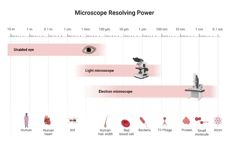
At first, microscope was a simple lens instrument with about 300x magnification. Now modern microscopes are highly developed instruments. Electron microscopes can magnify objects more than 250,000 times, and help to observe very fine cellular and sub-cellular structures.
Principle of Microscopes
Microscope is based on magnification of small object by using lens. The object is very small and it is not visible to naked eye. So lens system is used to make it large and clear.
In compound microscope, there are two lens system. One is objective lens and another is eyepiece lens. The light passes from the source and then through the specimen present on the slide.
After passing through the specimen, the light enters into the objective lens. The objective lens forms the first image of the object. This image is enlarged, real and inverted.
This first image then becomes object for the eyepiece lens. The eyepiece lens magnifies it again. It produces a large image which can be seen by eye.
So the microscope works by two time magnification. The first magnification is by objective lens. The second magnification is by eyepiece lens.
The total magnification of microscope is as follows-
Total magnification = Magnification of objective lens × Magnification of eyepiece lens
Parts of a Microscope
A. Structural parts
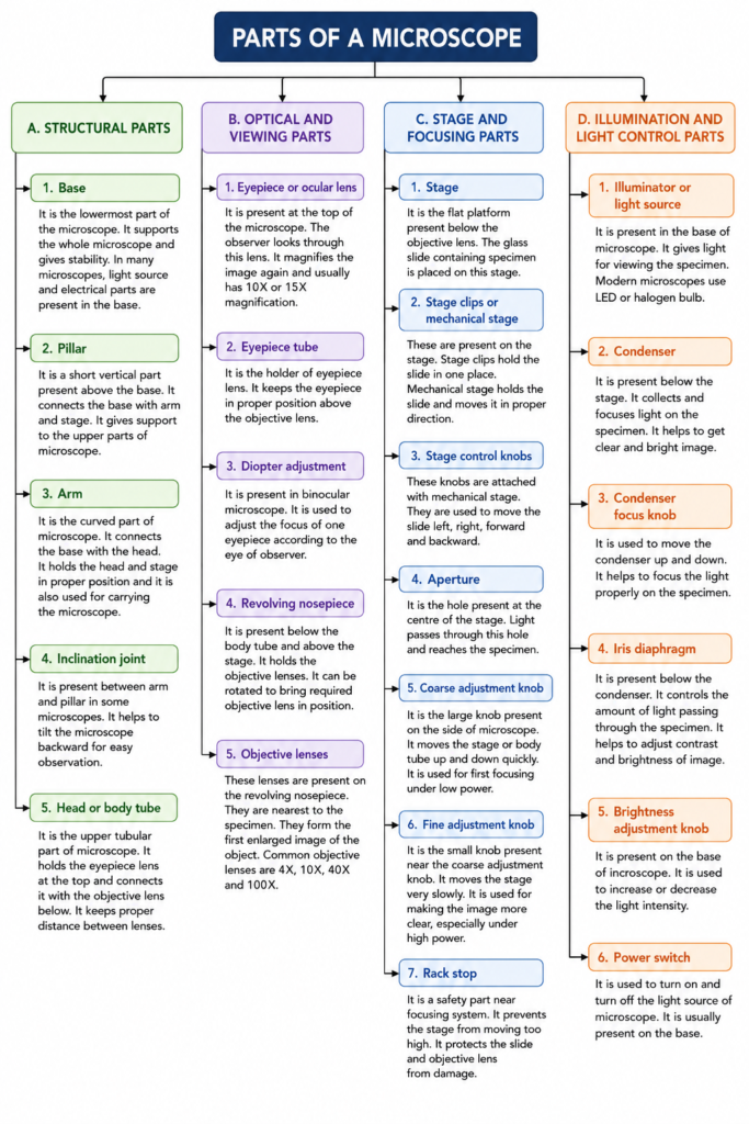
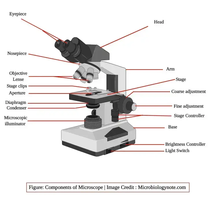
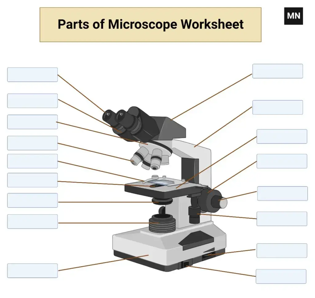
- Base – It is the lowermost part of the microscope. It supports the whole microscope and gives stability. In many microscopes, light source and electrical parts are present in the base.
- Pillar – It is a short vertical part present above the base. It connects the base with arm and stage. It gives support to the upper parts of microscope.
- Arm – It is the curved part of microscope. It connects the base with the head. It holds the head and stage in proper position and it is also used for carrying the microscope.
- Inclination joint – It is present between arm and pillar in some microscopes. It helps to tilt the microscope backward for easy observation.
- Head or body tube – It is the upper tubular part of microscope. It holds the eyepiece lens at the top and connects it with the objective lens below. It keeps proper distance between lenses.
B. Optical and viewing parts
- Eyepiece or ocular lens – It is present at the top of the microscope. The observer looks through this lens. It magnifies the image again and usually has 10X or 15X magnification.
- Eyepiece tube – It is the holder of eyepiece lens. It keeps the eyepiece in proper position above the objective lens.
- Diopter adjustment – It is present in binocular microscope. It is used to adjust the focus of one eyepiece according to the eye of observer.
- Revolving nosepiece – It is present below the body tube and above the stage. It holds the objective lenses. It can be rotated to bring required objective lens in position.
- Objective lenses – These lenses are present on the revolving nosepiece. They are nearest to the specimen. They form the first enlarged image of the object. Common objective lenses are 4X, 10X, 40X and 100X.
C. Stage and focusing parts
- Stage – It is the flat platform present below the objective lens. The glass slide containing specimen is placed on this stage.
- Stage clips or mechanical stage – These are present on the stage. Stage clips hold the slide in one place. Mechanical stage holds the slide and moves it in proper direction.
- Stage control knobs – These knobs are attached with mechanical stage. They are used to move the slide left, right, forward and backward.
- Aperture – It is the hole present at the centre of the stage. Light passes through this hole and reaches the specimen.
- Coarse adjustment knob – It is the large knob present on the side of microscope. It moves the stage or body tube up and down quickly. It is used for first focusing under low power.
- Fine adjustment knob – It is the small knob present near the coarse adjustment knob. It moves the stage very slowly. It is used for making the image more clear, especially under high power.
- Rack stop – It is a safety part near focusing system. It prevents the stage from moving too high. It protects the slide and objective lens from damage.
D. Illumination and light control parts
- Illuminator or light source – It is present in the base of microscope. It gives light for viewing the specimen. Modern microscopes use LED or halogen bulb.
- Condenser – It is present below the stage. It collects and focuses light on the specimen. It helps to get clear and bright image.
- Condenser focus knob – It is used to move the condenser up and down. It helps to focus the light properly on the specimen.
- Iris diaphragm – It is present below the condenser. It controls the amount of light passing through the specimen. It helps to adjust contrast and brightness of image.
- Brightness adjustment knob – It is present on the base of microscope. It is used to increase or decrease the light intensity.
- Power switch – It is used to turn on and turn off the light source of microscope. It is usually present on the base.
Applications of Microscopes
- Microscope is used in hospital and clinical laboratory for diagnosis of disease. It is used for observing blood cells, tissue section, urine sediments and other clinical samples.
- It is used in hematology, pathology and IVF related work. Special microscopes are also used in dentistry, eye study, gynecology and other medical fields.
- It is used for studying microscopic organisms and cell structures. Bacteria, fungi, protozoa, plant cells and animal tissues are observed by microscope.
- It helps to know the shape, size and arrangement of microorganisms. It also helps in study of cells and their small internal parts.
- It is used in research laboratory for cell study, tissue study and microbial study. Different cellular parts and small structures can be seen under microscope.
- It is used in industry for quality checking of materials. Semiconductors, circuit boards, metals, ceramics and polymers are examined under microscope.
- It helps to detect small cracks, defects and surface changes in industrial materials. This is useful in quality control and failure analysis.
- It is used in school, college and university laboratory. Students observe prepared slides, plant tissues, animal tissues and microorganisms for practical study.
- It is used in forensic laboratory for crime investigation. Hair, fibres, fingerprints, handwriting marks and bullet marks can be studied and compared.
- It is used for checking water and soil samples. Algae, planktons, microorganisms and small particles present in environment can be observed.
- It is used in agriculture and crop research. Fungal spores, plant cells, insect parts and soil microorganisms can be examined.
- It is used in veterinary laboratory for diagnosis of animal diseases. Parasites, bacteria, blood sample and semen sample are observed under microscope.
- It is used in geology and metallurgy for study of rocks, minerals and metals. Mineral grains, metal structure, gemstones and mining samples can be observed.
- It is used for examination of old materials and artifacts. Small surface marks, particles and structure of ancient samples can be studied.
Parts of a Microscope and Their Functions ppt
Parts of a Microscope and Their Functions Video
Parts of a Microscope Worksheet
1. parts of a compound microscope Worksheet
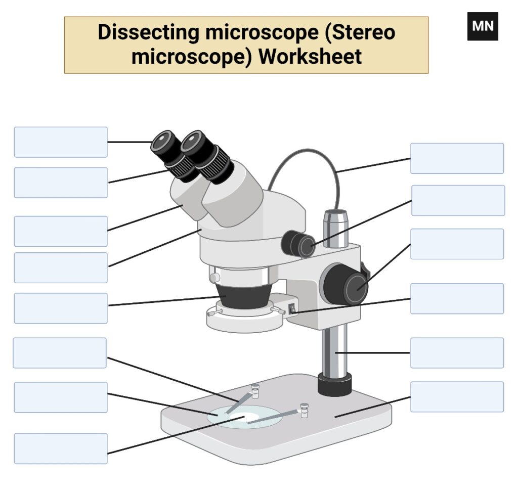
2. Dissecting microscope (Stereo microscope) Worksheet
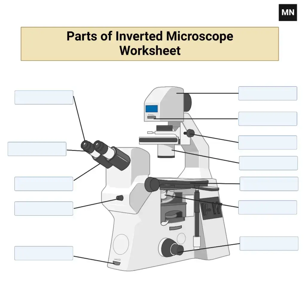
3. Inverted Microscope Worksheet
References
- Abramowitz, M., & Davidson, M. W. (2026). Photomicrography errors: Exposure, focus & color troubleshooting guide. Evident.
- Abramowitz, M., & Davidson, M. W. (2026). The concept of magnification. Evident.
- Abramowitz, M., Keller, H. E., Spring, K. R., Flynn, B. O., Long, J. C., Parry-Hill, M. J., Tchourioukanov, K. I., & Davidson, M. W. (2015). Molecular expressions microscopy primer: Anatomy of the microscope. The Florida State University.
- ACCU-SCOPE. (n.d.). Maintain your microscope to ensure longevity and precision.
- AL-Taei, A. J. (n.d.). Parts of a compound light microscope. Medical Laboratory Instrument.
- AmScope. (2025). The ultimate guide to microscope cleaning & maintenance.
- AmScope Team. (2022). How does a compound microscope work? AmScope.
- Bispo, G. (2026). How to maintain and clean your microscope for longevity. Precision Engineering Supply.
- Cappuccino, J. S., & Sherman, N. (n.d.). The microscope. In Microbiology: A laboratory manual (5th ed.). Benjamin/Cummings Science Publishing.
- Clayton, M. (n.d.). How to use a student compound microscope. Carolina.
- CodeLucky. (n.d.). Compound microscope explained: How magnification works | Light & optics [Video]. YouTube.
- Dahal, P. (2026). Parts of a microscope with functions and labeled diagram. Microbe Notes.
- Edmund Optics. (n.d.). Understanding microscopes and objectives.
- KEYENCE CORPORATION. (n.d.). Top 10 microscopy troubleshooting tips for clearer fluorescence images.
- Koenig, F. (2020). Compound microscope parts, functions, and labeled diagram. New York Microscope Company.
- Marks, K., & Hochman Adler, V. (2024). 3.2: The compound light microscope. Biology LibreTexts.
- Microscope Central. (n.d.). Troubleshooting common microscope issues.
- Microscope World. (n.d.). Troubleshooting microscope focusing issues.
- Nikon Instruments Inc. (2025). The microscope optical train. Nikon’s MicroscopyU.
- OpenStax College. (n.d.). Microscopes. Lumen Learning.
- Potts, C. (2025). Stop! Don’t touch your microscope until you read this guide (maintenance). Microscope Marketplace.
- Structural and optical architecture of the compound light microscope: A comprehensive analysis of component functionality and maintenance. (n.d.).
- The microscope. (n.d.). Microscope Lab.
- ZEISS Microscopy. (2025). Microscope cleaning & maintenance: The complete guide for optimal performance. ZEISS.
- Zumax. (2025). Common mistakes you should avoid when using a microscope. Albert Waeschle Ltd.