

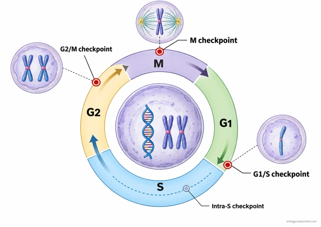
Checkpoints in the cell cycle are regulatory surveillance mechanisms that check whether a cell can proceed through important stages of the cell cycle. There are three major checkpoints, the G1/S checkpoint, G2/M checkpoint, and M checkpoint (spindle assembly checkpoint). In some classifications, the intra-S-phase checkpoint is also counted separately, making four checkpoints.
These checkpoints are not separate phases of the cell cycle. They control the progression of cells from one stage to another.
The G1/S checkpoint regulates the beginning of deoxyribonucleic acid (DNA) replication, while the G2/M checkpoint controls entry into mitosis. Separation of sister chromatids during mitosis is regulated by the M checkpoint. When the required conditions are not fulfilled, cell cycle progression can be delayed at these checkpoints.
Location of Checkpoints in the Cell Cycle
The cell cycle is divided into four phases, G1 (first gap), S (synthesis), G2 (second gap), and M (mitosis). These phases occur one after another in the following sequence-
G1 → S → G2 → M
Deoxyribonucleic acid (DNA) replication takes place during the S phase. There are three major checkpoints present at different positions of the cell cycle. These are the G1/S checkpoint, G2/M checkpoint, and M checkpoint (spindle assembly checkpoint).
The G1/S checkpoint is found near the end of G1 phase. The G2/M checkpoint occurs at the transition between G2 and M, while the M checkpoint operates during mitosis.
| Checkpoint name | Location | Primary conditions monitored | Progression controlled |
|---|---|---|---|
| G1/S checkpoint | Late G1 phase, before S phase | Cell size, nutrients, growth signals, and DNA damage | Entry into S phase for DNA replication |
| G2/M checkpoint | End of G2 phase, before mitosis | Completion of DNA replication and DNA damage | Entry into mitosis |
| M checkpoint (spindle assembly checkpoint) | M phase, before anaphase | Proper attachment of chromosome kinetochores to spindle microtubules | Separation of sister chromatids during anaphase |
An intra-S-phase checkpoint is also present during DNA replication. It responds to DNA damage and replication stress. DNA replication may be slowed down when these problems occur. In some classifications, this checkpoint is counted separately as the fourth checkpoint.
The term S/G2 checkpoint is used in some cases for surveillance near the transition between S and G2 phases. It is not universally considered as another major checkpoint. The completion of DNA replication may be checked by mechanisms operating during S phase and also before the cell enters mitosis.
G1 Checkpoint (G1/S Checkpoint)
The G1 checkpoint is the first major checkpoint of the cell cycle, which is present near the end of G1 phase before the beginning of S phase. It is also called the G1/S checkpoint. In this checkpoint, the cell is checked for its size, availability of nutrients, growth signals, and damage to deoxyribonucleic acid (DNA).
The following are the important features and functions of the G1 checkpoint-
- The G1 checkpoint checks whether the cell has reached sufficient size for division. The availability of nutrients and other cellular resources required for DNA replication is also checked.
- In many animal cells, growth factors are required for the progression through G1 phase. Without sufficient growth signals, the cell may stop progressing through the cell cycle.
- The restriction point (R point) is a regulatory point present in G1 phase. After passing this point, the cell becomes largely independent of further growth factor stimulation for completing the cell cycle. It is associated with G1 regulation but is not exactly the same as the DNA damage checkpoint.
- DNA is checked for any damage before the cell enters S phase. If DNA damage is detected, the cell cycle progression is delayed. This allows time for DNA repair before replication begins.
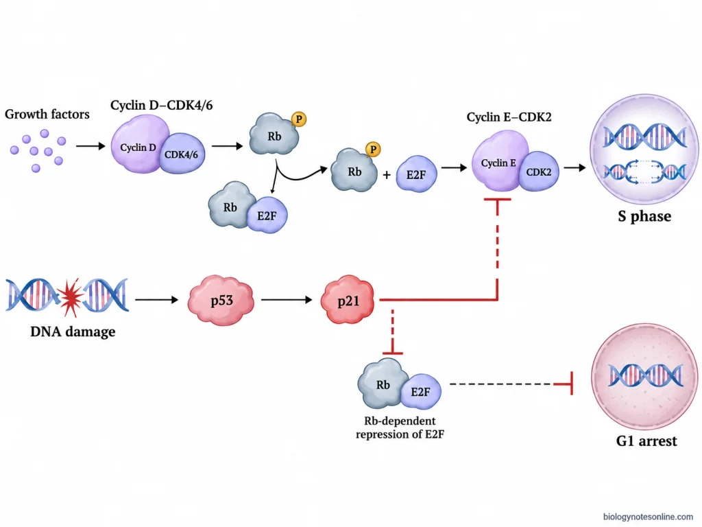
- In mammalian cells, DNA damage can activate the p53 protein, which increases the production of p21 protein. The p21 inhibits the activity of cyclin-dependent kinases (CDKs) and prevents the cell from entering S phase.
- The progression of G1 phase is also regulated by cyclin D-CDK4/6 and cyclin E-CDK2 complexes. Cyclin D-CDK4/6 promotes the phosphorylation of retinoblastoma protein (Rb). This helps in the release of E2F transcription factors which activate the genes required for entry into S phase.
- When all the required conditions are fulfilled, the cell enters S phase and DNA replication begins.
- In absence of sufficient growth signals, cells may enter the resting G0 phase. These cells remain in a non-dividing state. Some of the cells can again enter the cell cycle when suitable growth signals become available.
- Severe or persistent DNA damage may result in prolonged cell cycle arrest, cellular senescence, or programmed cell death (apoptosis). The response depends on the cell type and extent of DNA damage.
Intra-S-Phase Checkpoint
The intra-S-phase checkpoint is a regulatory mechanism that operates during the S (synthesis) phase of cell cycle. DNA replication takes place in this phase. It monitors damage to deoxyribonucleic acid (DNA) and problems in DNA replication, which is referred to as replication stress.
In some classifications, it is also counted as the fourth checkpoint of the cell cycle, apart from the three major checkpoints.
The important features and functions of intra-S-phase checkpoint are as follows-
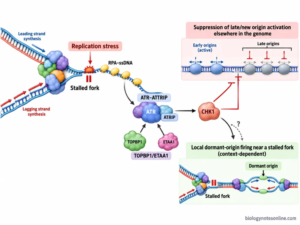
- During S phase, the DNA replication forks may become stalled due to DNA damage or problems in the replication process. In these conditions, the intra-S-phase checkpoint is activated.
- At the stalled replication forks, single-stranded DNA (ssDNA) may accumulate. These DNA regions are covered by replication protein A (RPA). The RPA helps in the recruitment and activation of ataxia telangiectasia and Rad3-related protein (ATR). The activated ATR then phosphorylates checkpoint kinase 1 (CHK1). This kinase is involved in the regulation of replication checkpoint response.
- The CHK1 inhibits the initiation of DNA replication at additional replication origins, especially those which are scheduled to become active later during S phase. The overall rate of DNA synthesis is reduced.
- DNA replication does not stop completely in every case. Some dormant replication origins located near the stalled replication forks may become active. DNA replication can continue in these regions.
- In case of DNA damage, the movement of replication forks may slow down. The checkpoint proteins help in maintaining the stability of stalled replication forks. They are also involved in the repair and restarting of damaged replication forks.
- After the replication stress is resolved, the stalled replication forks may start again and DNA synthesis continues.
- The intra-S-phase checkpoint also prevents the accumulation of DNA damage during replication.
- Defects in this checkpoint may result in incomplete or abnormal DNA replication. It can also increase genomic instability.
G2 Checkpoint (G2/M Checkpoint)
The G2 checkpoint is the second major checkpoint of the cell cycle. It is located at the end of G2 (second gap) phase, before the cell enters mitosis. It is also called the G2/M checkpoint.
The checkpoint checks whether deoxyribonucleic acid (DNA) replication is completed and if any DNA damage is present.
The important features and functions of G2 checkpoint are as follows-
- The DNA replication must be completed before the cell proceeds to M phase. When DNA replication remains incomplete or the replication forks are stalled, the entry into mitosis is delayed.
- Damage to DNA may occur during its replication. Radiation and chemical agents can also damage DNA. When this damage is detected, the cell cycle is stopped at G2 phase. The damaged DNA is given time for repair.
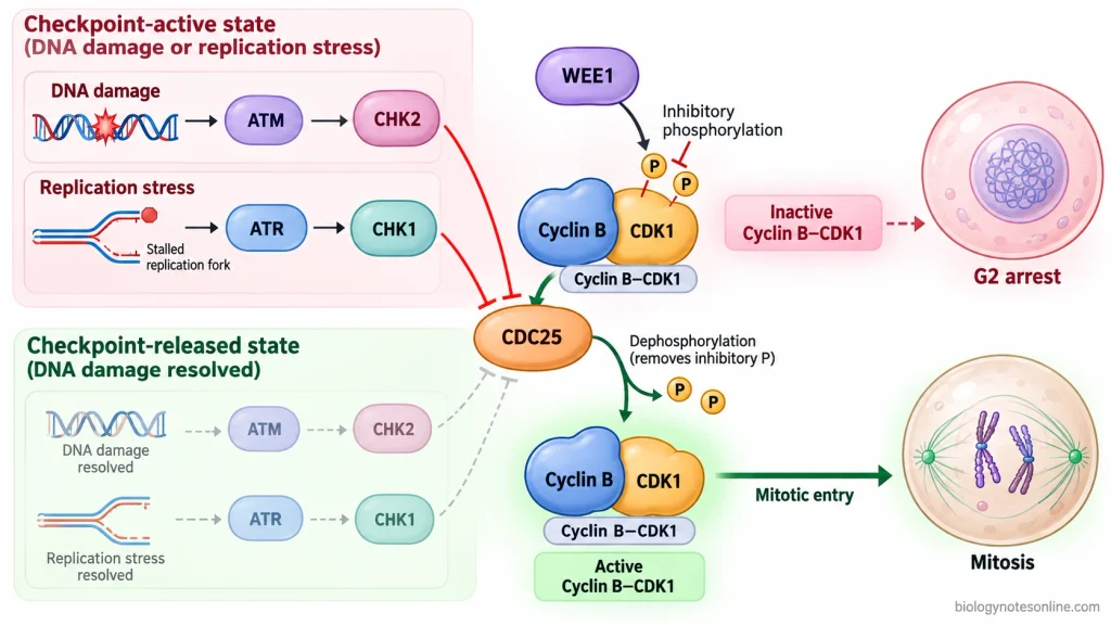
- The entry into mitosis is regulated by cyclin B and cyclin-dependent kinase 1 (CDK1) complex.
- The CDK1 remains inactive when inhibitory phosphate groups are attached to it. These phosphate groups are added by WEE1 kinase.
- In case of DNA damage, ataxia telangiectasia mutated (ATM) and ataxia telangiectasia and Rad3-related (ATR) kinases activate checkpoint kinase 2 (CHK2) and checkpoint kinase 1 (CHK1), respectively. The CHK1 and CHK2 inhibit the activity of cell division cycle 25 (CDC25) phosphatases. The inhibitory phosphate groups on CDK1 are not removed. The cyclin B-CDK1 complex remains inactive. Mitosis does not begin.
- After DNA repair, the checkpoint signalling is reduced. The CDC25 phosphatases remove the inhibitory phosphate groups from CDK1. This activates the cyclin B-CDK1 complex and the cell enters mitosis.
- In cells with defective G2 checkpoint, mitosis may begin even when DNA is damaged or its replication is not completed.
M Checkpoint (Spindle Assembly Checkpoint)
The M checkpoint is the third major checkpoint of the cell cycle. It is present during mitosis, before the beginning of anaphase. It is also known as the spindle assembly checkpoint (SAC).
The separation of sister chromatids is controlled by this checkpoint.
The important features and functions of M checkpoint are as follows-
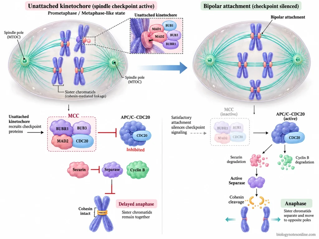
- The M checkpoint checks whether the chromosomes are properly attached to spindle microtubules. These microtubules are attached to chromosomes at protein structures called kinetochores. The kinetochores are present at the centromere region of chromosomes.
- During mitosis, the sister kinetochores are normally attached to spindle microtubules extending from opposite poles of the cell. The chromosomes are aligned at the metaphase plate.
- If any kinetochore remains unattached to spindle microtubules, the checkpoint is activated. Even a single unattached kinetochore can delay the beginning of anaphase.
- Some of the proteins involved in this checkpoint are mitotic arrest deficient 2 (MAD2) and budding uninhibited by benzimidazole-related 1 (BUBR1). These proteins, along with other checkpoint proteins, form the mitotic checkpoint complex (MCC).
- The MCC inhibits the activity of anaphase-promoting complex/cyclosome (APC/C) associated with its activator, cell division cycle 20 (CDC20). The sister chromatids remain attached. Anaphase is delayed.
- When all the kinetochores are properly attached to spindle microtubules, the checkpoint signalling is switched off. The APC/C-CDC20 complex becomes active.
- The activated APC/C causes the degradation of securin and cyclin B proteins. Securin normally keeps the separase enzyme inactive. After the degradation of securin, the separase becomes active and cleaves the cohesin proteins which hold the sister chromatids together. The sister chromatids are then separated. They move towards the opposite spindle poles.
- The degradation of cyclin B also helps in the exit from mitosis.
- The checkpoint mainly responds to the unattached kinetochores. It does not directly check the alignment of chromosomes. Some incorrect attachments of spindle microtubules may remain undetected.
- In cells with defective M checkpoint, the sister chromatids may separate before all chromosomes are properly attached to spindle microtubules. The daughter cells may contain abnormal numbers of chromosomes. This condition is called aneuploidy.
Molecular Regulation of Cell Cycle Checkpoints
The molecular regulation of cell cycle checkpoints is carried out by different proteins, such as cyclins, cyclin-dependent kinases (CDKs), checkpoint kinases and other regulatory proteins. These proteins control the progression of cell cycle. Some of them activate the CDKs, while others inhibit their activity when problems are detected.
The important proteins and mechanisms involved in the regulation of cell cycle checkpoints are as follows-
- Cyclins and CDKs- Cyclins are regulatory proteins that bind with CDKs to form active cyclin-CDK complexes. Different cyclins are involved in the regulation of different phases of cell cycle. The cyclin D-CDK4/6 complex controls G1 phase, while cyclin E-CDK2 regulates the entry into S phase. During S phase, cyclin A-CDK2 is involved in DNA replication. The entry into mitosis is regulated by cyclin B-CDK1 complex.
- Retinoblastoma protein (Rb)- The Rb regulates the activity of E2F transcription factors during G1 phase. It normally binds with E2F and inhibits its activity. When Rb is phosphorylated by cyclin-CDK complexes, the E2F becomes active. These transcription factors activate the genes required for deoxyribonucleic acid (DNA) replication.
- ATM and ATR kinases- In case of DNA damage, checkpoint protein kinases are activated. The ataxia telangiectasia mutated (ATM) kinase mainly responds to DNA double-strand breaks. Ataxia telangiectasia and Rad3-related (ATR) kinase is mainly involved in the response to replication stress and single-stranded DNA regions. These kinases activate other checkpoint proteins such as checkpoint kinase 1 (CHK1) and checkpoint kinase 2 (CHK2).
- p53 and p21 proteins- The p53 protein is activated and stabilized after DNA damage. It increases the production of p21 protein. The p21 inhibits the activity of cyclin-CDK complexes, including cyclin E-CDK2. The cell may remain arrested at G1 phase. The Rb also remains in its growth-inhibitory state when CDK activity is reduced.
- Intra-S-phase regulation- During S phase, DNA replication forks may become stalled due to replication stress. Single-stranded DNA (ssDNA) regions are formed and covered by replication protein A (RPA). The ATR is activated at these regions and phosphorylates CHK1. The activated CHK1 reduces the initiation of DNA replication at additional replication origins. It also helps in maintaining the stability of stalled replication forks.
- Dormant replication origins- Some dormant replication origins located near the stalled forks may become active during replication stress, while the activation of new replication regions is reduced.
- CDC25 phosphatases- The cell division cycle 25 (CDC25) phosphatases are involved in the activation of CDKs. They remove the inhibitory phosphate groups from CDKs.
- WEE1 kinase- The WEE1 kinase adds inhibitory phosphate groups to CDK1. It keeps the CDK1 in an inactive state.
- G2 checkpoint regulation- The entry into mitosis requires the activation of cyclin B-CDK1 complex. When DNA damage occurs, the CHK1 and CHK2 inhibit the activity of CDC25 phosphatases. The inhibitory phosphate groups on CDK1 are not removed. The cyclin B-CDK1 complex remains inactive and the cell is arrested at G2 phase. The damaged DNA is given time for repair.
- Activation of CDK1- After DNA repair, the checkpoint signalling is reduced. The CDC25 phosphatases remove the inhibitory phosphate groups from CDK1. The cyclin B-CDK1 complex becomes active and promotes the entry into mitosis.
- Spindle assembly checkpoint (SAC)- During mitosis, the checkpoint is activated when the kinetochores remain unattached to spindle microtubules. Some of the proteins involved in this checkpoint are mitotic arrest deficient 2 (MAD2) and BUB1-related 1 (BUBR1). These proteins, along with other checkpoint proteins and cell division cycle 20 (CDC20), form the mitotic checkpoint complex (MCC).
- Inhibition of APC/C- The MCC inhibits the activity of anaphase-promoting complex/cyclosome (APC/C) associated with CDC20. The securin and cyclin B proteins are not degraded. The sister chromatids remain attached and anaphase is delayed.
- Activation of anaphase- When all the kinetochores are properly attached to spindle microtubules, the checkpoint signalling is switched off. The APC/C-CDC20 complex becomes active. It promotes the degradation of securin and cyclin B. After securin degradation, the separase enzyme is activated. The separase cleaves the cohesin proteins which hold the sister chromatids together. The sister chromatids are separated. The degradation of cyclin B also causes the inactivation of CDK1 during mitotic exit.
Cellular Responses to Checkpoint Activation
Cellular responses to checkpoint activation are the changes that take place in a cell when problems are detected during the cell cycle. These problems include deoxyribonucleic acid (DNA) damage, incomplete DNA replication, replication stress and improper attachment of chromosomes to spindle microtubules.
Different cellular responses may occur after checkpoint activation. The response depends on the checkpoint involved, type and duration of stress, and the condition of cell.
The important cellular responses to checkpoint activation are as follows-
- Cell Cycle Arrest- The cell cycle progression is stopped temporarily when a checkpoint detects any problem. In G1 phase, the entry into S phase may be delayed due to DNA damage or insufficient growth signals. The G2 checkpoint stops the cell from entering mitosis when DNA replication is incomplete or DNA damage is present. This arrest may be temporary.
- DNA Repair- The DNA repair mechanisms are activated when DNA damage is detected. The cell cycle is delayed. During this period, the damaged DNA is given time for repair before the cell enters the next phase.
- Changes in DNA Replication- During S phase, DNA replication forks may become stalled due to replication stress. The intra-S-phase checkpoint reduces the initiation of DNA replication at additional replication origins. The movement of replication forks may also slow down. Checkpoint proteins help in maintaining the stability of stalled replication forks. Some dormant replication origins present near the stalled forks may become active.
- Mitotic Arrest- The spindle assembly checkpoint (SAC) is activated when kinetochores are not properly attached to spindle microtubules. The separation of sister chromatids is delayed. Anaphase can begin after the proper attachments are formed and the checkpoint is switched off.
- Resumption of Cell Cycle- After DNA repair or resolution of replication stress, the checkpoint signalling is reduced. The cell cycle regulatory proteins become active again and the cell may continue its progression through the cell cycle.
- Cellular Quiescence- In absence of sufficient growth signals, some cells may stop dividing and enter the resting G0 phase. It is a non-dividing state. These cells may again enter the cell cycle when suitable growth signals become available.
- Cellular Senescence- The cells may undergo long-term cell cycle arrest due to persistent DNA damage or prolonged cellular stress. This condition is referred to as cellular senescence. The cells remain alive but do not continue normal cell division. Some of the proteins involved in cellular senescence are p53, p21 and p16. Their involvement depends on the cell type and stress condition.
Causes of Checkpoint Dysfunction
Checkpoint dysfunction is a condition in which the proteins involved in the regulation of cell cycle checkpoints do not function properly. It may occur due to mutations in checkpoint genes, abnormal activity of regulatory proteins or changes in their expression.
In cells with defective checkpoints, cell division may continue even when deoxyribonucleic acid (DNA) is damaged or other cell cycle problems are present.
The important causes of checkpoint dysfunction are as follows-
- Gene Mutations- Mutations in the genes controlling cell cycle checkpoints may affect their normal function. The TP53 gene is responsible for the production of p53 protein, which is involved in cell cycle arrest after DNA damage. In cells with mutated TP53, the G1 checkpoint may not function properly.
- Loss of Rb Function- The loss of normal retinoblastoma protein (Rb) function can affect the regulation of G1 to S phase transition.
- Defective Checkpoint Kinases- The checkpoint signalling involves different protein kinases such as ataxia telangiectasia mutated (ATM), ataxia telangiectasia and Rad3-related (ATR), checkpoint kinase 1 (CHK1) and checkpoint kinase 2 (CHK2). Mutations or defects in these proteins may affect the cellular response to DNA damage and replication stress. The cell cycle arrest may not occur properly.
- Loss of CDK Inhibitors- The p16 and p21 proteins are involved in controlling the activity of cyclin-dependent kinases (CDKs). The loss or reduced activity of these proteins may allow the CDKs to remain active. In cells with defective p53, the production of p21 after DNA damage may be reduced.
- Increased Cyclin-CDK Activity- The abnormal increase in cyclin or CDK activity may disturb the normal regulation of cell cycle. Excessive cyclin E activity can promote the entry into S phase and affect G1 checkpoint control. In some cancer cells, the activity of cyclin-CDK complexes is increased due to changes in growth regulatory genes.
- Epigenetic Changes- The checkpoint regulatory genes may become inactive due to changes in DNA methylation, without any change in their DNA sequence. The CDKN2A gene produces p16 protein. In some cells, this gene is silenced by promoter hypermethylation. The production of p16 is reduced, affecting the regulation of CDK4/6 activity during G1 phase.
- Defects in G2 Regulatory Proteins- The entry into mitosis is controlled by cell division cycle 25 (CDC25) phosphatases and WEE1 kinase. The WEE1 adds inhibitory phosphate groups to CDK1 and keeps it inactive. These phosphate groups are removed by CDC25 phosphatases. The loss of WEE1 activity or abnormal activation of CDC25 may allow the cell to enter mitosis before the proper completion of checkpoint control.
- Replication Stress- Excessive activation of growth-promoting genes may cause replication stress during S phase. The DNA replication forks may become stalled or damaged. In cells with defective replication checkpoint, these stalled forks may not be protected properly and DNA damage can accumulate. Persistent replication stress also increases the demand on DNA repair and checkpoint mechanisms.
- Spindle Checkpoint Defects- The spindle assembly checkpoint involves proteins such as mitotic arrest deficient 2 (MAD2) and BUB1-related protein 1 (BUBR1). The reduced activity or abnormal expression of these proteins may weaken the checkpoint. The sister chromatids may separate before all chromosomes are properly attached to spindle microtubules. Abnormal chromosome segregation may occur.
- Viral Proteins- Some viruses produce proteins that interfere with the regulation of cell cycle checkpoints. For example, high-risk human papillomavirus (HPV) produces E6 and E7 proteins. The E6 promotes the degradation of p53, while E7 inactivates the Rb protein. These changes affect normal cell cycle arrest and may allow abnormal cell cycle progression.
Consequences of Checkpoint Failure
Checkpoint failure occurs when the cell cycle continues even though problems such as deoxyribonucleic acid (DNA) damage, incomplete DNA replication or improper chromosome attachment are present. The normal control of cell cycle is affected.
In cells with defective checkpoints, damaged DNA may be passed to daughter cells. Some cells may also undergo abnormal division or cell death.
The important consequences of checkpoint failure are as follows-
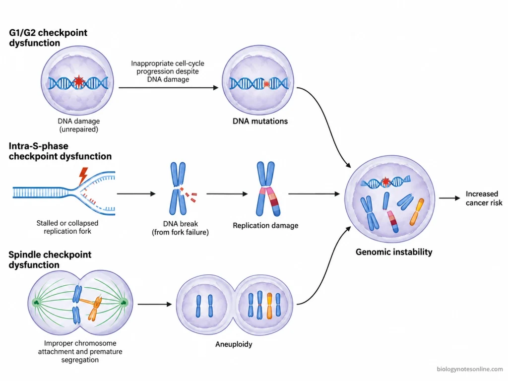
- Accumulation of DNA Damage- When the G1 or G2 checkpoint does not function properly, the cell may proceed to the next phase without repairing the damaged DNA. The unrepaired DNA may remain in the cell and can also be passed to daughter cells during division.
- Increased Mutations- The damaged DNA may be replicated without proper repair. During this process, changes in the DNA sequence may occur, which are known as mutations. These mutations can be inherited by daughter cells if the cell continues dividing.
- Incomplete DNA Replication- In case of defective intra-S-phase checkpoint, the stalled DNA replication forks may not be regulated properly. Some replication forks may collapse and produce DNA double-strand breaks. The DNA replication may remain incomplete. If the G2 checkpoint also fails to prevent mitosis, the cell may enter mitosis with incompletely replicated DNA.
- Chromosomal Abnormalities- The chromosomes may undergo structural changes due to DNA breakage or incorrect repair. These changes include deletion, duplication and translocation of chromosome segments.
- Aneuploidy- When the spindle assembly checkpoint fails, the sister chromatids may separate before all chromosomes are properly attached to spindle microtubules. The chromosomes may not be distributed equally between the daughter cells. Some cells receive extra chromosomes, while others may lose chromosomes. This condition is known as aneuploidy.
- Genomic Instability- The failure of checkpoints may increase the rate of changes in DNA sequence, chromosome structure and chromosome number. This condition is referred to as genomic instability. The genetic changes may continue to accumulate during subsequent cell divisions.
- Abnormal Mitosis- The cell may enter mitosis with damaged or incompletely replicated DNA when the G2 checkpoint is defective. Chromosome breakage and abnormal chromosome segregation may occur during mitosis.
- Cancer Development- The accumulation of mutations in genes controlling cell growth and division may increase the risk of cancer development. Some cells with defective checkpoints continue dividing despite having genetic abnormalities. These cells may acquire further mutations and give rise to abnormal cell populations. Checkpoint failure does not cause cancer in every cell.
- Cell Death and Senescence- Severe DNA damage and abnormal cell division may result in cell death. Some damaged cells may also undergo long-term cell cycle arrest, which is known as cellular senescence, when other protective mechanisms remain functional.
Significance of Checkpoints
Cell cycle checkpoints are regulatory mechanisms that control the progression of cell cycle. They prevent the cell from entering the next phase when deoxyribonucleic acid (DNA) damage, incomplete DNA replication or improper chromosome attachment is detected.
The important functions and significance of cell cycle checkpoints are as follows-
- Control of Cell Cycle Progression- The checkpoints check whether the required conditions are fulfilled before the cell enters the next phase. In case of any problem, the cell cycle progression is delayed.
- DNA Damage Repair- When DNA damage is detected, the cell cycle is stopped temporarily. The damaged DNA is given time for repair.
- Proper DNA Replication- During S phase, the checkpoint reduces the initiation of DNA replication at additional origins when replication stress occurs. It also helps in maintaining the stability of stalled replication forks. DNA replication may continue after the stress is resolved.
- Prevention of Premature Mitosis- The G2 checkpoint prevents the cell from entering mitosis when DNA replication is incomplete or DNA damage is present.
- Accurate Chromosome Segregation- The spindle assembly checkpoint checks the attachment of kinetochores to spindle microtubules. The sister chromatids are not separated until the proper attachments are formed. This reduces the formation of daughter cells with abnormal chromosome numbers, which is known as aneuploidy.
- Maintenance of Genetic Stability- The checkpoints help in reducing the accumulation of mutations and chromosomal abnormalities during cell division.
- Regulation of Cell Proliferation- In G1 phase, the progression of cell cycle depends on growth signals, nutrients and other cellular conditions. Some cells may enter the resting G0 phase in absence of sufficient growth signals. They can again enter the cell cycle when suitable conditions are available.
- Response to Severe DNA Damage- Persistent DNA damage may cause long-term cell cycle arrest, which is known as cellular senescence. Some cells may undergo programmed cell death (apoptosis). The response depends on the extent of damage, cell type and activity of regulatory proteins.
- Prevention of Cancer Development- The checkpoints prevent the division of cells containing damaged DNA. In cells with defective checkpoints, abnormal cell division may continue. More mutations can accumulate in these cells and the risk of cancer development increases.
Cell Cycle Checkpoints at a Glance
Cell cycle checkpoints are regulatory mechanisms that control the progression of cell cycle. They check DNA integrity, completion of DNA replication and proper chromosome attachment before allowing the cell to proceed.
| Checkpoint | Location | What It Checks | Major Regulatory Proteins | Response |
|---|---|---|---|---|
| G1 Checkpoint (G1/S) | End of G1 phase | DNA damage, growth signals and cellular conditions | p53, p21, Rb, cyclin-CDK complexes | Stops entry into S phase when conditions are not suitable. |
| Intra-S Checkpoint | During S phase | Replication stress, stalled replication forks and DNA damage | ATR, CHK1, RPA | Reduces new replication initiation and helps stabilize stalled forks. |
| G2 Checkpoint (G2/M) | End of G2 phase | Completion of DNA replication and DNA damage | ATM, ATR, CHK1, CHK2, WEE1, CDC25, cyclin B-CDK1 | Delays entry into mitosis until replication and repair requirements are met. |
| M Checkpoint (Spindle Assembly) | During mitosis, before anaphase | Attachment of kinetochores to spindle microtubules | MAD2, BUBR1, MCC, APC/C-CDC20 | Delays sister chromatid separation until all kinetochores are properly attached. |
Checkpoint failure may result in accumulation of mutations, abnormal chromosome segregation, genomic instability and increased risk of cancer development.
References
- Alberts, B., Heald, R., Johnson, A., Morgan, D., Raff, M., Roberts, K., & Walter, P. (2022). Molecular biology of the cell (7th ed.). W. W. Norton & Company. https://wwnorton.co.uk/books/9780393884852-molecular-biology-of-the-cell
- Alfieri, C., Chang, L., Zhang, Z., Yang, J., Maslen, S., Skehel, M., & Barford, D. (2016). Molecular basis of APC/C regulation by the spindle assembly checkpoint. Nature, 536(7617), 431–436. https://doi.org/10.1038/nature19083
- Bartek, J., Lukas, C., & Lukas, J. (2004). Checking on DNA damage in S phase. Nature Reviews Molecular Cell Biology, 5(10), 792–804. https://doi.org/10.1038/nrm1493
- Bartek, J., & Lukas, J. (2007). DNA damage checkpoints: From initiation to recovery or adaptation. Current Opinion in Cell Biology, 19(2), 238–245. https://doi.org/10.1016/j.ceb.2007.02.009
- Brito, D. A., & Rieder, C. L. (2006). Mitotic checkpoint slippage in humans occurs via cyclin B destruction in the presence of an active checkpoint. Current Biology, 16(12), 1194–1200. https://doi.org/10.1016/j.cub.2006.04.043
- Chen, C., Piano, V., Alex, A., Han, S. J. Y., Huis in ’t Veld, P. J., Roy, B., Fergle, D., Musacchio, A., & Joglekar, A. P. (2023). The structural flexibility of MAD1 facilitates the assembly of the mitotic checkpoint complex. Nature Communications, 14, Article 1529. https://doi.org/10.1038/s41467-023-37235-z
- Ciccia, A., & Elledge, S. J. (2010). The DNA damage response: Making it safe to play with knives. Molecular Cell, 40(2), 179–204. https://doi.org/10.1016/j.molcel.2010.09.019
- Cimprich, K. A., & Cortez, D. (2008). ATR: An essential regulator of genome integrity. Nature Reviews Molecular Cell Biology, 9(8), 616–627. https://doi.org/10.1038/nrm2450
- Coller, H. A. (2020). Regulation of cell cycle entry and exit: A single cell perspective. Comprehensive Physiology, 10(1), 317–344. https://doi.org/10.1002/cphy.c190014
- Diaz-Martinez, L. A., Tian, W., Li, B., Warrington, R., Jia, L., Brautigam, C. A., Luo, X., & Yu, H. (2015). The Cdc20-binding Phe box of the spindle checkpoint protein BubR1 maintains the mitotic checkpoint complex during mitosis. Journal of Biological Chemistry, 290(4), 2431–2443. https://doi.org/10.1074/jbc.M114.616490
- Engeland, K. (2022). Cell cycle regulation: p53-p21-RB signaling. Cell Death & Differentiation, 29(5), 946–960. https://doi.org/10.1038/s41418-022-00988-z
- Harper, J. W., & Elledge, S. J. (2007). The DNA damage response: Ten years after. Molecular Cell, 28(5), 739–745. https://doi.org/10.1016/j.molcel.2007.11.015
- Hartwell, L. H., & Weinert, T. A. (1989). Checkpoints: Controls that ensure the order of cell cycle events. Science, 246(4930), 629–634. https://doi.org/10.1126/science.2683079
- Jackson, S. P., & Bartek, J. (2009). The DNA-damage response in human biology and disease. Nature, 461(7267), 1071–1078. https://doi.org/10.1038/nature08467
- Kastan, M. B., & Bartek, J. (2004). Cell-cycle checkpoints and cancer. Nature, 432(7015), 316–323. https://doi.org/10.1038/nature03097
- Lara-Gonzalez, P., Westhorpe, F. G., & Taylor, S. S. (2012). The spindle assembly checkpoint. Current Biology, 22(22), R966–R980. https://doi.org/10.1016/j.cub.2012.10.006
- Massagué, J. (2004). G1 cell-cycle control and cancer. Nature, 432(7015), 298–306. https://doi.org/10.1038/nature03094
- Morgan, D. O. (2007). The cell cycle: Principles of control. New Science Press. https://search.worldcat.org/title/70173205
- Musacchio, A. (2015). The molecular biology of spindle assembly checkpoint signaling dynamics. Current Biology, 25(20), R1002–R1018. https://doi.org/10.1016/j.cub.2015.08.051
- Overlack, K., Primorac, I., Vleugel, M., Krenn, V., Maffini, S., Hoffmann, I., Kops, G. J. P. L., & Musacchio, A. (2015). A molecular basis for the differential roles of Bub1 and BubR1 in the spindle assembly checkpoint. eLife, 4, Article e05269. https://doi.org/10.7554/eLife.05269
- Recolin, B., van der Laan, S., Tsanov, N., & Maiorano, D. (2014). Molecular mechanisms of DNA replication checkpoint activation. Genes, 5(1), 147–175. https://doi.org/10.3390/genes5010147
- Stracker, T. H. (2024). Regulation of p53 by the mitotic surveillance/stopwatch pathway: Implications in neurodevelopment and cancer. Frontiers in Cell and Developmental Biology, 12, Article 1451274. https://doi.org/10.3389/fcell.2024.1451274
- Zeman, M. K., & Cimprich, K. A. (2014). Causes and consequences of replication stress. Nature Cell Biology, 16(1), 2–9. https://doi.org/10.1038/ncb2897
- Zhou, B.-B. S., & Elledge, S. J. (2000). The DNA damage response: Putting checkpoints in perspective. Nature, 408(6811), 433–439. https://doi.org/10.1038/35044005