

Apoptosis is a regulated cell death process by which unwanted, aged or damaged cells are removed from the body. It is commonly referred to as programmed cell death. This process is important during normal development and maintenance of tissues.
During this process, several changes occur in the cell. The cell shrinks. Chromatin becomes condensed and fragmentation of nucleus takes place, followed by formation of small membrane-bound structures called apoptotic bodies. These are then removed by phagocytic cells.
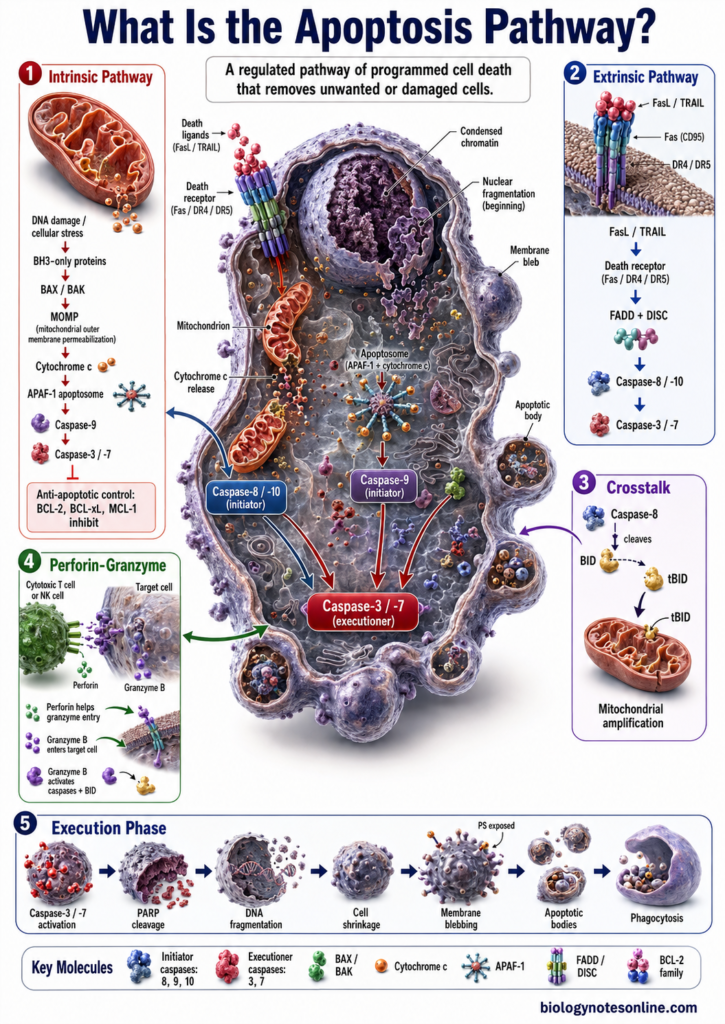
There are two major pathways of apoptosis, intrinsic pathway and extrinsic pathway. The intrinsic pathway is mainly associated with mitochondria, while the extrinsic pathway begins through specific death receptors present on cell membrane. Both pathways finally activate caspases, which cause breakdown of different cellular components.
Apoptosis is necessary for tissue homeostasis, embryonic development and normal immune function. Abnormal regulation of this process is associated with cancer and several degenerative diseases.
What Is the Apoptosis Pathway?
The apoptosis pathway is a group of regulated cellular reactions which finally results in death and removal of a cell. It is a controlled process. Different signals can start these reactions, followed by activation of specific proteins and enzymes inside the cell. The major enzymes involved are called caspases.
There are two major classical pathways of apoptosis, intrinsic pathway and extrinsic pathway. The intrinsic pathway mainly develops from signals arising within the cell and involves the mitochondria. DNA damage, severe cellular stress or loss of survival signals can initiate it. Extrinsic pathway starts from outside the cell. Here, death ligands bind with specific death receptors present on the cell membrane.
Both pathways finally activate initiator and then executioner caspases. These cleave different cellular proteins. The cell is then dismantled in an organized manner and undergoes apoptosis.
Why Do Cells Undergo Apoptosis?
Cells undergo apoptosis when a cell is no longer required or its continued survival may become harmful to the organism. It is not used only for damaged cells. During normal development and in adult tissues, many normal cells are also removed by this process.
The major reasons why cells undergo apoptosis are-
- Development and shaping of tissues – Apoptosis removes cells that are required only for a particular stage of development. The cells between developing digits are an example. They die. In different developing tissues, such removal of cells helps in formation of normal tissue shape and size.
- Maintenance of cell number – New cells are continuously formed in many tissues, and some cells have to be removed at the same time. Apoptosis provides this cell loss. Thus, the number of cells within a tissue can be maintained and unnecessary increase in tissue size is prevented.
- Removal of damaged cells – Cells may receive severe DNA damage, oxidative stress or other cellular injuries. Some damage can be repaired. If the damage becomes too severe, apoptosis may be initiated instead, preventing such damaged cell from continuing its division. The p53 pathway has an important role during apoptosis produced by several forms of DNA damage.
- Removal of unwanted immune cells – Apoptosis has an important role during development of immune system. Autoreactive lymphocytes, which can react against the body’s own components, are removed through apoptotic mechanisms during lymphocyte selection. Apoptosis also controls lymphocyte numbers after an immune response. Too many activated cells are not maintained permanently.
- Removal of infected cells – Some cells infected with viruses or other intracellular pathogens can be eliminated through apoptosis. Cytotoxic immune cells can also induce apoptotic death in infected target cells. This acts as one of the cellular defense mechanisms and helps in removal of infected cells.
- Removal of cells that are no longer needed – Some cells perform their function only for a certain period. After that, their survival is not required. Such cells can be removed by apoptosis without producing the extensive cellular rupture commonly associated with accidental cell injury.
- Protection against abnormal cell survival – Cells carrying harmful genetic alterations should not continue to divide indefinitely. Apoptosis can remove such potentially dangerous cells. Failure of apoptotic control, therefore, can allow abnormal cells to survive and is an important feature associated with development of many cancers.
Overview of the Apoptosis Pathway
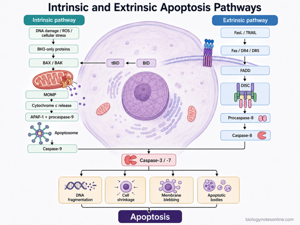
The process of apoptosis takes place mainly through two pathways, the intrinsic pathway and extrinsic pathway. Both are started by different types of signals. The events are also different at the beginning. Finally, activation of executioner caspases takes place, mainly caspase-3 and caspase-7, and the cell undergoes apoptosis.
Intrinsic pathway
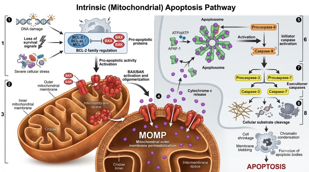
The intrinsic pathway is also referred to as the mitochondrial pathway. It is started by conditions arising within the cell, such as severe cellular stress and DNA damage. BCL-2 family proteins control an important part of this pathway.
Some BCL-2 family members inhibit apoptosis, while others promote the process. BAX and BAK are the major pore-forming proteins involved here. After their activation, mitochondrial outer membrane becomes permeable. This process is called mitochondrial outer membrane permeabilization (MOMP).
During this process, cytochrome c is released from mitochondria. It then takes part in formation of the apoptosome together with APAF-1 and procaspase-9. Caspase-9 becomes activated. After this, caspase-3 and caspase-7 are activated and degradation of different cellular components takes place.
The pathway is as follows-
Cellular stress
↓
BCL-2 family regulation
↓
BAX/BAK activation
↓
MOMP
↓
Cytochrome c
↓
Apoptosome
↓
Caspase-9
↓
Caspase-3/-7
↓
Apoptosis
Extrinsic pathway
The extrinsic pathway starts outside the cell. Here, a specific death ligand binds with its corresponding death receptor present on cell membrane. Receptor activation then allows recruitment of adaptor proteins.
FADD is an important adaptor protein in this pathway, and participates in formation of the death-inducing signaling complex (DISC). Procaspase-8 or procaspase-10 can be recruited to this complex. Their activation produces caspase-8/-10. These initiator caspases then activate the executioner caspases. The cell finally enters apoptosis.
The simple pathway is as follows-
Death ligand
↓
Death receptor
↓
FADD/DISC
↓
Caspase-8/-10
↓
Caspase-3/-7
↓
Apoptosis
Major Components of the Apoptosis Pathway
Different proteins and protein complexes are involved in the apoptosis pathway. Their functions are not same. Some receive the death signal, some control mitochondrial changes, while a group of enzymes carries out final destruction of the cell. Some of the important components are-
- Death Ligands and Death Receptors – Death ligands are extracellular signaling molecules. Fas ligand (FasL) and TRAIL are important examples. These bind with their corresponding death receptors present on cell membrane, such as Fas (CD95), DR4 and DR5. The receptors contain an intracellular death domain which takes part in further signaling after receptor activation.
- FADD and DISC – FADD (Fas-associated death domain) is an adaptor protein associated with the activated death receptor. It then recruits procaspase-8 through protein-protein interaction, and the proteins assembled at the receptor form a complex called death-inducing signaling complex (DISC). Caspase activation starts here.
- BCL-2 Family Proteins – The BCL-2 family is the major group controlling mitochondrial apoptosis. It contains proteins with opposite functions. BCL-2, BCL-XL and MCL-1 mainly prevent apoptosis. BAX and BAK promote it. There is another group called BH3-only proteins, including BIM, BID, PUMA and NOXA, which respond to different cell stresses and regulate the activity of other BCL-2 family members.
- BAX and BAK – These are pro-apoptotic pore-forming proteins. During intrinsic pathway, BAX and BAK become activated and oligomerize in mitochondrial outer membrane. The membrane becomes permeable. This is referred to as mitochondrial outer membrane permeabilization (MOMP).
- Cytochrome c – Cytochrome c is normally a component of mitochondrial electron transport chain. During apoptosis its function is different. After MOMP, it is released from mitochondrial intermembrane space into cytosol and binds with APAF-1, which is required for formation of apoptosome.
- APAF-1 and Apoptosome – APAF-1 (Apoptotic protease-activating factor 1) is present in cytosol. Cytochrome c binds to it. In the presence of nucleotide, APAF-1 molecules assemble and a large complex called apoptosome is formed. Procaspase-9 is recruited to this complex and activation of caspase-9 takes place.
- Caspases – Caspases are cysteine proteases and have the major role in apoptotic cell destruction. They are initially present as inactive procaspases. Apoptotic caspases are mainly of two types, initiator and executioner caspases. Caspase-8 and caspase-10 are mainly associated with extrinsic pathway, whereas caspase-9 functions in intrinsic pathway. Caspase-3 and caspase-7 are major executioner caspases. These cleave many cellular proteins after activation. Cell breakdown follows.
- IAPs and Smac/DIABLO – Inhibitor of apoptosis proteins (IAPs) regulate the caspase system. XIAP can directly inhibit caspase-9, caspase-3 and caspase-7. Smac/DIABLO, on the other hand, is a mitochondrial protein released during MOMP. It binds with XIAP and reduces its inhibitory action. Caspases can remain active and the apoptotic process continues.
Mechanisms of Apoptosis
1. Intrinsic Pathway of Apoptosis
The intrinsic pathway of apoptosis is also called the mitochondrial pathway. It is mainly activated by stress arising within the cell, such as DNA damage, oxidative stress, growth factor withdrawal and some other severe cellular injuries. The BCL-2 family proteins have the major role in controlling this pathway.
Some BCL-2 family proteins prevent apoptosis, while BH3-only proteins, BAX and BAK promote it. When the apoptotic signal becomes sufficient, BAX and BAK become activated and mitochondrial outer membrane is permeabilized. This is called MOMP. An important step.
During this process, cytochrome c is released from mitochondria into the cytosol. It binds with APAF-1 and formation of the apoptosome takes place, which activates caspase-9. Caspase-9 then activates caspase-3 and caspase-7. Breakdown of different cellular components follows and the cell finally undergoes apoptosis.
What Triggers the Intrinsic Apoptosis Pathway?
The intrinsic apoptosis pathway is activated by different types of cellular stress and loss of normal survival signals. These signals finally affect the BCL-2 family proteins, resulting in activation of BAX and BAK when the pro-apoptotic signals become sufficient. The mitochondria are then involved. Some of the major triggers are as follows-
- DNA Damage – It is one of the major triggers of intrinsic apoptosis. DNA can be damaged by ionizing radiation, ultraviolet radiation, genotoxic chemicals or problems occurring during DNA replication. The cell initially tries to repair such damage. When damage is severe or remains for a longer period, proteins such as p53 can promote the pro-apoptotic response and mitochondrial apoptosis may take place.
- Growth Factor Withdrawal – Many cells depend on continuous growth and survival signals for their survival. When these signals are removed, the activity of pro-survival pathways decreases and BH3-only proteins can become active, which then changes the balance between anti-apoptotic and pro-apoptotic BCL-2 family proteins. BAX/BAK activation can follow.
- Oxidative Stress – Excess formation of reactive oxygen species (ROS) produces oxidative stress. Proteins, lipids, DNA and mitochondria can be damaged. At higher or prolonged levels, this stress can activate the intrinsic apoptotic pathway.
- Endoplasmic Reticulum Stress – Accumulation of unfolded or incorrectly folded proteins produces endoplasmic reticulum (ER) stress. The cell first activates the unfolded protein response in order to restore normal ER function. This response is mainly protective at the beginning. But when ER stress becomes severe and remains unresolved, pro-apoptotic signals are produced and mitochondrial apoptosis can be activated.
- Hypoxia and Metabolic Stress – Low oxygen supply or serious disturbance of cellular metabolism can also trigger intrinsic apoptosis. This is referred to as hypoxia and metabolic stress. If the cell cannot adapt to these conditions, mitochondrial apoptotic signaling may start.
- Replication Stress and Mitotic Defects – Problems during DNA replication are another trigger. Abnormal changes of microtubules or serious defects during mitosis can also produce the apoptotic signal, especially when these abnormalities cannot be corrected by normal cellular checkpoints. Such stresses can finally act through BCL-2 family regulation and lead towards mitochondrial outer membrane permeabilization.
- Toxic Cellular Damage – Different cytotoxic chemicals and some anticancer treatments produce strong cellular injury. DNA damage, oxidative stress or mitochondrial damage may occur together. When this damage becomes greater than the survival capacity of the cell, the intrinsic pathway can be activated.
Steps of the Intrinsic Apoptosis Pathway
The intrinsic apoptosis pathway mainly takes place through regulation of mitochondria. Different intracellular stresses can start the process. The BCL-2 family proteins, BAX/BAK, cytochrome c and caspases are mainly involved. The steps are as follows-
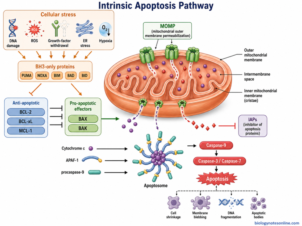
- Formation of Apoptotic Signal – The first step begins after serious stress inside the cell, such as DNA damage or loss of normal survival signals. The balance between pro-apoptotic and anti-apoptotic BCL-2 family proteins is changed. Pro-apoptotic signaling now increases.
- Activation of BAX and BAK – BAX and BAK are the main effector proteins of mitochondrial apoptosis. These proteins become activated and oligomerization takes place at mitochondrial outer membrane. Their activation is an important step. Cells lacking both BAX and BAK are strongly resistant to mitochondrial cytochrome c release after many apoptotic signals.
- Mitochondrial Outer Membrane Permeabilization – Activated BAX and BAK form large openings in mitochondrial outer membrane. The membrane becomes permeable. This process is called mitochondrial outer membrane permeabilization (MOMP).
- Release of Cytochrome c – After MOMP, cytochrome c is released from mitochondrial intermembrane space into the cytosol. Other mitochondrial proteins can also be released during this process. Cytochrome c now takes part in apoptotic signaling instead of its normal mitochondrial function.
- Formation of Apoptosome – Cytochrome c binds with APAF-1 (apoptotic protease-activating factor 1) in the presence of nucleotide. APAF-1 molecules then assemble into a large protein complex. This is referred to as the apoptosome.
- Activation of Caspase-9 – Procaspase-9 is recruited to the apoptosome and activation of caspase-9 takes place. Caspase-9 is the major initiator caspase of this pathway. The apoptotic protease cascade is now started.
- Activation of Executioner Caspases – Activated caspase-9 further activates downstream executioner caspases, mainly caspase-3 and caspase-7. These enzymes have the major role in cellular breakdown.
- Execution of Apoptosis – Caspase-3 and caspase-7 cleave different cellular proteins. Structural and biochemical changes of apoptosis then take place, and the cell is finally dismantled. The apoptotic process is completed.
Regulation of the Intrinsic Apoptosis Pathway
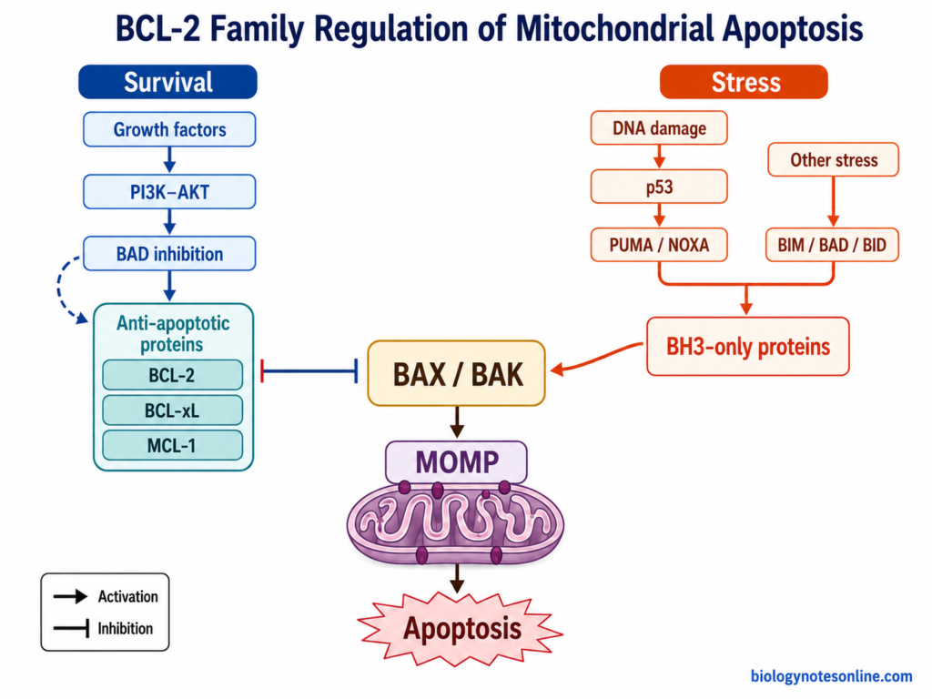
The intrinsic apoptosis pathway is regulated at several levels. The major control is provided by the BCL-2 family proteins before mitochondrial permeabilization. Some proteins prevent the process. Others promote it. After release of cytochrome c, apoptosome and caspase activity are also regulated.
The following are the important regulatory mechanisms-
- BCL-2 Family Regulation – The BCL-2 family contains both anti-apoptotic and pro-apoptotic proteins and their relative activity controls mitochondrial apoptosis. BCL-2, BCL-XL and MCL-1 mainly maintain cell survival. They prevent activation or action of the pro-apoptotic proteins BAX and BAK. When this protection is overcome, mitochondrial permeabilization can occur. BCL-2 itself has been shown to prevent mitochondrial release of cytochrome c during apoptosis.
- BH3-Only Proteins – These are the stress-responsive members of BCL-2 family. BIM, BID, PUMA, NOXA and BAD are some important members. They act mainly by neutralizing anti-apoptotic BCL-2 proteins, and some BH3-only proteins can also promote BAX/BAK activation. Thus, apoptotic stress is passed towards the mitochondrial machinery.
- BAX and BAK Regulation – BAX and BAK are the main effector proteins of this pathway. Normally their pore-forming activity is kept under control. After sufficient pro-apoptotic signaling, these proteins become activated and oligomerization takes place at mitochondrial outer membrane. MOMP follows. BAX and BAK together form an important gateway for mitochondrial apoptosis because cells lacking both proteins show strong resistance to cytochrome c release after many death stimuli.
- p53 Regulation – p53 has an important role particularly after DNA damage and some other cellular stresses. It can increase expression of pro-apoptotic BH3-only genes such as PUMA and NOXA. PUMA can promote BAX/BAK-dependent mitochondrial apoptosis. Noxa also participates in p53-mediated apoptotic response, although its importance can vary with cell type.
- Survival Signaling – Growth and survival signals can suppress the intrinsic pathway. One important mechanism involves the PI3K-AKT pathway. Akt phosphorylates the pro-apoptotic protein BAD, reducing its death-promoting activity. In this way, normal survival signals keep mitochondrial apoptosis under control. Loss of these signals can shift the cell towards apoptosis.
- Apoptosome Regulation – After MOMP, cytochrome c comes into cytosol and binds with APAF-1. In the presence of ATP or dATP, APAF-1 oligomerization takes place and the apoptosome is formed. Procaspase-9 is recruited to this complex. Activation of caspase-9 then starts the downstream caspase reactions.
- IAPs and Smac/DIABLO – Caspase activity is also controlled after mitochondrial permeabilization. XIAP, a member of inhibitor of apoptosis proteins (IAPs), can inhibit caspase-9, caspase-3 and caspase-7. Smac/DIABLO does the opposite work. It is released from mitochondria during apoptosis and binds with IAP proteins, reducing their inhibition of caspases. Caspase activity can now continue.
2. Extrinsic Pathway of Apoptosis
The extrinsic pathway of apoptosis is started by death signals coming from outside the cell. It is also referred to as the death receptor pathway. Here, specific ligands bind with receptors present on cell membrane. Fas ligand (FasL) and TRAIL are common examples, while Fas (CD95), DR4 and DR5 act as their death receptors.
After ligand binding, the receptor becomes activated. FADD is now recruited to its cytoplasmic death domain, followed by recruitment of procaspase molecules and formation of death-inducing signaling complex (DISC). This is an important signaling complex. Caspase-8 activation takes place here.
The activated caspase-8 then activates caspase-3 and caspase-7. Cell destruction follows. In some cells, however, this signal is not sufficient and mitochondrial pathway is also involved. Caspase-8 cleaves BID into tBID. It then acts on mitochondria and helps in increasing the apoptotic signal.
What Triggers the Extrinsic Apoptosis Pathway?
The extrinsic apoptosis pathway is started when extracellular death signals bind with specific death receptors present on cell surface. These receptors belong to the TNF receptor superfamily. All members of this family do not cause apoptosis. Fas (CD95), TNFR1, DR4 and DR5 are some of the important death receptors. The major triggers are as follows-
- Fas Ligand (FasL) – It is one of the major death ligands involved in extrinsic apoptosis. FasL binds with Fas (CD95/APO-1) present on the target cell. The receptor is now activated. After this, FADD and procaspase-8 are recruited and formation of the death-inducing signaling complex (DISC) takes place. Caspase-8 activation follows.
- TRAIL – TRAIL (TNF-related apoptosis-inducing ligand) is another death ligand of the TNF family. In humans, its apoptosis-producing receptors are DR4 (TRAIL-R1) and DR5 (TRAIL-R2). These receptors contain intracellular death domains. TRAIL binds with them, followed by recruitment of FADD and procaspase-8 to the receptor complex. This can start the caspase pathway.
- Tumor Necrosis Factor (TNF) – TNF acts through TNFR1 and can produce different cellular responses. Apoptosis is one of them. TNFR1 signaling can also activate inflammatory and survival pathways, therefore binding of TNF does not always result in death of the cell. Under apoptosis-favouring condition, a cytosolic death complex containing FADD and caspase-8 is formed. This is referred to as complex II. Apoptotic signaling is then started.
- Death Signals from Immune Cells – Cytotoxic immune cells can provide the extracellular signal for this pathway. Cytotoxic T lymphocytes can express FasL which binds with Fas on the target cell. The Fas death pathway is activated. Natural killer (NK) cells and other immune cells can also use death-receptor ligands under different conditions. These cells have another killing system, the perforin-granzyme pathway, but it is not itself a death receptor trigger.
Steps of the Extrinsic Apoptosis Pathway
The extrinsic apoptosis pathway starts after activation of specific death receptors present on cell surface. Death ligands, adaptor proteins and caspases are mainly involved. The steps are as follows-
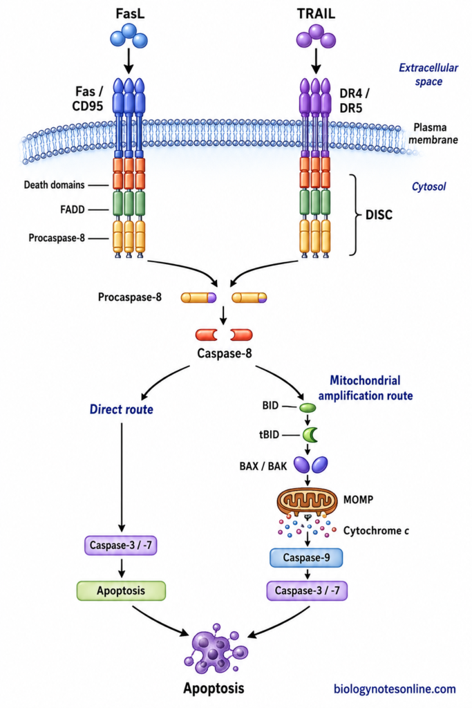
- Binding of Death Ligand – The first step is binding of a death ligand with its corresponding death receptor. FasL binds with Fas (CD95), while TRAIL binds mainly with DR4 and DR5. The receptors become activated. Receptor clustering also takes place.
- Formation of DISC – After receptor activation, adaptor proteins are recruited to the intracellular death domain. FADD is the major adaptor protein involved here. It further recruits procaspase-8 and, in human cells, procaspase-10. A protein complex is now formed at the receptor. This is called the death-inducing signaling complex (DISC).
- Activation of Initiator Caspase – Procaspase-8 molecules are brought close together at the DISC and activation takes place. Caspase-8 is the major initiator caspase of extrinsic pathway. Caspase-10 can also participate in some human death receptor complexes. The caspase pathway now starts.
- Activation of Executioner Caspases – Activated caspase-8 acts on downstream caspases. Caspase-3 and caspase-7 are the major executioner caspases. These cleave different structural and regulatory proteins of the cell. Cell breakdown begins.
- Mitochondrial Amplification – In some cells, direct activation of executioner caspases is not sufficient. Here, caspase-8 cleaves the BH3-only protein BID into tBID. tBID then acts on mitochondria and promotes mitochondrial outer membrane permeabilization. Release of cytochrome c follows and the apoptotic signal becomes stronger.
- Execution of Apoptosis – Executioner caspases cleave many cellular substrates. The cell shrinks. Chromatin condensation, nuclear fragmentation and membrane blebbing take place, followed by formation of apoptotic bodies. The cell finally undergoes apoptosis.
Intrinsic vs Extrinsic Pathways of Apoptosis
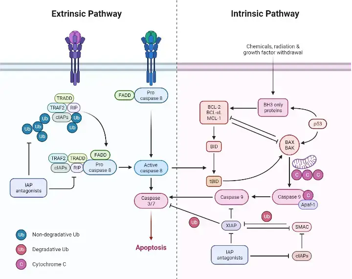
| Features | Intrinsic Pathway | Extrinsic Pathway |
|---|---|---|
| Other name | It is also called the mitochondrial pathway. | It is also called the death receptor pathway. |
| Origin of signal | The apoptotic signal mainly arises from inside the cell. | The signal comes mainly from outside the cell. |
| Major triggers | DNA damage, oxidative stress, growth factor withdrawal, ER stress and other cellular stresses. | Binding of death ligands such as FasL and TRAIL with their receptors. |
| Major site involved | Mitochondria have the central role. | The pathway starts at death receptors present on cell membrane. |
| Important regulatory proteins | Mainly controlled by BCL-2 family proteins. | Mainly regulated through death receptors, FADD, DISC and proteins such as c-FLIP. |
| BAX and BAK | BAX and BAK become activated and promote mitochondrial outer membrane permeabilization. | These are not required for the initial death receptor signal, but can become involved through BID-mediated mitochondrial amplification. |
| MOMP | Mitochondrial outer membrane permeabilization (MOMP) is a major step. | MOMP is not necessary for the direct pathway, although it can occur in type II cells. |
| Cytochrome c | Cytochrome c is released from mitochondria into cytosol. | Cytochrome c is not part of the initial pathway. It can become involved after mitochondrial amplification. |
| Major protein complex | Apoptosome is formed from APAF-1, cytochrome c and procaspase-9. | Death-inducing signaling complex (DISC) is formed at activated death receptors. |
| Initiator caspase | Caspase-9 is the major initiator caspase. | Caspase-8 is the major initiator caspase. Caspase-10 can also participate in humans. |
| Executioner caspases | Caspase-9 activates caspase-3 and caspase-7. | Caspase-8 activates caspase-3 and caspase-7. |
| Connection between pathways | Can receive a death signal from the extrinsic pathway through tBID. | Caspase-8 can cleave BID into tBID, which activates the mitochondrial pathway. |
| Final result | Cellular proteins are cleaved and the cell undergoes apoptosis. | Executioner caspases produce the same final apoptotic changes. |
3. Crosstalk Between the Intrinsic and Extrinsic Apoptosis Pathways
The intrinsic and extrinsic apoptosis pathways can interact with each other during apoptotic signaling. The major connection occurs through the BID protein and mitochondria. In some cells this connection is necessary for sufficient activation of the executioner caspases. The following are the major events-
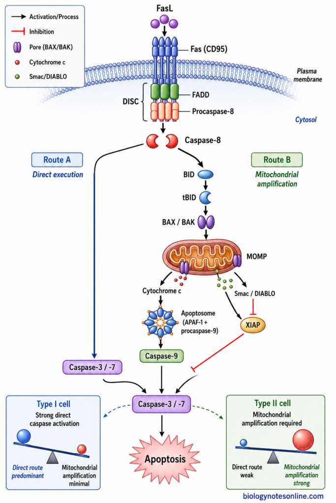
- Cleavage of BID by Caspase-8 – During extrinsic apoptosis, activation of death receptors results in formation of active caspase-8. Caspase-8 can cleave the BH3-only protein BID. A shorter active form, called tBID (truncated BID), is produced. This provides an important connection between death receptor signaling and mitochondria.
- Activation of Mitochondrial Pathway – tBID moves towards mitochondria and promotes the activity of BAX and BAK. The mitochondrial outer membrane becomes permeable. MOMP takes place. Cytochrome c and other mitochondrial apoptotic proteins can now be released into cytosol.
- Amplification of the Death Signal – After release of cytochrome c, apoptosome formation and caspase-9 activation take place. Mitochondria can also release Smac/DIABLO, which reduces inhibition of caspases by XIAP. Therefore, an initially weaker death receptor signal can produce much greater activation of the executioner caspases through this mitochondrial branch.
- Type I and Type II Signaling – The requirement of this mitochondrial connection is not same in all cells. In classical type I cells, strong DISC formation produces sufficient caspase-8 activity and downstream caspases can be activated with less dependence on mitochondria. Type II cells form lower amounts of active caspase-8 at the DISC. They depend more strongly on BID and mitochondrial amplification for apoptosis.
- Role of XIAP – XIAP (X-linked inhibitor of apoptosis protein) can suppress the activity of downstream caspases. This becomes especially important in type II signaling. Mitochondrial events help to overcome this inhibition, and studies have shown XIAP as an important factor separating type I and type II Fas-mediated apoptosis.
- Common Execution Pathway – Both pathways finally reach the executioner caspases, mainly caspase-3 and caspase-7. Cellular proteins are then cleaved. The characteristic changes of apoptosis follow and the cell is finally removed.
4. Perforin-Granzyme Mechanism
The perforin-granzyme mechanism is an important pathway used by cytotoxic T lymphocytes (CTLs) and natural killer (NK) cells for killing infected, transformed or other abnormal target cells. Cytotoxic granules are mainly involved. These contain perforin and different granzymes, of which granzyme B is one of the best characterized for induction of apoptosis.
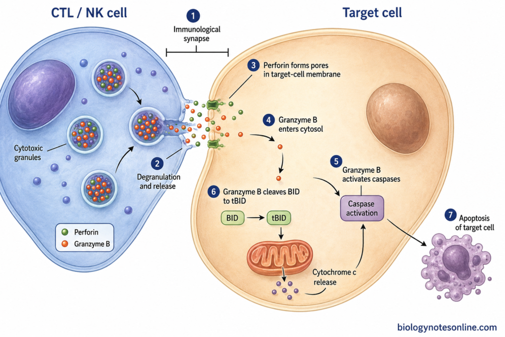
- Recognition of Target Cell – The cytotoxic lymphocyte first recognizes and comes in close contact with its target cell. An immunological synapse is formed between the two cells. Cytotoxic granules then move towards this region and their contents are released into the synaptic space.
- Release of Perforin and Granzymes – The cytotoxic granules fuse with plasma membrane of CTL or NK cell. Perforin and granzymes are released. The release is directed towards the target cell, therefore the cytotoxic molecules become concentrated at the contact region.
- Action of Perforin – Perforin is a pore-forming protein and has an essential role in granule-mediated cytotoxicity. It acts on the target-cell membrane and allows efficient delivery of granzymes into the target-cell cytosol. Perforin-dependent membrane damage and uptake processes are involved in this delivery. Granzymes then reach the cytoplasm.
- Action of Granzyme B – Granzyme B is a serine protease. After reaching the cytosol it cleaves several proteins involved in apoptotic cell death. It can act on the caspase system. Granzyme B also directly cleaves the BH3-only protein BID, producing a truncated form which acts on mitochondria.
- Mitochondrial Apoptosis – Cleaved BID promotes mitochondrial changes and release of cytochrome c. The mitochondrial pathway is now involved. Studies have shown an important role of BID and mitochondrial cytochrome c release during granzyme B-induced apoptosis.
- Activation of Executioner Caspases – Granzyme B-mediated signaling results in activation of executioner caspases, particularly caspase-3. These enzymes cleave different cellular proteins. Cell destruction follows.
- Death of Target Cell – The target cell finally shows the characteristic changes of apoptosis. DNA fragmentation and other cellular changes take place. The infected or abnormal cell is then eliminated by the cytotoxic immune response.
5. Execution Phase of Apoptosis
The execution phase of apoptosis starts after activation of downstream executioner caspases. Caspase-3 and caspase-7 have the major role. A number of cellular proteins are now cleaved and characteristic changes of apoptotic cell take place. The major events are as follows-
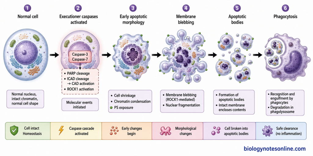
- Activation of Executioner Caspases – Initiator caspases activate the executioner caspases, mainly caspase-3 and caspase-7. These enzymes have many cellular substrates. Once activated, degradation of different structural and regulatory proteins begins.
- Cleavage of Cellular Proteins – Executioner caspases cleave proteins present in cytoplasm and nucleus. PARP-1, which is associated with DNA repair, is one of the well-known caspase substrates. Cytoskeletal proteins are also affected. Normal organization of the cell is gradually lost.
- DNA Fragmentation – CAD (caspase-activated DNase) is normally kept inactive by ICAD. During apoptosis, caspase-3 cleaves ICAD. CAD becomes free and active. It then causes fragmentation of nuclear DNA, one of the characteristic biochemical changes during the execution phase.
- Cell Shrinkage and Membrane Blebbing – Changes now occur in cytoskeleton. Caspase-3 cleaves ROCK1, producing an active form which increases actomyosin contraction. The cell shrinks. Membrane blebs are formed and fragmented nuclear material can also move into these blebs.
- Exposure of Phosphatidylserine – Phosphatidylserine (PS), which is normally concentrated on the inner leaflet of plasma membrane, becomes exposed on the outer cell surface during apoptosis. The phospholipid scramblase XKR8 has an important role in this process and can be activated by caspase cleavage. Exposed PS acts as an important recognition signal for removal of the dying cell.
- Formation and Removal of Apoptotic Bodies – The dying cell finally breaks into small membrane-covered fragments called apoptotic bodies. Nuclear and cytoplasmic materials remain packed within them. These are recognized and engulfed by macrophages or other neighboring phagocytic cells. The apoptotic cell is then removed.
Regulation of the Apoptosis Pathway
The apoptosis pathway is controlled by several pro-apoptotic and anti-apoptotic proteins. Some promote the process. Others prevent it. Regulation occurs at the death receptors, mitochondria and also during activation of caspases. The following are some of the important regulatory mechanisms-
- BCL-2 Family Proteins – The BCL-2 family has a major role in regulation of intrinsic apoptosis. BCL-2, BCL-XL and MCL-1 mainly prevent apoptosis. BAX and BAK promote mitochondrial outer membrane permeabilization. The activity of these opposite groups determines whether the mitochondrial death pathway will proceed or remain inhibited.
- BH3-Only Proteins – BIM, BID, PUMA, NOXA and BAD are some members of this group. They respond to different cellular stresses. Some bind and inhibit the anti-apoptotic BCL-2 proteins, while some can also promote activation of BAX or BAK. Mitochondrial apoptosis is then favored.
- Regulation at Death Receptors – Extrinsic apoptosis is also controlled before caspase-8 becomes fully activated. c-FLIP is an important regulator present at the death receptor signaling complex. It can limit activation of procaspase-8 at the DISC and therefore suppress death receptor-induced apoptosis.
- IAP Proteins – Inhibitor of apoptosis proteins (IAPs) regulate the caspase system. XIAP is particularly important. It directly inhibits caspase-9, and the executioner caspases caspase-3 and caspase-7. Caspase activity is therefore kept under control.
- Smac/DIABLO – Smac/DIABLO is stored within mitochondria and is released during mitochondrial permeabilization. Its action is opposite to IAP-mediated inhibition. Smac binds with IAP proteins and reduces their inhibition of caspases. The caspases can now remain active and apoptotic process proceeds.
- p53 Regulation – p53 has an important role after DNA damage and some other cellular stresses. It can increase expression of pro-apoptotic proteins such as PUMA, NOXA and BAX. The balance moves towards mitochondrial apoptosis when damage cannot be properly corrected.
- Survival Signaling – Growth factors can produce signals which keep apoptosis suppressed. One important pathway is PI3K-AKT. Akt can phosphorylate pro-apoptotic proteins such as BAD, reducing their apoptotic activity. Thus, continuous survival signaling helps the cell to remain alive, while loss of such signals may favor apoptosis.
- NF-κB-Mediated Regulation – NF-κB can also promote cell survival in many conditions. It increases expression of several anti-apoptotic proteins, including some BCL-2 family members, IAP proteins and c-FLIP. The effect is therefore seen at more than one level of apoptosis pathway.
What Are the Assays Used for Detection of Apoptosis?
Different methods are used for detection of apoptosis. The changes occurring in cell membrane, nucleus, mitochondria and caspase system can be detected by different assays. One assay does not detect every event. Some of the commonly used assays are-
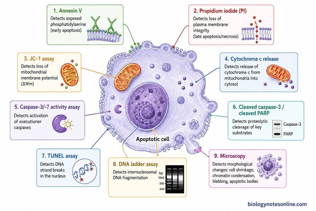
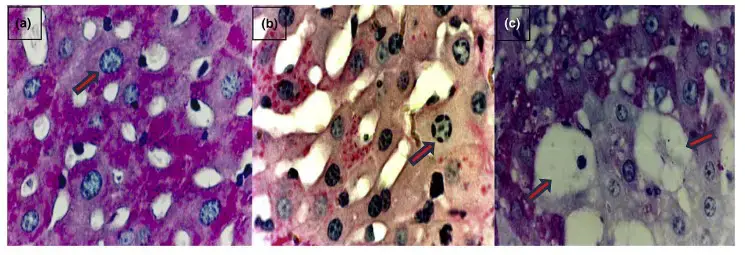
- Morphological Examination – Apoptotic cells show cell shrinkage, chromatin condensation and membrane blebbing. Later, nuclear fragmentation and apoptotic bodies may be seen. These changes can be observed by light, fluorescence or electron microscopy. It is one of the basic methods for identification of apoptotic morphology.
- Annexin V Assay – During early apoptosis, phosphatidylserine (PS) becomes exposed on outer surface of plasma membrane. Annexin V binds with this phospholipid. It is commonly used together with propidium iodide (PI). Annexin V-positive/PI-negative cells generally have exposed PS with an intact membrane, while PI also enters after membrane integrity is lost.
- TUNEL Assay – TUNEL (Terminal deoxynucleotidyl transferase dUTP nick end labeling) detects DNA strand breaks. In this method, terminal deoxynucleotidyl transferase adds labeled nucleotides at free DNA ends. The fragmented DNA can then be detected in individual cells or tissue sections. TUNEL positivity, however, is not completely specific only for apoptosis.
- Caspase Activity Assay – Caspase activation is an important event of apoptotic process. Caspase-3/-7 activity is commonly measured. Fluorogenic, chromogenic or luminescent substrates can be used, which are cleaved after activation of the corresponding caspase. The signal is then measured.
- DNA Ladder Assay – During many forms of apoptosis, chromosomal DNA is cleaved at internucleosomal regions. The isolated DNA is separated by agarose gel electrophoresis. A characteristic ladder-like pattern can be obtained. But all apoptotic cells do not necessarily produce a clear DNA ladder.
- Mitochondrial Membrane Potential Assay – This assay is mainly useful for studying mitochondrial changes during apoptosis. Fluorescent dyes such as JC-1 can be used. The distribution and fluorescence of the dye depend on mitochondrial membrane potential. When the potential changes or collapses, a change in fluorescence is observed.
- Detection of Cleaved Caspases and PARP – Western blotting or immunostaining can be used for detection of activated apoptotic proteins. Cleaved caspase-3 is commonly examined. Cleaved PARP is another frequently used marker because PARP is cleaved during caspase-dependent apoptosis. These markers are generally used together with other assays.
- Cytochrome c Release Assay – During intrinsic apoptosis, cytochrome c comes out from mitochondria into cytosol. Its redistribution can be studied after separation of mitochondrial and cytosolic fractions followed by immunoblotting. Imaging methods can also be used. This assay mainly indicates involvement of the mitochondrial apoptosis pathway.
Biological Importance of the Apoptosis Pathway
The apoptosis pathway has an important role in normal development and maintenance of multicellular organisms. Cells are continuously formed and also removed. Apoptosis provides a controlled process for this removal. Some of the important biological functions are-
- Embryonic Development – Apoptosis is required during development of different tissues and organs. Unwanted cells are removed. It also helps in tissue remodeling and formation of normal body structures during embryonic development.
- Maintenance of Tissue Homeostasis – New cells are continuously produced in many tissues, while aged or unnecessary cells have to be removed. Apoptosis balances this cell production with cell loss. The normal number of cells is therefore maintained.
- Removal of Damaged and Obsolete Cells – Cells having serious cellular damage or cells that are no longer required can be eliminated through apoptosis. Damaged cells do not remain permanently within the tissue. This is important for normal tissue function.
- Development of Immune System – Apoptosis has a major role during development of B and T lymphocytes. Self-reactive lymphocytes can be removed during immune-cell selection. This prevents many potentially harmful lymphocytes from entering the functional immune-cell population.
- Control of Immune Responses – Activated lymphocytes increase greatly during an immune response. They are not all maintained after the response is completed. Apoptotic removal of excess activated cells helps in returning the immune-cell population towards its normal level.
- Protection Against Cancer – Apoptosis can remove cells carrying severe damage or potentially dangerous alterations before their continued survival and proliferation. Failure of apoptotic control is common in cancer and can favor survival of malignant cells. Thus, apoptosis acts as an important anti-cancer defense mechanism.
- Elimination of Infected or Abnormal Cells – Cytotoxic T lymphocytes and natural killer cells can induce apoptosis in infected or altered target cells. This is one of the mechanisms used by the immune system for cellular defense. The unwanted target cell is destroyed.
- Controlled Removal of Dying Cells – Apoptotic cells are generally recognized and removed by phagocytic cells before extensive leakage of their intracellular contents occurs. Tissue disturbance is therefore limited under normal conditions. This makes apoptosis suitable for continuous cell removal in healthy tissues.
Apoptosis Pathway in Disease
Abnormal regulation of the apoptosis pathway is associated with different human diseases. The process may become reduced or excessively activated. Both are harmful. Reduced apoptosis allows abnormal or unwanted cells to remain alive, whereas excessive apoptosis can result in loss of normal functional cells. Some of the important conditions are-
- Cancer – Reduced apoptosis is an important feature of many cancers. Abnormal cells which normally should die can continue to survive and divide. Alterations in p53, increased activity of anti-apoptotic BCL-2 family proteins, or defects in other apoptotic proteins can contribute to this resistance. BCL-2 overexpression is particularly important in some lymphoid cancers. Cancer cells therefore become more difficult to remove by the normal cell death machinery.
- Autoimmune and Lymphoproliferative Diseases – Apoptosis normally removes many autoreactive and excess lymphocytes. If this removal is defective, such cells can remain in the body. Autoimmunity may follow. A clear example is autoimmune lymphoproliferative syndrome (ALPS), where defective Fas-mediated apoptosis causes abnormal accumulation of lymphocytes together with autoimmune manifestations.
- Neurodegenerative Diseases – Excessive loss of neurons is associated with several neurodegenerative conditions. Apoptotic signaling has been implicated in Alzheimer’s disease, Parkinson’s disease, Huntington’s disease and amyotrophic lateral sclerosis. Mitochondrial dysfunction, oxidative stress and caspase activation can take part. However, neuronal death in these diseases is complex and apoptosis is not the only mechanism involved.
- Ischemia-Reperfusion Injury – Apoptosis can also occur when blood supply is reduced and then restored. This is seen during ischemia-reperfusion injury. Oxidative stress, mitochondrial damage and calcium disturbance can activate cell death pathways during this condition. Both apoptosis and other forms of cell death contribute to tissue injury.
- Viral Infections – Viruses can affect apoptosis in different ways. Some viruses inhibit apoptosis in order to keep the infected cell alive for replication, while others can induce apoptotic death and tissue damage. The host also uses apoptosis for removal of infected cells. Thus, the same pathway can act in host defense as well as viral pathogenesis.
- HIV Infection and Loss of T Cells – Excessive death of CD4+ T lymphocytes has an important role in HIV disease. Apoptotic mechanisms contribute to this progressive loss of immune cells, although several forms of cell death and different viral and host factors are involved. The immune system becomes progressively weakened.
- Disease Due to Imbalance of Cell Survival and Death – Therefore, apoptosis must remain properly regulated. Too little apoptosis favors survival and accumulation of abnormal cells. Too much can destroy cells which are difficult to replace, especially neurons and some other differentiated cells. The effect depends on tissue and disease condition.
How Is Apoptosis Inhibited?
Inhibition of apoptosis occurs when the proteins or signals required for cell death are blocked. It can occur at different stages. Mitochondrial changes may be prevented, death receptor signaling can be reduced or caspase activity may remain inhibited. The important mechanisms are-
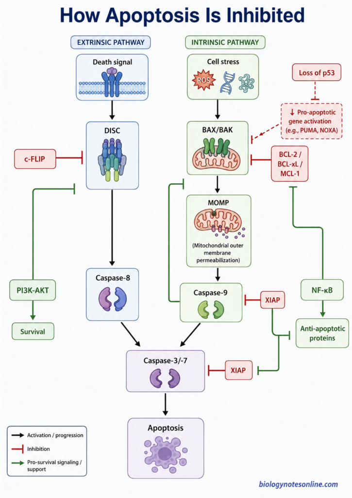
- Anti-apoptotic BCL-2 Family Proteins – BCL-2, BCL-XL and MCL-1 are major anti-apoptotic proteins. These prevent the activity of pro-apoptotic BCL-2 family members and reduce activation of BAX and BAK. MOMP is prevented. Cytochrome c release is therefore reduced or does not take place.
- IAP Proteins – Inhibitor of apoptosis proteins (IAPs) regulate caspase activity. XIAP is an important member of this group. It can directly inhibit caspase-9, caspase-3 and caspase-7. The caspases remain inactive.
- c-FLIP – c-FLIP is mainly associated with regulation of death receptor-mediated apoptosis. It acts at the DISC and affects activation of procaspase-8. Increased inhibitory action of c-FLIP can reduce caspase-8 activation. Extrinsic apoptotic signaling is then suppressed.
- PI3K-AKT Survival Pathway – Growth factors activate different survival pathways in the cell. PI3K-AKT is one of the important pathways. Akt phosphorylates several proteins involved in cell survival, including BAD. Its pro-apoptotic activity becomes reduced.
- NF-κB-Mediated Survival – NF-κB can increase the expression of anti-apoptotic proteins. These include c-FLIP, IAP proteins and some anti-apoptotic members of BCL-2 family. Thus, apoptosis can be blocked at more than one level.
- Loss of p53 Activity – p53 normally responds to DNA damage and other cellular stresses. It can promote expression of pro-apoptotic proteins when damage becomes severe. Loss or mutation of p53 reduces this response. Damaged cells may continue to survive instead of entering apoptosis.
Examples of Apoptosis
Apoptosis occurs normally during development, tissue turnover and different immune processes. It is also used for removal of damaged or infected cells. Some of the common examples are-
- Removal of Interdigital Cells – During embryonic development, cells present between the developing digits undergo programmed cell death. The interdigital tissue is removed. This helps in separation and shaping of individual digits.
- Removal of Autoreactive T Cells – Developing T lymphocytes that strongly recognize self-antigens can be eliminated by apoptosis in the thymus. This process is called negative selection. Autoreactive T cells are therefore removed before entering the peripheral immune system.
- Death of Activated Lymphocytes – During an immune response, large numbers of lymphocytes are produced. Most are not required after the antigen has been controlled. A large proportion of these effector cells then undergo apoptosis during the contraction phase of immune response.
- Neutrophil Turnover – Neutrophils have a short functional life and commonly undergo apoptosis after completing their activity. The apoptotic neutrophils are removed by macrophages. This is important during normal turnover and resolution of inflammation.
- Mammary Gland Involution – After weaning, milk-producing epithelial cells of mammary gland are no longer required in large numbers. Many of these cells undergo apoptosis during mammary gland involution, together with extensive tissue remodeling.
- Removal of Severely DNA-Damaged Cells – Severe DNA damage can result in apoptosis when the damage cannot be properly repaired. p53 has an important role in many such responses. The damaged cell is eliminated instead of continuing its proliferation.
- Killing of Infected or Abnormal Cells – Cytotoxic T lymphocytes (CTLs) can cause apoptosis of infected target cells. They use perforin and granzymes, and can also use the Fas-FasL pathway. Natural killer (NK) cells use similar cell-killing mechanisms against some infected or transformed cells.
Apoptosis Pathway vs Necrosis
Apoptosis and necrosis are two different patterns of cell death. Apoptosis is generally regulated and occurs during both normal physiological processes and disease. Classical necrosis, on the other hand, is mainly associated with severe cellular injury and loss of membrane integrity.
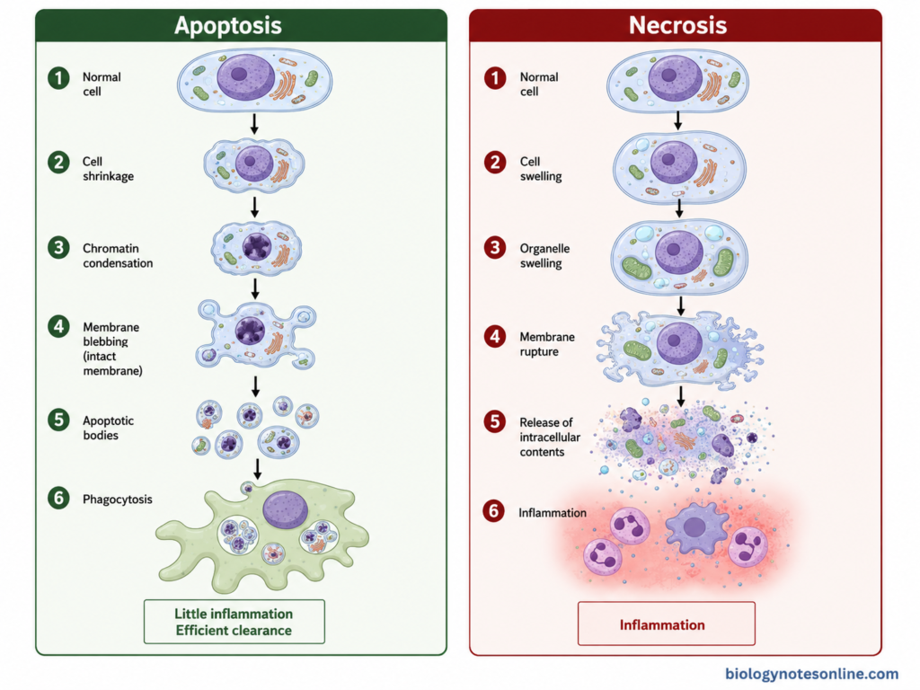
| Features | Apoptosis | Necrosis |
|---|---|---|
| Type of cell death | It is a regulated form of cell death. | Classical necrosis occurs mainly after severe and irreversible cell injury. |
| Occurrence | Occurs during normal development, tissue turnover and also under pathological conditions. | It is mainly associated with pathological injury. |
| Energy requirement | It is an active process and involves energy-dependent biochemical reactions. | Severe ATP depletion is commonly associated with necrotic injury. |
| Cell size | The cell becomes smaller. Cell shrinkage occurs. | The cell and its organelles generally swell. |
| Plasma membrane | Plasma membrane remains intact during the main apoptotic process, although membrane blebbing occurs. | Plasma membrane loses its integrity and finally ruptures. |
| Cellular contents | Cellular components remain enclosed and are packed into membrane-bound fragments. | Intracellular contents are released into surrounding tissue after membrane rupture. |
| Nuclear changes | Chromatin condensation and nuclear fragmentation take place. | Nuclear changes occur with progressive cellular injury and degradation. |
| DNA fragmentation | Controlled DNA fragmentation is characteristic. | DNA degradation is generally less orderly. |
| Caspases | Caspases have the major role in classical apoptosis. | Classical accidental necrosis is not normally dependent on apoptotic caspase activation. |
| Apoptotic bodies | Small membrane-bound apoptotic bodies are formed. | Apoptotic bodies are not formed. |
| Inflammation | Usually produces little or no inflammation when apoptotic cells are rapidly removed. | Membrane rupture and release of cellular contents generally produce inflammation. |
| Removal of dead cells | Apoptotic cells and bodies are recognized and engulfed by phagocytic cells. | Necrotic material is cleared after cellular disruption and inflammatory recruitment. |
| Number of cells affected | Frequently affects individual cells. | Severe injury can affect groups of neighboring cells or areas of tissue. |
| Major role | Development, tissue homeostasis and removal of damaged or unwanted cells. | Mainly occurs following severe injury such as toxins, trauma, ischemia or other damaging conditions. |
Note: Necrosis is not always completely “unregulated.” Different forms of regulated necrotic cell death, including necroptosis, are now recognized. Thus, the table mainly compares apoptosis with classical necrosis.
References
- Alberts, B., Johnson, A., Lewis, J., Raff, M., Roberts, K., & Walter, P. (2002). Programmed cell death (apoptosis). In Molecular biology of the cell (4th ed.). Garland Science. https://www.ncbi.nlm.nih.gov/books/NBK26873/
- Algeciras-Schimnich, A., Barnhart, B. C., & Peter, M. E. (2013). Apoptosis dependent and independent functions of caspases. In Madame Curie Bioscience Database. Landes Bioscience. https://www.ncbi.nlm.nih.gov/books/NBK6198/
- Bálint, É. E., & Vousden, K. H. (2001). Activation and activities of the p53 tumour suppressor protein. British Journal of Cancer, 85(12), 1813–1823. https://doi.org/10.1054/bjoc.2001.2128
- Baumann, S., Krueger, A., Kirchhoff, S., & Krammer, P. H. (2002). Regulation of T cell apoptosis during the immune response. Current Molecular Medicine, 2(3), 257–272. https://pubmed.ncbi.nlm.nih.gov/12041729/
- Coleman, M. L., Sahai, E. A., Yeo, M., Bosch, M., Dewar, A., & Olson, M. F. (2001). Membrane blebbing during apoptosis results from caspase-mediated activation of ROCK I. Nature Cell Biology, 3(4), 339–345. https://doi.org/10.1038/35070009
- Collins, M. (1995). Potential roles of apoptosis in viral pathogenesis. American Journal of Respiratory and Critical Care Medicine, 152(4 Pt 2), S20–S24. https://pubmed.ncbi.nlm.nih.gov/7551407/
- Datta, S. R., Dudek, H., Tao, X., Masters, S., Fu, H., Gotoh, Y., & Greenberg, M. E. (1997). Akt phosphorylation of BAD couples survival signals to the cell-intrinsic death machinery. Cell, 91(2), 231–241. https://doi.org/10.1016/S0092-8674(00)80405-5
- Dho, S. H., Cho, M., Woo, W., Jeong, S., & Kim, L. K. (2025). Caspases as master regulators of programmed cell death: Apoptosis, pyroptosis and beyond. Experimental & Molecular Medicine, 57, 1121–1132. https://doi.org/10.1038/s12276-025-01470-9
- Eefting, F., Rensing, B., Wigman, J., Pannekoek, W. J., Liu, W. M., Cramer, M. J., Lips, D. J., & Doevendans, P. A. (2004). Role of apoptosis in reperfusion injury. Cardiovascular Research, 61(3), 414–426. https://doi.org/10.1016/j.cardiores.2003.12.023
- Elmore, S. (2007). Apoptosis: A review of programmed cell death. Toxicologic Pathology, 35(4), 495–516. https://doi.org/10.1080/01926230701320337
- Février, M., Dorgham, K., & Rebollo, A. (2011). CD4+ T cell depletion in human immunodeficiency virus (HIV) infection: Role of apoptosis. Viruses, 3(5), 586–612. https://doi.org/10.3390/v3050586
- Fulda, S. (2018). Cell death-based treatment of glioblastoma. Cell Death & Disease, 9, 121. https://doi.org/10.1038/s41419-017-0021-8
- Galluzzi, L., Vitale, I., Aaronson, S. A., Abrams, J. M., Adam, D., Agostinis, P., Alnemri, E. S., Altucci, L., Amelio, I., Andrews, D. W., Annicchiarico-Petruzzelli, M., Antonov, A. V., Arama, E., Baehrecke, E. H., Barlev, N. A., Bazan, N. G., Bernassola, F., Bertrand, M. J. M., Bianchi, K., … Kroemer, G. (2018). Molecular mechanisms of cell death: Recommendations of the Nomenclature Committee on Cell Death 2018. Cell Death & Differentiation, 25, 486–541. https://doi.org/10.1038/s41418-017-0012-4
- Gavrieli, Y., Sherman, Y., & Ben-Sasson, S. A. (1992). Identification of programmed cell death in situ via specific labeling of nuclear DNA fragmentation. Journal of Cell Biology, 119(3), 493–501. https://doi.org/10.1083/jcb.119.3.493
- Kale, J., Osterlund, E. J., & Andrews, D. W. (2018). BCL-2 family proteins: Changing partners in the dance towards death. Cell Death & Differentiation, 25, 65–80. https://doi.org/10.1038/cdd.2017.186
- Kalkavan, H., & Green, D. R. (2018). MOMP, cell suicide as a BCL-2 family business. Cell Death & Differentiation, 25, 46–55. https://doi.org/10.1038/cdd.2017.179
- Kerr, J. F. R., Wyllie, A. H., & Currie, A. R. (1972). Apoptosis: A basic biological phenomenon with wide-ranging implications in tissue kinetics. British Journal of Cancer, 26(4), 239–257. https://doi.org/10.1038/bjc.1972.33
- Lakhani, S. A., Masud, A., Kuida, K., Porter, G. A., Jr., Booth, C. J., Mehal, W. Z., Inayat, I., & Flavell, R. A. (2006). Caspases 3 and 7: Key mediators of mitochondrial events of apoptosis. Science, 311(5762), 847–851. https://doi.org/10.1126/science.1115035
- Li, H., Zhu, H., Xu, C.-J., & Yuan, J. (1998). Cleavage of BID by caspase 8 mediates the mitochondrial damage in the Fas pathway of apoptosis. Cell, 94(4), 491–501. https://doi.org/10.1016/S0092-8674(00)81590-1
- Li, P., Nijhawan, D., Budihardjo, I., Srinivasula, S. M., Ahmad, M., Alnemri, E. S., & Wang, X. (1997). Cytochrome c and dATP-dependent formation of Apaf-1/caspase-9 complex initiates an apoptotic protease cascade. Cell, 91(4), 479–489. https://doi.org/10.1016/S0092-8674(00)80434-1
- Mattson, M. P. (2000). Apoptosis in neurodegenerative disorders. Nature Reviews Molecular Cell Biology, 1(2), 120–129. https://doi.org/10.1038/35040009
- Merheb, E., Zubair, M., & Bokhari, S. R. A. (2026). Apoptosis and cell death: Signaling in health and diseases. In StatPearls. StatPearls Publishing. https://www.ncbi.nlm.nih.gov/books/NBK499821/
- Ow, Y.-L. P., Green, D. R., Hao, Z., & Mak, T. W. (2008). Cytochrome c: Functions beyond respiration. Nature Reviews Molecular Cell Biology, 9(7), 532–542. https://doi.org/10.1038/nrm2434
- Reed, J. C. (2003). Protein domains involved in apoptosis regulation. In D. W. Kufe, R. E. Pollock, R. R. Weichselbaum, et al. (Eds.), Holland-Frei cancer medicine (6th ed.). BC Decker. https://www.ncbi.nlm.nih.gov/books/NBK13247/
- Scaffidi, C., Fulda, S., Srinivasan, A., Friesen, C., Li, F., Tomaselli, K. J., Debatin, K.-M., Krammer, P. H., & Peter, M. E. (1998). Two CD95 (APO-1/Fas) signaling pathways. EMBO Journal, 17(6), 1675–1687. https://doi.org/10.1093/emboj/17.6.1675
- Scott, F. L., Denault, J.-B., Riedl, S. J., Shin, H., Renatus, M., & Salvesen, G. S. (2005). XIAP inhibits caspase-3 and -7 using two binding sites: Evolutionarily conserved mechanism of IAPs. EMBO Journal, 24(3), 645–655. https://doi.org/10.1038/sj.emboj.7600544
- Sharp, D. A., Lawrence, D. A., & Ashkenazi, A. (2005). Selective knockdown of the long variant of cellular FLICE inhibitory protein augments death receptor-mediated caspase-8 activation and apoptosis. Journal of Biological Chemistry, 280(19), 19401–19409. https://doi.org/10.1074/jbc.M413962200
- Suzuki, J., Denning, D. P., Imanishi, E., Horvitz, H. R., & Nagata, S. (2013). Xk-related protein 8 and CED-8 promote phosphatidylserine exposure in apoptotic cells. Science, 341(6144), 403–406. https://doi.org/10.1126/science.1236758
- Trapani, J. A., & Smyth, M. J. (2002). Functional significance of the perforin/granzyme cell death pathway. Nature Reviews Immunology, 2(10), 735–747. https://doi.org/10.1038/nri911
- Vermes, I., Haanen, C., Steffens-Nakken, H., & Reutelingsperger, C. (1995). A novel assay for apoptosis: Flow cytometric detection of phosphatidylserine expression on early apoptotic cells using fluorescein-labelled Annexin V. Journal of Immunological Methods, 184(1), 39–51. https://doi.org/10.1016/0022-1759(95)00072-I
- Vogler, M., Braun, Y., Smith, V. M., Westhoff, M.-A., et al. (2025). The BCL2 family: From apoptosis mechanisms to new advances in targeted therapy. Signal Transduction and Targeted Therapy, 10, 91. https://doi.org/10.1038/s41392-025-02176-0
- Wei, M. C., Zong, W.-X., Cheng, E. H.-Y., Lindsten, T., Panoutsakopoulou, V., Ross, A. J., Roth, K. A., MacGregor, G. R., Thompson, C. B., & Korsmeyer, S. J. (2001). Proapoptotic BAX and BAK: A requisite gateway to mitochondrial dysfunction and death. Science, 292(5517), 727–730. https://doi.org/10.1126/science.1059108
- Wolf, B. B., Schuler, M., Echeverri, F., & Green, D. R. (1999). Caspase-3 is the primary activator of apoptotic DNA fragmentation via DNA fragmentation factor-45/inhibitor of caspase-activated DNase inactivation. Journal of Biological Chemistry, 274(43), 30651–30656. https://doi.org/10.1074/jbc.274.43.30651
- Yang, J., Liu, X., Bhalla, K., Kim, C. N., Ibrado, A. M., Cai, J., Peng, T.-I., Jones, D. P., & Wang, X. (1997). Prevention of apoptosis by Bcl-2: Release of cytochrome c from mitochondria blocked. Science, 275(5303), 1129–1132. https://doi.org/10.1126/science.275.5303.1129
- Zhang, J., Huang, K., O’Neill, K. L., Pang, X., & Luo, X. (2016). Bax/Bak activation in the absence of Bid, Bim, Puma, and p53. Cell Death & Disease, 7, e2266. https://doi.org/10.1038/cddis.2016.167
- Zou, H., Henzel, W. J., Liu, X., Lutschg, A., & Wang, X. (1997). Apaf-1, a human protein homologous to C. elegans CED-4, participates in cytochrome c-dependent activation of caspase-3. Cell, 90(3), 405–413. https://doi.org/10.1016/S0092-8674(00)80501-2