

Reverse transfection is a transfection method in which the nucleic acid-transfection reagent complexes are prepared first, and the cells are then added to these complexes. This is opposite to conventional or forward transfection, where the transfection mixture is added to already plated cells. The method was first described with cDNA in transfected-cell microarrays and was later adapted for siRNA and other gene-function studies. It is commonly used in cell arrays and high-throughput screening, where many genes can be studied at the same time.
What Is Reverse Transfection?
Reverse transfection is a transfection method in which the transfection complexes are prepared before the cells are seeded. These complexes commonly contain DNA, siRNA, or another nucleic acid together with a transfection reagent. The cells are then added and they come in contact with the complexes at or near the time of cell seeding.
The term “reverse” refers to the reversed order of adding cells and the transfection material. In conventional or forward transfection, cells are generally plated first and the nucleic acid-transfection complexes are added afterward. In reverse transfection, the complexes are placed or formed first, followed by addition of the cells. The original transfected-cell microarray method was named reverse transfection for this reversed order of addition.
Reverse transfection can be performed in a liquid multiwell format. Here, the nucleic acid and transfection reagent are mixed in wells of a multiwell plate, and the cell suspension is then added. Such reverse transfection has been used with siRNA in 96-well and other microtiter plate formats.
Another form is solid-phase reverse transfection. In this method, the nucleic acid-transfection material is immobilized, dried or printed on a solid surface before the cells are seeded over it. This approach is commonly used for transfected cell microarrays, where different DNA or siRNA samples can be present at separate positions on the same surface.
Why Is It Called Reverse Transfection?
It is called reverse transfection because the normal order of adding the nucleic acid and cells is reversed. In conventional transfection, cells are present in the culture vessel first and the transfection material is added to them. In reverse transfection, the nucleic acid-transfection material is prepared or placed first, and cells are seeded afterward.
The term was introduced by Ziauddin and Sabatini during development of transfected-cell microarrays. In their method, DNA was printed at defined positions on a glass slide and mammalian cells were then cultured over the printed DNA areas. Thus, cells encounter the transfection material after they are added to the surface. This reversed order of DNA and cell addition is the basic reason for the name “reverse transfection”.
The same reverse arrangement is now also used in different cell-based transfection formats. The important point is the order. Transfection material first, followed by the cells, rather than adding the transfection complexes to previously plated cells. Reverse-transfected cell microarrays are also described as an established method in Cell-Based Microarrays: Methods and Protocols.
Types of Reverse Transfection
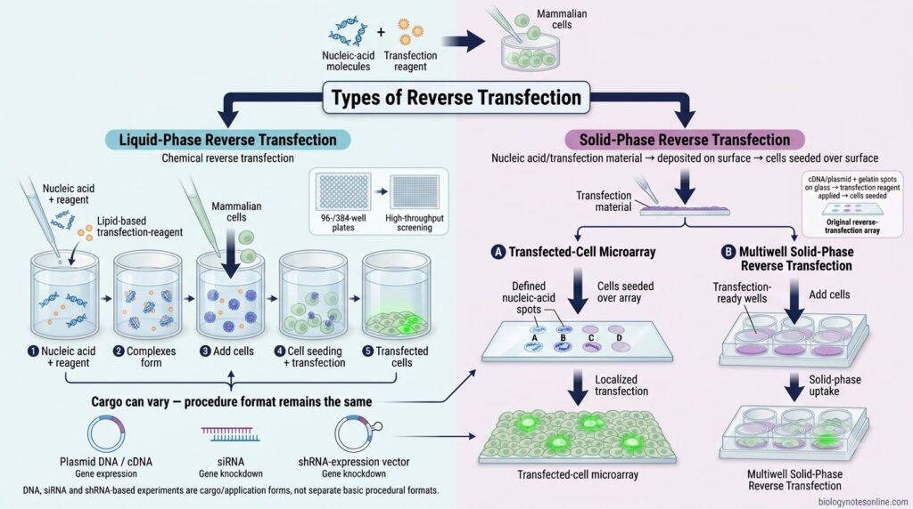
Reverse transfection is commonly performed in two main formats, depending on how the nucleic acid-transfection material is present before addition of cells. These are liquid-phase reverse transfection and solid-phase reverse transfection. This division reflects the major formats described in reverse-transfection studies and cell-based array methods.
- Liquid-Phase Reverse Transfection
- In this type, the nucleic acid and transfection reagent are mixed in the liquid medium of a culture well.
- The transfection complexes are formed first. Cells in suspension are then added into the wells containing these complexes.
- Thus, the cells are seeded and exposed to the transfection complexes at approximately the same time.
- It is commonly performed in 96-well or 384-well plates, especially for siRNA-based screening.
- This format is also referred to as chemical reverse transfection when lipid or other chemical transfection reagents are used.
- Solid-Phase Reverse Transfection
- In solid-phase reverse transfection, the nucleic acid-transfection material is deposited, printed or attached onto a solid culture surface before addition of cells.
- Cells are then seeded over this prepared surface. The cells growing at or over these regions take up the transfection material.
- The original reverse-transfection method used DNA-containing spots on glass slides, producing separate clusters of transfected cells.
- This type is widely used for transfected-cell microarrays and siRNA arrays, where many different nucleic acids can be arranged at defined positions on one surface.
- Solid-phase reverse transfection has also been adapted to multiwell plates. Here, the transfection mixture is attached within individual wells before the cells are added.
- The method can therefore be present as cell-array solid-phase transfection or multiwell solid-phase transfection, depending on the experimental arrangement.
DNA reverse transfection, siRNA reverse transfection or shRNA reverse transfection describe the nucleic acid being delivered. They are better considered applications or cargo-based forms, rather than separate basic types of the reverse-transfection procedure. This distinction is supported by studies using the same solid-phase principle with DNA, siRNA and shRNA.
Principle of Reverse Transfection
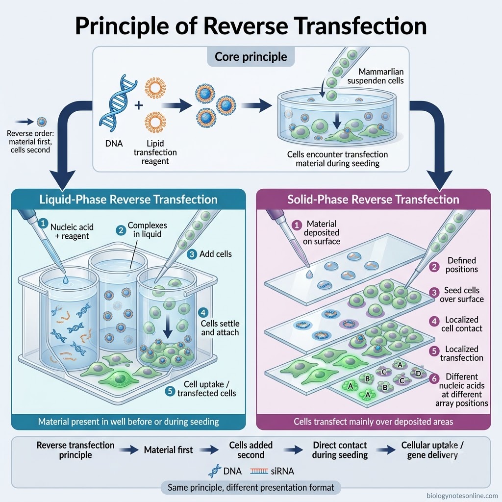
The principle of reverse transfection is based on exposing cells to the nucleic acid-transfection reagent complexes at the time of cell seeding. Unlike conventional transfection, the transfection material is prepared or placed first. The cells are then added over or into the transfection mixture. This reversed order allows the newly seeded cells to come in direct contact with the complexes.
In liquid-phase reverse transfection, the nucleic acid and transfection reagent complexes are present in the culture well before or while the cell suspension is added. The cells settle and attach while being exposed to these complexes. The nucleic acid is then taken up by the cells through the usual mechanism of the particular transfection reagent being used.
In solid-phase reverse transfection, DNA, siRNA or their transfection mixture is deposited at defined areas of a solid surface. Cells are seeded over the surface and transfection takes place mainly in cells growing over these areas. Thus, different nucleic acids can be transfected at separate positions on the same cell array. This forms the basic principle of reverse-transfection cell microarrays.
How Does Reverse Transfection Work?
The following is the general procedure of liquid-phase reverse transfection used in multiwell plates. The exact amount of nucleic acid, transfection reagent, cell number and incubation period depends on the cell type and transfection reagent used.
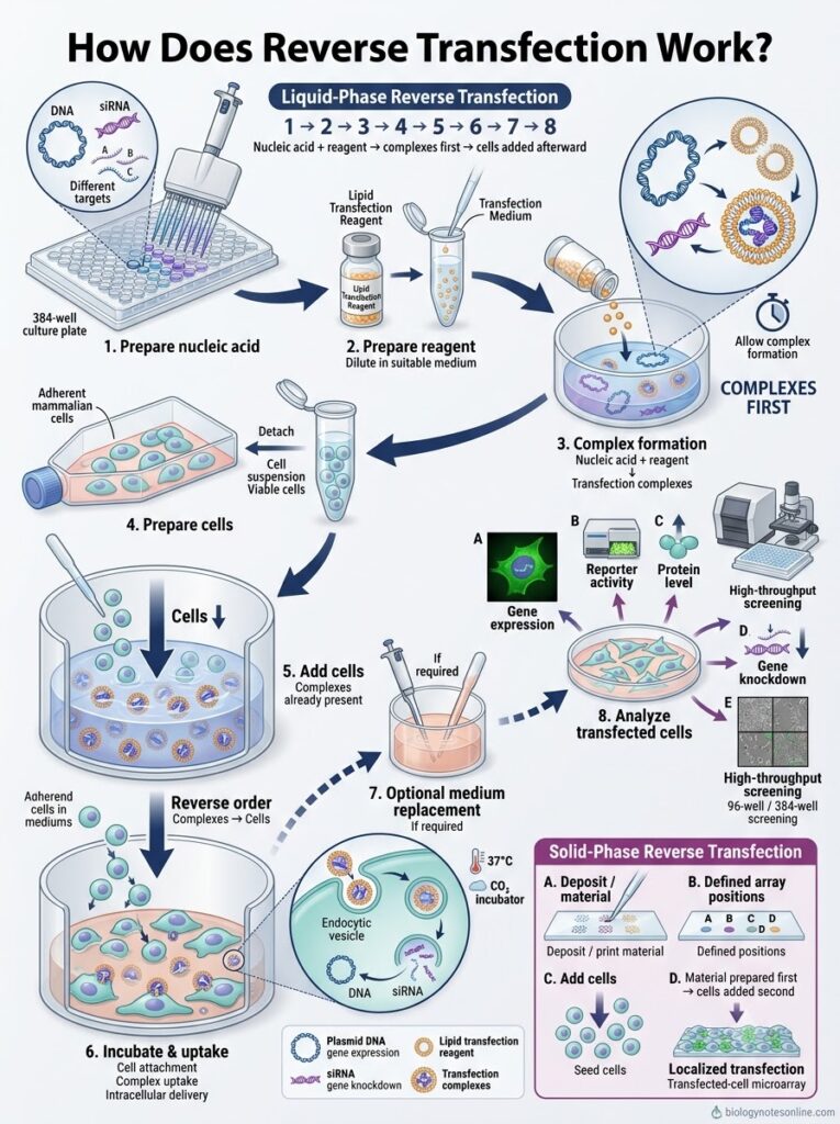
Step 1- Preparation of nucleic acid
The required DNA, siRNA or other nucleic acid is taken in the wells of a culture plate. For screening experiments, different nucleic acids can be placed in separate wells. The required concentration is selected according to the experiment.
Step 2- Preparation of transfection reagent
The transfection reagent is diluted in a suitable transfection medium. Lipid-based reagents are commonly used for reverse transfection. The reagent amount needs optimization because excess reagent can affect cell viability.
Step 3- Formation of transfection complexes
The diluted transfection reagent is mixed with the nucleic acid present in each well. It is allowed to form nucleic acid-transfection reagent complexes for the required period. These complexes are formed before addition of cells, which is the important feature of reverse transfection.
Step 4- Preparation of cells
The cells are harvested from the culture vessel. Adherent cells are generally detached and suspended in fresh growth medium. A suitable number of viable cells is then prepared for each well.
Step 5- Addition of cells to the complexes
The prepared cell suspension is directly added into wells already containing the transfection complexes. Thus, unlike conventional transfection, cells are not plated before preparation of the transfection complexes. Cells come in contact with the complexes during their attachment to the culture surface.
Step 6- Incubation
The plate is kept under suitable cell-culture conditions. During this period, the cells attach to the surface and take up the delivered nucleic acid. The incubation conditions and time depend on the cell line and the type of experiment.
Step 7- Medium replacement, if required
The culture medium can be replaced when prolonged exposure to the transfection reagent causes cytotoxicity or when required by the particular protocol. It is not necessary in every reverse transfection experiment.
Step 8- Analysis of transfected cells
After the required post-transfection period, cells are examined for the expected effect. Gene expression, reporter activity, protein expression, gene knockdown, cell viability or another cellular phenotype can be measured depending on the nucleic acid used. Reverse transfection in 96-well and 384-well plates is especially suitable for such screening experiments.
In solid-phase reverse transfection, the procedure is slightly different. Here, DNA, siRNA or another transfection material is first deposited or printed at defined regions of a solid surface. Cells are then seeded over the prepared surface, and localized groups of cells become transfected at the corresponding regions. This method forms the basis of transfected-cell microarrays.
Reverse Transfection Protocol
The reverse transfection protocol can vary considerably with the cell type, nucleic acid or cargo, and the transfection reagent used. Therefore, one fixed reagent-to-cargo ratio should not be used for every experiment. Cell density, amount of reagent and cargo concentration need to be optimized for the particular experimental system.
Materials Required
The following are the general materials required for reverse transfection-
- Cultured cells suitable for the transfection experiment.
- Suitable cell culture medium required for maintenance and growth of the selected cells.
- Nucleic acid/cargo, such as plasmid DNA, siRNA or other material depending on the experiment.
- A compatible transfection reagent selected according to the cargo and cell type.
- Sterile culture plate, commonly a multiwell plate for liquid-phase reverse transfection.
- Cell-counting system for determining the required number of viable cells.
- Appropriate experimental controls. These may include untreated cells, reagent-only or mock control, negative-control nucleic acid and a positive control when required.
General Procedure
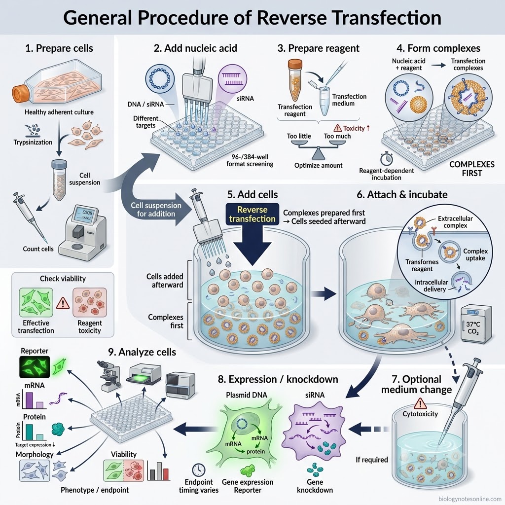
- Preparation of cells
Healthy cultured cells are selected for the experiment. Adherent cells are detached when required and a uniform cell suspension is prepared. The cells are counted before seeding. - Addition of nucleic acid/cargo
The required DNA, siRNA or other cargo is placed into the culture wells. Different cargo can be placed in separate wells during screening experiments. Reverse transfection has been widely performed in 96-well and 384-well formats. - Preparation of transfection reagent
The compatible transfection reagent is prepared according to its particular instructions. The required amount is not same for all cell lines or reagents. Too much reagent can increase cellular toxicity, whereas insufficient reagent may reduce transfection. - Formation of transfection complexes
The transfection reagent is combined with the nucleic acid or other compatible cargo. The mixture is allowed to form transfection complexes as required for that reagent. The conditions and complex-formation period are reagent dependent. - Addition of cells
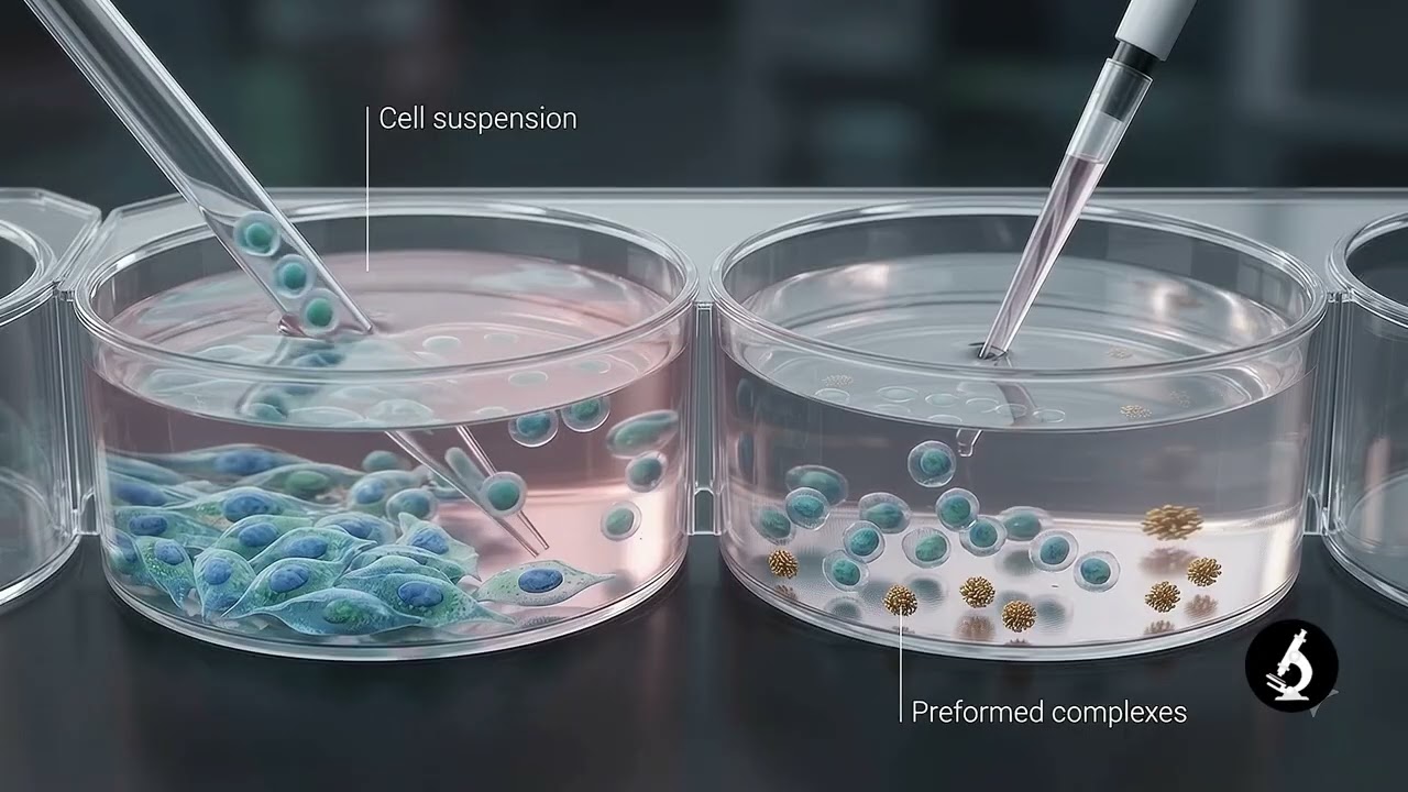
The prepared cell suspension is added directly into wells containing the transfection complexes. This is the important step of reverse transfection. Thus, cell plating and exposure to the transfection complexes take place together or very close to the same time. - Cell attachment and incubation
The plate is placed under the normal culture conditions required for the selected cells. During this period, cells attach and remain exposed to the transfection complexes. Incubation conditions should be selected according to the cell type, transfection system and required endpoint. - Medium change, when required
A medium replacement is not required for every transfection reagent. For sensitive cells, prolonged exposure or excessive amount of a reagent can affect cell health. The medium can therefore be replaced when recommended for the reagent or when cytotoxicity becomes a problem. - Incubation for expression or knockdown
Cells are maintained until sufficient gene expression, gene silencing or another expected cellular response has developed. The required period is dependent on the cargo, target and type of assay. Thus, endpoint timing should be established experimentally. - Analysis of cells
The required endpoint is finally measured. Depending on the experiment, this may include reporter expression, mRNA level, protein expression, gene knockdown, cell morphology or another phenotype. Cell viability should also be considered because reagent-associated toxicity can interfere with interpretation of transfection results.
Important Parameters That Must Be Optimized
- Cell density
A suitable number of cells should be used in each well. Very high cell numbers can reduce the effective amount of cargo available per cell, whereas very low density can affect normal growth of some cell cultures. The suitable density therefore depends on the cell line and assay. - Transfection reagent amount
The amount of transfection reagent has to be optimized. Insufficient reagent can result in poor delivery, while excessive reagent may produce cytotoxicity. Different cell lines can also show different responses to the same reagent. - Cargo concentration
The amount of DNA, siRNA or other cargo should be determined experimentally. For siRNA, both insufficient and excessive concentrations can create problems, including weak silencing or unwanted effects. Optimal concentration is dependent on the cell and target. - Complex formation
Proper formation of cargo-transfection reagent complexes is necessary for chemical transfection. The mixing conditions and complex-formation period should follow the requirement of the particular reagent rather than using a single condition for all systems. - Culture medium
The selected medium must be compatible with both cells and the transfection reagent. Serum, antibiotics and other medium components can have different effects depending on the reagent used. Thus, the reagent-specific protocol should be followed for medium conditions. - Incubation conditions
The period of exposure to the transfection complexes needs adjustment for sensitive cells. Prolonged exposure to some transfection reagents can reduce cell health. - Cell viability
Cell viability should be monitored together with transfection efficiency. A high signal is not sufficient if substantial reagent-associated cell death is also present. Transfection efficiency and toxicity can vary between different cell lines. - Endpoint timing
The time of analysis is selected according to the biological endpoint. Gene knockdown, protein reduction and reporter expression do not necessarily reach their useful measurement point at the same time. Therefore, endpoint timing is also optimized for the particular assay.
Exact conditions of reverse transfection are not universal. Current technical guidance specifically recommends empirical optimization of cell conditions, transfection reagent amount and cargo concentration for the selected cell type.
Reverse Transfection of siRNA
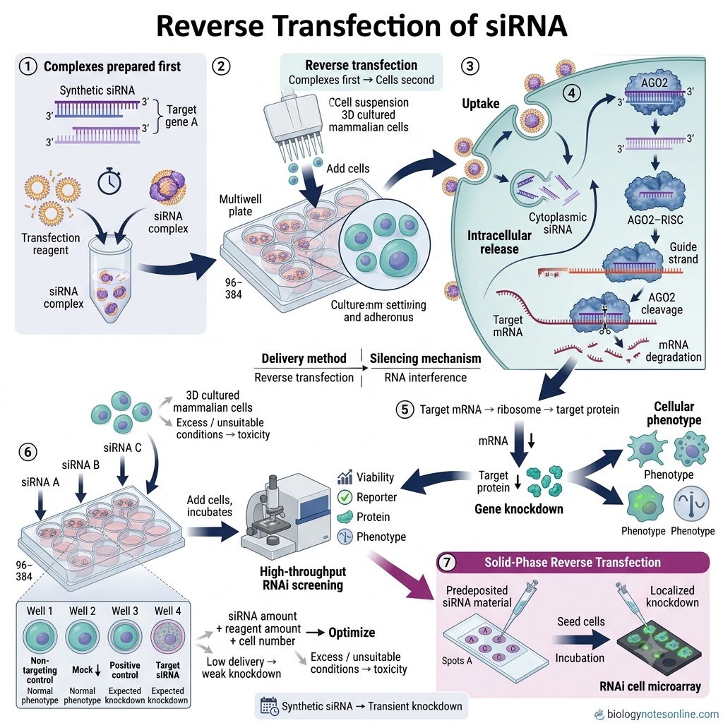
- siRNA reverse transfection is a method used to introduce small interfering RNA (siRNA) into cultured cells for gene silencing. The siRNA-transfection reagent complexes are prepared in the culture wells first. Cells are added afterward, so they encounter the complexes during or shortly after seeding.
- In this method, a synthetic siRNA duplex targeting a particular gene is generally mixed with a compatible lipid or another transfection reagent. The complexes are formed before addition of the cell suspension. This is different from forward siRNA transfection where cells are plated before the transfection complexes are added.
- After cells are added, the siRNA-containing complexes are taken up by the cells. The delivered siRNA then enters the cellular RNA interference pathway. One strand acts as the guide strand in the RNA-induced silencing complex (RISC) and directs the complex toward complementary target mRNA. Target RNA is then silenced, commonly through AGO2-mediated cleavage and degradation when the complementarity is suitable.
- Thus, reverse transfection changes mainly the order of transfection and cell seeding. It does not represent a different mechanism of siRNA-mediated RNA interference.
- Reverse siRNA transfection is frequently performed in 96-well and 384-well plates. siRNAs can be pre-arrayed in individual wells and cells are subsequently added to them. For this reason, the method is useful for testing many gene targets under similar experimental conditions.
- It is particularly used in high-throughput RNAi screening. Separate siRNAs or siRNA pools can target different genes in separate wells, followed by measurement of cell viability, reporter activity, protein level or another cellular phenotype. Genome-wide screens have also been performed using this arrangement.
- Reverse transfection of siRNA can also be carried out by a solid-phase method. Here, siRNA-transfection material is deposited or printed at defined areas of a surface before cells are seeded over it. Cells present on these regions become transfected, which forms RNA interference microarrays or cell spot microarrays.
- The amount of siRNA, transfection reagent and the number of cells cannot be kept as one universal condition. These have to be optimized for the particular cell type and assay. Low transfection can produce weak knockdown, while unsuitable siRNA or lipid conditions can increase toxicity or unwanted effects. Optimization and suitable controls are therefore important during siRNA screening.
- Common controls include a non-targeting siRNA, mock-transfected cells and, when suitable, a positive-control siRNA that gives a known phenotype. Confirmation of gene knockdown at the mRNA or protein level is also used to determine whether the siRNA has produced the expected effect.
- Reverse siRNA transfection is therefore mainly used for transient gene knockdown, functional genomic studies and high-throughput screening. Synthetic siRNA usually produces transient silencing, which distinguishes it from vector-based shRNA systems capable of more sustained knockdown.
Why Reverse Transfection Is Used for siRNA Screening
Reverse transfection is commonly used for siRNA screening because it fits well with arrayed and high-throughput experiments. A large number of siRNAs can be placed in separate wells or defined positions first, followed by addition of the cells. Thus, different genes can be knocked down in parallel under the same screening workflow.
- Suitable for high-throughput screening
- Reverse transfection can be performed in 96-well, 384-well and even highly miniaturized multiwell formats.
- Each well can contain an siRNA against a different gene.
- Large groups of genes can therefore be screened in parallel. A published colorectal cancer RNAi screen used reverse transfection in 1,536-well plates.
- siRNAs can be pre-arrayed
- The siRNA library can be dispensed into the assay plate before addition of cells.
- Transfection reagent is then added and the complexes are formed. Cells are finally seeded into these wells.
- This arrangement is convenient when hundreds or thousands of individual siRNAs have to be tested.
- Fewer separate cell-handling steps
- In conventional transfection, cells are generally seeded first and transfected later.
- During reverse transfection, cells are added directly to wells containing the prepared siRNA-transfection complexes.
- Cell seeding and exposure to siRNA therefore occur at approximately the same stage of the experiment. This makes the workflow suitable for large screening plates.
- Compatible with automation
- High-throughput siRNA screening requires repeated dispensing of siRNA, reagent and cells.
- Reverse transfection can be carried out using automated liquid-handling and dispensing systems.
- Automated systems have been used for siRNA transfer, reagent dispensing, cell addition and later phenotype analysis.
- Useful for systematic gene knockdown
- RNA interference (RNAi) allows loss-of-function analysis of individual genes.
- When an arrayed siRNA library is combined with reverse transfection, many genes can be silenced separately and the resulting phenotype can be measured.
- It is therefore used for functional genomics, pathway studies and identification of genes affecting a selected cellular response.
- Suitable for high-content screening
- Reverse-transfected siRNA arrays can be combined with fluorescence microscopy, immunofluorescence and time-lapse imaging.
- Changes in cell division, morphology, protein localization and other cellular phenotypes can then be studied after gene knockdown.
- Automated time-lapse RNAi screening has also been performed using transfection-ready siRNA microarrays.
- Allows transfection-ready arrays
- In solid-phase reverse transfection, siRNA-containing transfection mixtures can be printed on a surface before the cells are added.
- Such arrays can be prepared beforehand. Erfle and coworkers reported prefabricated siRNA arrays that retained transfection activity during storage and were used for genome-wide screening.
- This is useful when the same type of screening experiment has to be repeated.
- Can reduce reagent use in solid-phase arrays
- Solid-phase reverse-transfection arrays use very small spots of siRNA-containing material.
- This considerably decreases the amount of screening reagent required compared with larger multiwell assays and permits a high number of experiments on a small surface.
Reverse transfection is therefore mainly selected for scale, parallel processing, miniaturization and automation of siRNA experiments. It should not be considered automatically more efficient for every cell type. The cell density, siRNA concentration and transfection conditions still require optimization before a large screening experiment. Published high-throughput studies specifically perform this optimization before screening.
Reverse Transfection of Plasmid DNA
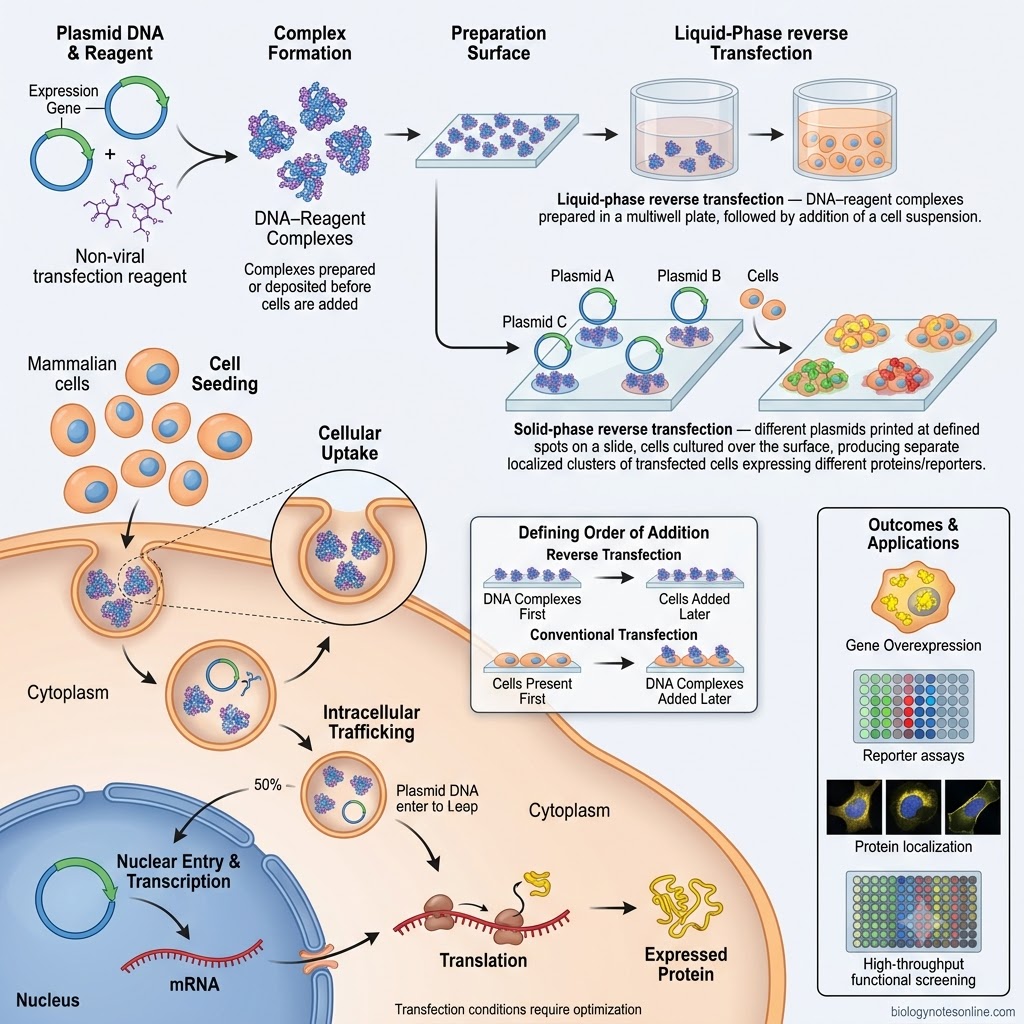
- Plasmid DNA reverse transfection is a method used to introduce plasmid DNA into cultured mammalian cells. The plasmid-transfection material is prepared or placed first and cells are seeded afterward. It is commonly used for transient expression of cloned genes and reporter constructs.
- In this method, the plasmid DNA generally carries a gene or cDNA under a suitable mammalian expression system. After successful transfection, cells containing the plasmid can express the encoded protein. The original reverse-transfection study used cDNAs cloned into expression plasmids and produced localized groups of cells expressing different gene products.
- The plasmid is combined with a compatible transfection reagent, such as a lipid-based or another non-viral DNA delivery reagent. Cells are then brought into contact with the prepared DNA-transfection material. Thus, the order is different from conventional transfection, where adherent cells are normally present before addition of the DNA complexes.
- After cellular uptake, plasmid DNA has to become available in the nucleus for transcription of the introduced gene. The encoded mRNA is produced and subsequently translated into the required protein. Nuclear delivery is therefore an important barrier during non-viral plasmid DNA transfection.
- Liquid-phase reverse transfection can also be used with plasmid DNA. Here, DNA-transfection complexes are prepared in the culture vessel and the cell suspension is added during the reverse-transfection workflow. This arrangement is suitable for multiwell experiments and automated plasmid transfection.
- In solid-phase reverse transfection, plasmid DNA is deposited or printed at defined positions of a solid surface. Cells are then cultured over the prepared surface. Cells growing over the DNA-containing regions become transfected, producing separate clusters of cells carrying different plasmids.
- Different plasmid constructs can therefore be arranged on the same reverse-transfection array. This has been used for gene overexpression, protein localization, reporter assays and functional screening. Reporter plasmids have also been reverse transfected into hundreds of independent cell clusters on a single slide.
- Reverse transfection of plasmid DNA does not require one fixed DNA-to-reagent ratio for every experiment. Transfection efficiency depends on the cell type, plasmid preparation, delivery reagent, cell density and experimental format. These conditions need optimization for the selected cells and assay. Studies with plasmid reverse transfection also show that different cell systems need modification of the transfection conditions.
- The method is particularly useful when many plasmids have to be tested in parallel. The Springer Cell-Based Microarrays: Methods and Protocols also describes cell-based arrays for high-throughput overexpression and functional analysis of proteins.
Plasmid DNA vs siRNA Reverse Transfection
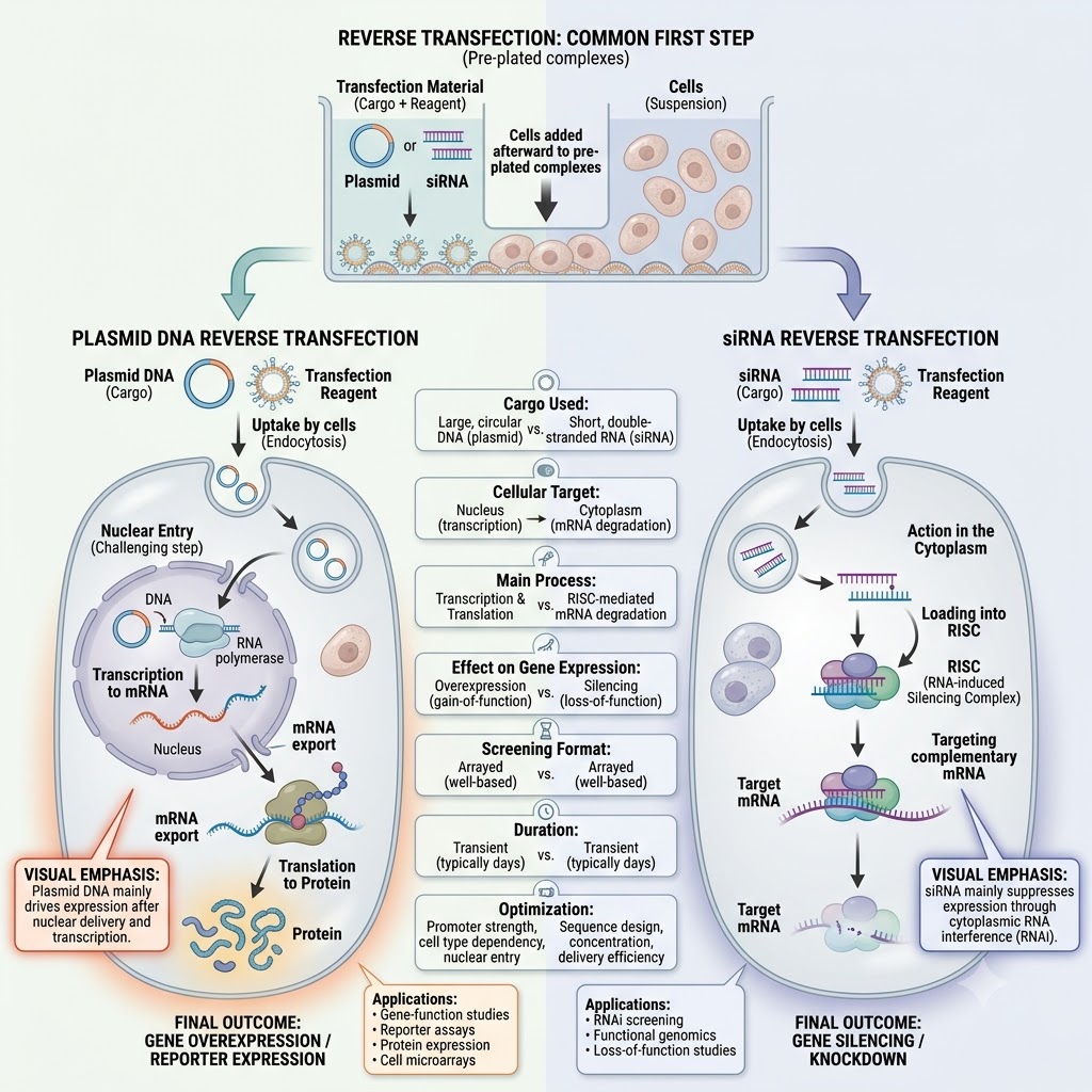
| Basis | Plasmid DNA Reverse Transfection | siRNA Reverse Transfection |
|---|---|---|
| Cargo used | Uses plasmid DNA containing a gene or reporter construct. | Uses small interfering RNA (siRNA) specific to a target gene. |
| Main purpose | Mainly used for gene overexpression or reporter expression. | Mainly used for gene silencing or knockdown. |
| Cellular target | Plasmid DNA generally needs to reach the nucleus for transcription. | siRNA acts mainly in the cytoplasm through the RNA interference pathway. |
| Major cellular process | Introduced DNA is transcribed into mRNA and then translated into protein. | siRNA is loaded into RISC, which targets complementary mRNA. |
| Effect on gene expression | Usually increases or introduces expression of a selected gene. | Decreases expression of the selected gene. |
| Common applications | Gene-function studies, reporter assays, protein expression and transfected cell microarrays. | RNAi screening, functional genomics and loss-of-function studies. |
| Screening format | Frequently used in plasmid or cDNA expression arrays. | Very commonly used in 96-well, 384-well and siRNA screening arrays. |
| Duration of effect | Usually transient with non-integrating plasmids, depending on the system. | Generally produces transient gene knockdown with synthetic siRNA. |
| Optimization | DNA amount, reagent amount, cell density and plasmid properties need optimization. | siRNA concentration, reagent amount, cell density and knockdown time need optimization. |
In both methods, the transfection material is prepared first and cells are added afterward. The major difference is the purpose. Plasmid DNA is mainly used for gene expression, whereas siRNA is used for gene silencing.
Factors Affecting Reverse Transfection Efficiency
The efficiency of reverse transfection is affected by several experimental conditions. It does not remain same for every cell line or transfection reagent. Cell density, amount of nucleic acid and reagent conditions are some of the major factors and usually require optimization.
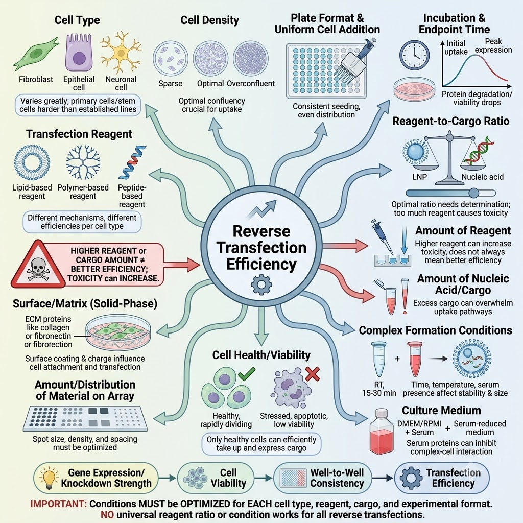
- Cell Type
- Different cell types do not show the same transfection efficiency.
- Some established cell lines are readily transfected, whereas primary cells and some differentiated cells can be difficult to transfect.
- The suitable transfection reagent and conditions therefore depend strongly on the cells being used. Studies comparing different cells and reagents have shown large differences in lipid-mediated transfection efficiency.
- Cell Density
- The number of cells added during reverse transfection is an important factor.
- Too many or too few cells can change the amount of transfection complex available to each cell and also change growth conditions.
- A uniform seeding density is particularly important in multiwell screening. Cell density is therefore usually optimized before the actual experiment.
- Transfection Reagent
- The type of transfection reagent affects both nucleic acid delivery and cell viability.
- A reagent suitable for one cell line may give low efficiency in another cell type.
- During siRNA screening, testing different lipid reagents produced considerably different levels of knockdown and viability.
- Amount of Transfection Reagent
- An insufficient amount of reagent can produce poor complex formation or low cellular delivery.
- Excess reagent, on the other hand, may increase cytotoxicity.
- Thus, increasing reagent amount does not always mean increasing transfection efficiency. The useful amount has to maintain both transfection and cell survival.
- Amount of Nucleic Acid or Cargo
- The concentration of plasmid DNA, siRNA or other cargo also affects the final response.
- Too little cargo may give weak expression or gene knockdown. Excess amount can increase unwanted effects and, for siRNA, may increase off-target responses.
- Reverse-transfection microarray studies have shown that changing siRNA concentration can considerably change the degree of gene silencing.
- Reagent-to-Cargo Proportion
- The proportion between the nucleic acid and transfection reagent affects formation of the transfection complexes.
- There is no one universal ratio suitable for all reverse transfections.
- Different ratios can produce different efficiencies and different levels of cell toxicity. The ratio should be optimized for the selected reagent, cargo and cell type.
- Complex Formation Conditions
- Proper formation of the lipid-DNA, lipid-siRNA or other nucleic acid-reagent complexes is required before the cells are added.
- Composition of the complex, mixing conditions and the matrix used can influence its delivery.
- In reverse-transfection arrays, changing the composition of lipid-DNA mixtures produced marked differences in transfection efficiency.
- Culture Medium
- The medium present during complex formation and cell seeding can affect the transfection process.
- Serum and other medium components may interact differently with different transfection systems. For this reason, the medium conditions should follow the requirements of the particular reagent and cell type.
- Screening studies generally establish the medium and transfection conditions together during optimization.
- Cell Health and Viability
- Healthy growing cells generally provide more reliable reverse transfection.
- Excessive toxicity caused by the reagent can reduce the number of viable transfected cells and may also interfere with the experimental phenotype.
- Therefore, transfection efficiency and cell viability should be measured together during optimization.
- Incubation and Endpoint Time
- Sufficient time is required after reverse transfection for plasmid expression or siRNA-mediated gene knockdown to appear.
- The suitable time is not same for every target.
- A measurement performed too early may therefore show apparently poor efficiency even when nucleic acid delivery has occurred. RNAi screening studies commonly select their analysis time according to the required protein or phenotypic endpoint.
- Plate Format and Uniform Cell Addition
- Reverse transfection is commonly performed in multiwell plates, especially during high-throughput experiments.
- Unequal addition of cells or reagents between wells can produce variations in the transfection result.
- Automated dispensing and uniform cell density are therefore used in many 384-well screening assays to reduce well-to-well variation.
- Surface and Matrix in Solid-Phase Reverse Transfection
- This factor is especially important for solid-phase reverse transfection and transfected-cell microarrays.
- The surface must retain the nucleic acid-transfection material and still permit cell attachment. Coatings such as poly-L-lysine (PLL) or gamma-aminopropyl silane (GAPS) have been used for this purpose.
- Surface coating and composition of the spotting mixture can cause large differences in transfection efficiency.
- Amount and Distribution of Material on the Array
- In solid-phase methods, the amount of material printed at each spot also affects transfection.
- The concentration of siRNA or DNA, spot composition and local distribution determine how much transfection material is available to cells growing over that region.
- These conditions need optimization while keeping toxicity and non-specific effects low.
Controls Used in Reverse Transfection
Different controls are used in reverse transfection to separate the actual effect of the delivered nucleic acid from the effects of cell handling, transfection reagent and non-specific responses. The required controls depend on whether siRNA, plasmid DNA or another cargo is being used. The following are some of the important controls-
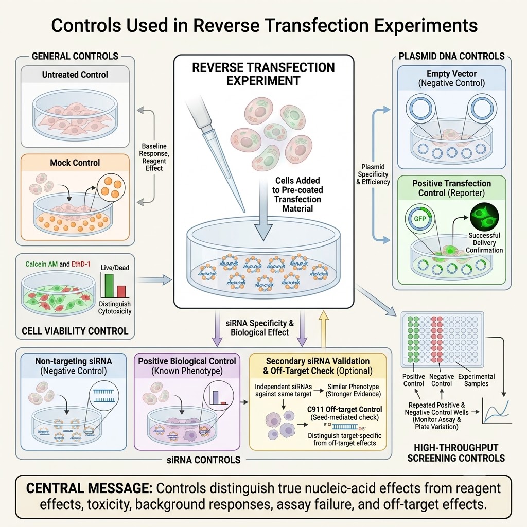
- Untreated Control
- It contains cells that do not receive the transfection reagent or nucleic acid.
- This control shows the normal condition of the cells without transfection.
- Changes in growth, viability or phenotype can be compared with this group. Untreated cells are commonly included while evaluating transfection-associated effects.
- Mock Transfection Control
- In this control, cells receive the transfection reagent but no experimental nucleic acid is added.
- It is used to determine whether the reagent or transfection procedure itself produces an effect.
- Mock or sham controls have also been included in reverse-transfection RNAi experiments.
- Non-Targeting siRNA Control
- This is one of the major negative controls during siRNA reverse transfection.
- A non-targeting siRNA is processed through the same transfection procedure but is not intended to silence the experimental target gene.
- It provides a background for comparison with gene-specific siRNA. Genome-wide reverse-transfection screens commonly include such negative-control wells.
- The control sequence should also be selected carefully. Some apparently non-targeting or scrambled siRNAs can produce unintended cellular effects.
- Empty Vector Control
- For plasmid DNA reverse transfection, an empty plasmid vector can be used as the negative control.
- It contains the vector backbone but lacks the experimental gene insert.
- This helps to distinguish the effect of the expressed gene from effects caused by the plasmid backbone and transfection itself. Empty-vector transfected cells have been used as controls in reverse-transfection cell-array studies.
- Positive Transfection Control
- A known transfectable nucleic acid can be included to check whether nucleic acid delivery is working.
- A fluorescent reporter or another detectable transfection indicator is often suitable for this purpose.
- Such a control helps identify a failure in transfection separately from absence of the expected biological effect. Fluorescent transfection indicators have been used for this purpose in cultured-cell RNAi methods.
- Positive Biological Control
- This control produces a known biological response after successful transfection.
- For an siRNA viability screen, an siRNA targeting an essential gene can serve as a positive control. PLK1, UBB or UBC have been used as positive controls in published reverse-transfection screens, depending on the assay.
- The positive control is assay dependent. It should produce a clear and expected phenotype.
- Cell Viability Control
- Transfection reagent and nucleic acid treatment can sometimes reduce cell viability independently of the intended gene effect.
- Cell number or viability is therefore checked along with the experimental endpoint when cytotoxicity can interfere with the result.
- This is especially important in siRNA screening because transfection itself can produce growth-related effects.
- Plate Positive and Negative Controls
- During high-throughput reverse transfection, positive and negative control wells are normally included on each screening plate.
- These controls are used to monitor plate-to-plate variation and whether the assay can separate a real positive response from the negative background.
- Published 384-well reverse-transfection screens have placed several positive and negative control wells on every plate.
- Independent siRNA Validation Control
- A phenotype obtained with one siRNA should not immediately be considered a gene-specific effect.
- Candidate genes from a primary screen are commonly tested again using additional independent siRNAs against the same target.
- Similar phenotypes from independent sequences give stronger evidence that the result is related to knockdown of the intended gene rather than an off-target effect.
- Off-Target Control
- Additional sequence controls can be used when specificity of an siRNA phenotype is important.
- For example, the C911 control changes bases in the central region of an siRNA while retaining much of its seed-region sequence. It has been developed to help distinguish target-specific effects from seed-dependent off-target effects.
- This is mainly a validation control and is not required in every basic reverse-transfection experiment.
Advantages of Reverse Transfection
The following are some of the important advantages of reverse transfection–
- Suitable for high-throughput screening
A large number of DNA or siRNA samples can be tested at the same time. - Allows parallel transfection
Different nucleic acids can be transfected in separate wells or different positions of an array. - Compatible with multiwell plates
It can be performed in 96-well, 384-well and other miniaturized plate formats. - Easy to miniaturize
Large numbers of transfection experiments can be arranged in a small area. - Requires less reagent in cell arrays
Solid-phase methods use very small amounts of DNA, siRNA and other reagents. - Suitable for automation
Cell addition, reagent dispensing and analysis can be carried out using automated systems. - Allows localized transfection
In solid-phase reverse transfection, cells are mainly transfected over the area containing the deposited nucleic acid. - Useful for high-content screening
Transfected cells can be studied by fluorescence microscopy and imaging-based assays. - Used for gene expression and gene silencing
Plasmid DNA can be used for gene overexpression, while siRNA is used for gene knockdown. - Useful in functional genomics
Different genes can be studied separately to determine their effects on cellular functions and phenotype.
Limitations and Disadvantages of Reverse Transfection
The following are some of the important limitations of reverse transfection–
- Cell-type dependent efficiency
Transfection efficiency can vary considerably between cell types. Primary cells and some difficult-to-transfect cells may give poor results. - Requires optimization
Cell density, nucleic acid amount and transfection conditions need optimization. One condition does not work equally for all cells. - Possible cytotoxicity
Excess transfection reagent can reduce cell viability. This is especially important with lipid-based transfection. - Variable transfection efficiency
Reverse-transfection efficiency is not always uniform. Standard reverse-transfection arrays have shown considerable variation between experimental systems. - Off-target effects in siRNA screening
Higher siRNA concentrations can increase unwanted off-target effects. Low siRNA and lipid concentrations are therefore preferred during optimization. - Cross-contamination between array spots
In open solid-phase arrays, neighbouring transfection spots are not physically separated. Movement of cells or material can therefore cause cross-contamination. - Problem with highly motile cells
Traditional reverse-transfection cell arrays work better with cells having limited movement. Highly migrating cells can move away from the original transfection spot. - Limited use with some non-adherent cells
Conventional solid-phase cell arrays mainly depend on cell attachment to the surface. Thus, poorly adherent or suspension cells can be difficult to use without modification of the system. - Surface conditions are important
In solid-phase reverse transfection, cell attachment and transfection depend on the surface, matrix and printed transfection mixture. These conditions may require separate optimization for different cells. - Long-term experiments can be difficult on open arrays
Lack of physical separation between cell spots makes control of the local cell environment more difficult. Specialized microfluidic systems have been developed to reduce this problem. - Requires validation in screening experiments
A phenotype obtained after siRNA reverse transfection may result from incomplete knockdown or non-specific effects. Suitable controls and independent validation are therefore required.
Some of these limitations are mainly related to solid-phase reverse-transfection arrays and are not equally present in liquid multiwell reverse transfection. The methods and common experimental pitfalls are also covered in Cell-Based Microarrays: Methods and Protocols.
Applications of Reverse Transfection
The following are some of the important applications of reverse transfection–
- High-throughput screening
A large number of DNA or siRNA samples can be tested in parallel. - Functional genomics
It is used to study the functions of different genes by gene overexpression or gene silencing. - siRNA screening
Reverse transfection is widely used for RNA interference (RNAi) screening and identification of genes producing a particular phenotype. - Gene overexpression studies
Different plasmid DNA or cDNA constructs can be expressed in separate groups of cells. - High-content screening
Reverse-transfected cell arrays are used with automated microscopy for analysis of cellular phenotypes. - Protein-protein interaction studies
Cell arrays can be combined with mammalian two-hybrid assays to study protein-protein interactions. - Reporter gene studies
Reporter plasmids can be reverse transfected to study promoter activity and gene expression in living cells. - Cellular phenotype screening
Changes in apoptosis, cell growth and other cellular functions can be detected after transfection of different genes. - Live-cell imaging
Reverse-transfected cells can be continuously observed to study changes in gene expression and cellular responses with time. - Synthetic biology studies
Reverse-transfection platforms can be used for testing synthetic gene circuits and different genetic constructs. - Miniaturized cell-based assays
It is used in cell microarrays and microfluidic systems, where many independent experiments can be carried out in a small area.
Solid-Phase Reverse Transfection and Transfected Cell Microarrays
- Solid-phase reverse transfection is a form of reverse transfection where nucleic acid-transfection material is deposited on a solid surface before cells are added. Glass slides are commonly used for making these arrays.
- The DNA, siRNA or other transfection material is printed at defined positions on the surface. These spots are allowed to remain attached or dried before cell seeding.
- A suspension of cells is then spread over the prepared surface. Cells growing above the printed regions come in contact with the transfection material and become transfected.
- Each printed spot can contain a different plasmid DNA or siRNA. Therefore, separate groups of cells on the same surface can express or silence different genes.
- The resulting arrangement is called a transfected cell microarray (TCM). It contains many spatially separated clusters of transfected cells on a single solid support.
- Transfected cell microarrays were first demonstrated by Ziauddin and Sabatini in 2001 using expression plasmids containing different cDNAs. They used the method for parallel analysis of gene function in mammalian cells.
- The same solid-phase principle was later adapted for siRNA arrays. These arrays allow many genes to be knocked down separately and are useful for genome-wide RNA interference screening.
- The method can be combined with immunofluorescence, automated microscopy and time-lapse imaging. Cellular morphology, protein localization and other phenotypes can then be examined.
- Transfected cell microarrays permit a large number of experiments on a small surface. They also reduce the amount of nucleic acid and transfection reagent required in many array formats.
- The efficiency of solid-phase transfection depends on cell type, surface coating, nucleic acid concentration, transfection reagent and composition of the printed mixture. These conditions need optimization before large-scale screening.
Forward Transfection vs Reverse Transfection
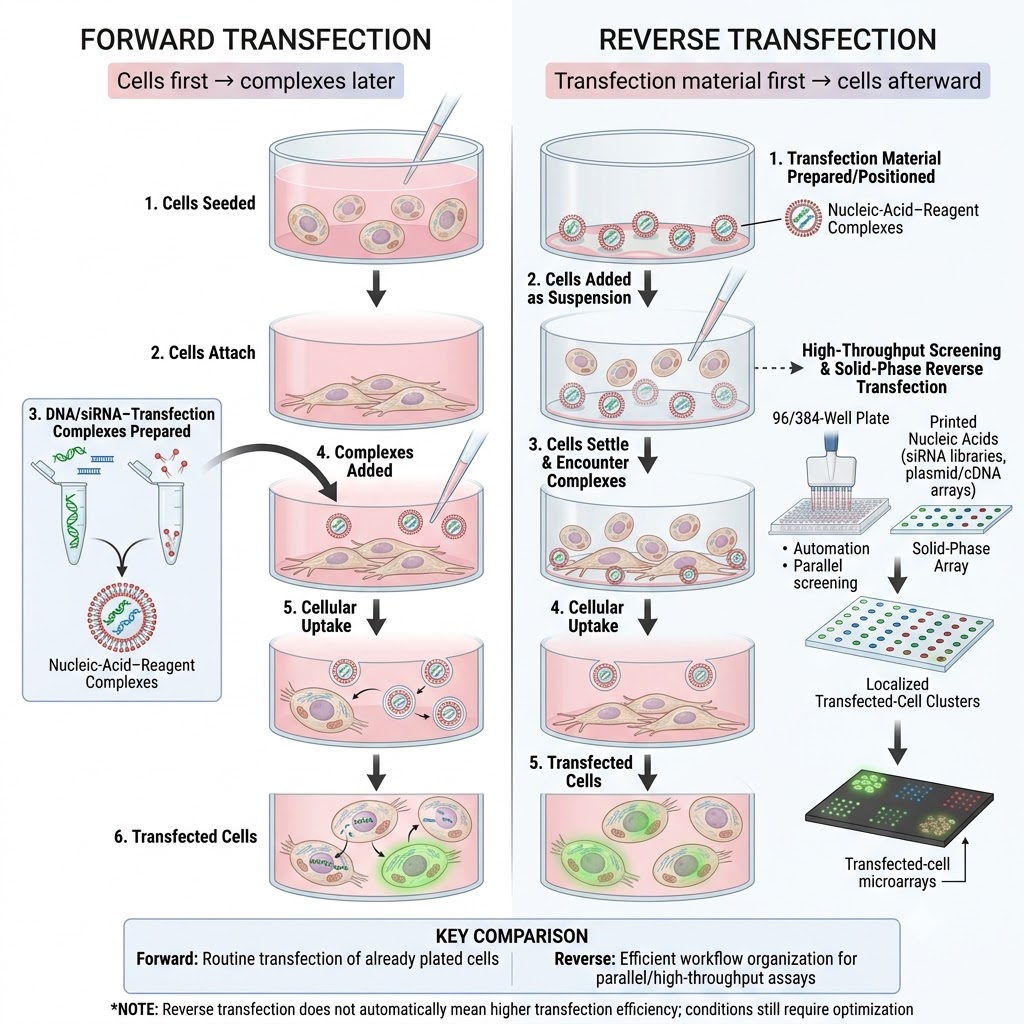
The major difference between forward transfection and reverse transfection is the order in which cells and transfection material are added. In reverse transfection, this normal order is reversed. Ziauddin and Sabatini originally used this difference for naming the method.
| Basis | Forward Transfection | Reverse Transfection |
|---|---|---|
| Definition | A conventional transfection method where cells are generally seeded first. The transfection complexes are added afterward. | A transfection method where the transfection material is prepared or positioned first. Cells are then added to it. |
| Order of addition | Cells first → transfection complexes later. | Transfection material first → cells afterward. This reversed order gives the method its name. |
| Cell condition during transfection | Cells are usually already attached to the culture surface when complexes are added. | Cells are generally added as a suspension and encounter the complexes during or near the time of seeding. |
| Cell seeding | Cell seeding and transfection are separate operations. | Cell seeding and exposure to transfection complexes take place together or very close in time. |
| Complex preparation | DNA/siRNA-reagent complexes are prepared and then added to previously seeded cells. | Complexes are prepared in the wells, or transfection material is deposited on a surface, before cells are added. |
| Cell attachment | Cells commonly attach before transfection begins. | Cells can interact with the complexes while settling and attaching to the plate. |
| Basic workflow | Cell preparation → cell seeding → attachment → complex formation → addition of complexes. | Cargo/reagent preparation → complex formation or deposition → cell addition → attachment and transfection. |
| Time between plating and transfection | Usually requires a separate period between cell plating and transfection. | A separate pre-plating period is generally avoided because cells are added directly to the prepared complexes. |
| Multiwell format | Can be performed in multiwell plates. | Particularly suitable for 96-well and 384-well plates, where siRNAs or other cargo can be pre-arranged before cell addition. |
| High-throughput screening | Possible, but requires handling of already seeded cells followed by another transfection step. | Commonly used for high-throughput RNAi screening because cargo can be pre-arrayed and cells are then dispensed across the plate. |
| Automation | Can be automated, but cell plating and later transfection remain separate operations. | Well suited for automated screening because pre-arrayed cargo, reagent dispensing and cell addition can be organized in the same plate workflow. |
| Solid-phase format | It is not the usual arrangement of conventional forward transfection. | Can be carried out as solid-phase reverse transfection, where DNA or siRNA material is printed or deposited on a surface before cells are seeded. |
| Transfected cell microarrays | Not the basic method used for making classical transfected-cell microarrays. | It forms the basis of transfected cell microarrays, where different nucleic acids are printed at defined positions and cells are cultured over them. |
| Localized transfection | Transfection normally takes place throughout the treated well or culture area. | Solid-phase reverse transfection can produce localized groups of transfected cells over individual printed spots. |
| Use with plasmid DNA | Commonly used for transfection of plasmid DNA into previously cultured cells. | Plasmid DNA can be reverse transfected. The original transfected-cell microarray used expression plasmids containing defined cDNAs. |
| Use with siRNA | siRNA complexes are added to already plated cells. | Widely used for siRNA library screening, particularly in 96-well and 384-well formats. |
| Reagent consumption | Depends on plate size and experimental format. | Solid-phase microarray versions can use much smaller amounts of nucleic acid and reagent because individual transfection spots are very small. |
| Miniaturization | Can be miniaturized in multiwell systems. | Especially suitable for high-density arrays. Thousands of separate reverse-transfection spots can be arranged on a single slide. |
| Main advantage | Simple and well established for routine transfection of already cultured cells. | Useful when many different nucleic acids have to be tested in parallel, especially functional genomics and RNAi screening. |
| Main limitation | Requires handling of cells again after they have been seeded. | Efficiency remains dependent on cell type, cargo and reagent conditions. Solid-phase arrays can also face spot cross-contamination and cell migration. |
| Transfection efficiency | Efficiency depends on cell type, reagent, cargo amount and experimental conditions. | Reverse transfection does not automatically give higher efficiency. Its conditions also have to be optimized for each experimental system. |
| Best suited for | Routine transfection, smaller experiments and experiments using already plated cells. | High-throughput screening, siRNA libraries, cell microarrays, gene overexpression arrays and other parallel cell-based assays. |
In simple form, the difference is-
Forward transfection: Cells are seeded first → transfection complexes are added later.
Reverse transfection: Transfection material is prepared first → cells are added afterward.
References
- Bian, S., Zhou, Y., Hu, Y., Cheng, J., Chen, X., Xu, Y., & Liu, P. (2017). High-throughput in situ cell electroporation microsystem for parallel delivery of single guide RNAs into mammalian cells. Scientific Reports, 7, 42512. https://doi.org/10.1038/srep42512
- Buehler, E., Chen, Y.-C., & Martin, S. (2012). C911: A bench-level control for sequence specific siRNA off-target effects. PLoS ONE, 7(12), e51942. https://doi.org/10.1371/journal.pone.0051942
- Chang, K., Elledge, S. J., & Hannon, G. J. (2012). RNAi in cultured mammalian cells using synthetic siRNAs. Cold Spring Harbor Protocols, 2012(9), 957–961. https://doi.org/10.1101/pdb.prot071076
- Cheng, A., Gonçalves, K. A., & Golomb, M. (2011). Optimization of transfection conditions and analysis of siRNA potency using real-time PCR. In S. R. Mitra (Ed.), RNA interference: Methods and protocols (Methods in Molecular Biology, Vol. 764, pp. 199–213). Humana Press. https://doi.org/10.1007/978-1-61779-188-8_13
- Daga, N., Eicher, S., Kannan, A., Larsson, E., & Lehmann, K. V. (2018). Growth-restricting effects of siRNA transfections: A largely deterministic combination of off-target binding and hybridization-independent competition. Nucleic Acids Research, 46(18), 9309–9320. https://doi.org/10.1093/nar/gky798
- Echeverri, C. J., & Perrimon, N. (2006). High-throughput RNAi screening in cultured cells: A user’s guide. Nature Reviews Genetics, 7(5), 373–384. https://doi.org/10.1038/nrg1836
- Erfle, H., Neumann, B., Liebel, U., Rogers, P., Held, M., Walter, T., Ellenberg, J., & Pepperkok, R. (2007). Reverse transfection on cell arrays for high content screening microscopy. Nature Protocols, 2(2), 392–399. https://doi.org/10.1038/nprot.2006.483
- Erfle, H., Neumann, B., Rogers, P., Bulkescher, J., Ellenberg, J., & Pepperkok, R. (2008). Work flow for multiplexing siRNA assays by solid-phase reverse transfection in multiwell plates. Journal of Biomolecular Screening, 13(6), 575–580. https://doi.org/10.1177/1087057108320133
- Fiebitz, A., Nyarsik, L., Haendler, B., Hu, Y.-H., Wagner, F., Thamm, S., Lehrach, H., Janitz, M., & Vanhecke, D. (2008). High-throughput mammalian two-hybrid screening for protein-protein interactions using transfected cell arrays. BMC Genomics, 9, 68. https://doi.org/10.1186/1471-2164-9-68
- Fujita, S., Ota, E., Sasaki, C., Takano, K., Miyake, M., & Miyake, J. (2007). Highly efficient reverse transfection with siRNA in multiple wells of microtiter plates. Journal of Bioscience and Bioengineering, 104(4), 329–333. https://doi.org/10.1263/jbb.104.329
- Henderson, M. C., Gonzales, I. M., Arora, S., Choudhary, A., Trent, J. M., Von Hoff, D. D., Mousses, S., & Azorsa, D. O. (2013). High-throughput RNAi screening for the identification of novel targets for cancer therapy. Methods in Molecular Biology, 986, 87–98. https://doi.org/10.1007/978-1-62703-311-4_6
- Hu, Y.-H., Warnatz, H.-J., Vanhecke, D., Wagner, F., Fiebitz, A., Thamm, S., Kahlem, P., Lehrach, H., Yaspo, M.-L., & Janitz, M. (2006). Cell array-based intracellular localization screening reveals novel functional features of human chromosome 21 proteins. BMC Genomics, 7, 155. https://doi.org/10.1186/1471-2164-7-155
- Klanert, G., Fernandez, D. J., Weinguny, M., Eisenhut, P., Bühler, E., Melcher, M., Titus, S. A., Borth, N., & Grillari, J. (2019). A cross-species whole genome siRNA screen in suspension-cultured Chinese hamster ovary cells identifies novel engineering targets. Scientific Reports, 9, 8689. https://doi.org/10.1038/s41598-019-45159-2
- Li, N., Sun, J., Benet, Z. L., Wang, Z., Al-Khodor, S., John, S. P., Lin, B., Sung, M.-H., & Fraser, I. D. C. (2015). Development of a cell system for siRNA screening of pathogen responses in human and mouse macrophages. Scientific Reports, 5, 9559. https://doi.org/10.1038/srep09559
- Munkonge, F. M., Amin, V., Hyde, S. C., Green, A.-M., Pringle, I. A., Gill, D. R., & Colledge, W. H. (2003). Emerging significance of plasmid DNA nuclear import in gene therapy. Advanced Drug Delivery Reviews, 55(6), 749–760. https://doi.org/10.1016/S0169-409X(03)00050-4
- Neumann, B., Held, M., Liebel, U., Erfle, H., Rogers, P., Pepperkok, R., & Ellenberg, J. (2006). High-throughput RNAi screening by time-lapse imaging of live human cells. Nature Methods, 3(5), 385–390. https://doi.org/10.1038/nmeth876
- Ooi, A., Wong, A., Esau, L., Lemtiri-Chlieh, F., & Gehring, C. (2016). A guide to transient expression of membrane proteins in HEK-293 cells for functional characterization. Frontiers in Physiology, 7, 300. https://doi.org/10.3389/fphys.2016.00300
- Ovcharenko, D., Jarvis, R., Hunicke-Smith, S., Kelnar, K., & Brown, D. (2005). High-throughput RNAi screening in vitro: From cell lines to primary cells. RNA, 11(6), 985–993. https://doi.org/10.1261/rna.7288405
- Pai, G., Chattopadhyay, S., Hoebe, R. A., van Beusechem, V. W., Brakenhoff, R. H., & Leemans, C. R. (2023). Genome-wide siRNA screens identify RBBP9 function as a critical determinant of survival in Fanconi anemia pathway-deficient head and neck cancer. Communications Biology, 6, 21. https://doi.org/10.1038/s42003-022-04389-3
- Palmer, E. L. (Ed.). (2011). Cell-based microarrays: Methods and protocols (Methods in Molecular Biology, Vol. 706). Humana Press. https://doi.org/10.1007/978-1-61737-970-3
- Palmer, E. L., Miller, A. D., & Freeman, T. C. (2006). Identification and characterisation of human apoptosis inducing proteins using cell-based transfection microarrays and expression analysis. BMC Genomics, 7, 145. https://doi.org/10.1186/1471-2164-7-145
- Rajan, S., Djambazian, H., Pham Dang, H. C., Sladek, R., & Hudson, T. J. (2011). The living microarray: A high-throughput platform for measuring transcription dynamics in single cells. BMC Genomics, 12, 115. https://doi.org/10.1186/1471-2164-12-115
- Rantala, J. K., Mäkelä, R., Aaltola, A.-R., Laasola, P., Mpindi, J.-P., Nees, M., Saviranta, P., & Kallioniemi, O. (2011). A cell spot microarray method for production of high density siRNA transfection microarrays. BMC Genomics, 12, 162. https://doi.org/10.1186/1471-2164-12-162
- Shiokawa, M., Miura, R., Okubo, A., Hagita, Y., Yoshimura, I., & Aoki, H. (2021). Bovine endometrium-derived cultured cells are suitable for lipofection. Scientific Reports, 11, 16207. https://doi.org/10.1038/s41598-021-95848-0
- Silva, J. M., Mizuno, H., Brady, A., Lucito, R., & Hannon, G. J. (2004). RNA interference microarrays: High-throughput loss-of-function genetics in mammalian cells. Proceedings of the National Academy of Sciences of the United States of America, 101(17), 6548–6552. https://doi.org/10.1073/pnas.0400165101
- Stojic, L., Lun, A. T. L., Mascalchi, P., Ernst, C., Redmond, A. M., Mangei, J., Barr, A. R., Bousgouni, V., Bakal, C., Marioni, J. C., Odom, D. T., & Gergely, F. (2020). A high-content RNAi screen reveals multiple roles for long noncoding RNAs in cell division. Nature Communications, 11, 1851. https://doi.org/10.1038/s41467-020-14978-7
- Sun, J., Katz, S., Dutta, B., Wang, Z., & Fraser, I. D. C. (2017). Genome-wide siRNA screen of genes regulating the LPS-induced TNF-α response in human macrophages. Scientific Data, 4, 170007. https://doi.org/10.1038/sdata.2017.7
- Tan, S., Tao, Z., Loo, S., Su, L., Chen, X., & Ye, L. (2019). Non-viral vector based gene transfection with human induced pluripotent stem cells derived cardiomyocytes. Scientific Reports, 9, 14404. https://doi.org/10.1038/s41598-019-50980-w
- Tschuch, C., Schulz, A., Pscherer, A., Werft, W., Benner, A., Hotz-Wagenblatt, A., Barrionuevo, L. S., Lichter, P., & Mertens, D. (2008). Off-target effects of siRNA specific for GFP. BMC Molecular Biology, 9, 60. https://doi.org/10.1186/1471-2199-9-60
- Villa-Diaz, L. G., Garcia-Perez, J. L., Krebsbach, P. H., & Smith, G. D. (2010). Enhanced transfection efficiency of human embryonic stem cells by the incorporation of DNA liposomes in extracellular matrix. Stem Cells and Development, 19(12), 1949–1957. https://doi.org/10.1089/scd.2009.0505
- Williams, S. P., Barthorpe, A. S., Lightfoot, H., Garnett, M. J., & McDermott, U. (2017). High-throughput RNAi screen for essential genes and drug synergistic combinations in colorectal cancer. Scientific Data, 4, 170139. https://doi.org/10.1038/sdata.2017.139
- Woodruff, K., & Maerkl, S. J. (2016). A high-throughput microfluidic platform for mammalian cell transfection and culturing. Scientific Reports, 6, 23937. https://doi.org/10.1038/srep23937
- Ziauddin, J., & Sabatini, D. M. (2001). Microarrays of cells expressing defined cDNAs. Nature, 411(6833), 107–110. https://doi.org/10.1038/35075114