

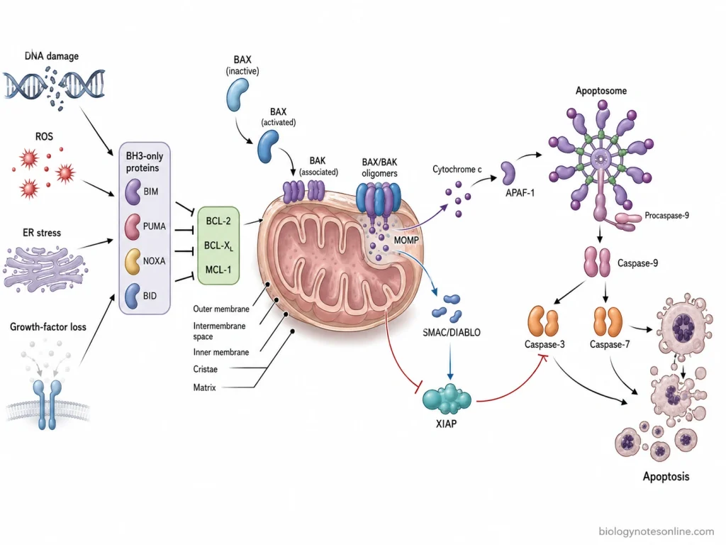
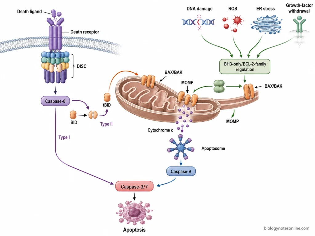
The intrinsic pathway of apoptosis is a type of programmed cell death which takes place through the mitochondria. It is also known as the mitochondrial pathway of apoptosis. DNA damage, endoplasmic reticulum (ER) stress, deprivation of growth factors and other severe cellular stresses can activate this pathway. During this process, the pro-apoptotic BH3-only proteins become active.
These proteins regulate the members of the BCL-2 protein family. Some of the important anti-apoptotic proteins are BCL-2, BCL-XL and MCL-1, which are inhibited, while BAX and BAK become activated. BAX and BAK then form oligomers in the outer mitochondrial membrane. As a result, permeability of the mitochondrial outer membrane increases.
This is referred to as mitochondrial outer membrane permeabilization (MOMP). After this, cytochrome c and other apoptotic protein such as SMAC/DIABLO are released from the mitochondrial intermembrane space into the cytoplasm. Cytochrome c combines with APAF-1 in the presence of dATP.
A large multiprotein complex is formed, which is called the apoptosome. The apoptosome recruits procaspase-9 and activates it to form caspase-9. Caspase-9 now activates the executioner caspases, mainly caspase-3 and caspase-7. These caspases act on different cellular proteins and cause their cleavage.
The characteristic changes of apoptosis are produced and finally death of the cell occurs. SMAC/DIABLO also takes part in this process. It inhibits the inhibitor of apoptosis proteins (IAPs), which normally restrict the activity of caspases.
What Triggers the Intrinsic Apoptotic Pathway?
The intrinsic apoptotic pathway is activated due to different stress conditions produced inside the cell. The following are some of the important triggers-
- DNA damage- The major trigger of intrinsic apoptosis is severe damage of DNA. It may occur due to ionizing radiation, errors during DNA replication or DNA-damaging agents. If the damage is not repaired, apoptotic proteins are activated. p53-associated proteins such as PUMA can also take part during this process.
- Growth factor withdrawal- Growth factors and cytokines are required for survival of many cells. Their removal causes loss of the normal survival signals. During this condition, BH3-only proteins can become active and changes occur in the activity of BCL-2 family proteins, leading to intrinsic apoptosis.
- Oxidative stress- It is caused by excess formation of reactive oxygen species (ROS) inside the cell. ROS can damage DNA and proteins and also affect different cellular components. Mitochondrial function is also disturbed when oxidative stress becomes severe.
- Endoplasmic reticulum (ER) stress- Accumulation of improperly folded proteins or disturbance in normal functioning of ER causes ER stress. Different mechanisms are initially activated by the cell for correcting this condition. If the stress remains for a longer period and cannot be corrected, pro-apoptotic signaling is started and mitochondrial apoptosis can occur.
- Replication stress and mitotic defects- Abnormal DNA replication is another trigger of the intrinsic pathway. Defects during mitosis and alteration of microtubules can also produce cellular stress. Severe and prolonged defects can activate the apoptotic process.
- Hypoxia, nutrient deprivation and calcium overload- Low level of oxygen and deficiency of nutrients disturb the normal metabolism of the cell. Excess intracellular calcium also affects mitochondrial homeostasis. These conditions when become severe can act as internal signals for activation of the intrinsic apoptotic pathway.
Key Molecular Components of the Intrinsic Pathway
Some of the important molecular components of the intrinsic apoptotic pathway are given below-
| Molecular group | Major components | Cellular location | Role in the pathway |
|---|---|---|---|
| BH3-only proteins | BIM, BID, PUMA, NOXA, BAD, BMF | Cytoplasm and different cellular membranes | These are pro-apoptotic members of the BCL-2 family. They become active during cellular stress and inhibit anti-apoptotic BCL-2 proteins or help in activation of BAX and BAK. |
| Anti-apoptotic BCL-2 proteins | BCL-2, BCL-XL, MCL-1 | Mainly outer mitochondrial membrane and other intracellular membranes | These proteins prevent apoptosis by controlling the pro-apoptotic proteins. BAX and BAK activation is inhibited by them. |
| BAX and BAK | BAX, BAK | BAX occurs mainly in cytoplasm before activation, while BAK is present in outer mitochondrial membrane | They are the major pore-forming proteins of this pathway. After activation, BAX and BAK oligomerize in the outer mitochondrial membrane. This results in mitochondrial outer membrane permeabilization (MOMP). |
| Mitochondrial apoptotic proteins | Cytochrome c, SMAC/DIABLO | Mitochondrial intermembrane space. Released into cytoplasm after MOMP | Cytochrome c is required for formation of apoptosome. SMAC/DIABLO inhibits inhibitor of apoptosis proteins (IAPs), allowing caspase activity to continue. |
| Apoptosome components | APAF-1, cytochrome c, dATP and procaspase-9 | Cytoplasm | Cytochrome c binds with APAF-1 and the apoptosome is formed. Procaspase-9 is recruited into this complex and becomes activated. |
| Initiator caspase | Caspase-9 | Cytoplasm, associated with apoptosome during its activation | Caspase-9 is the main initiator caspase of intrinsic pathway. It activates downstream executioner caspases. |
| Executioner caspases | Caspase-3 and caspase-7 | Cytoplasm | These caspases are activated by caspase-9. They cleave different cellular proteins during the apoptotic process. |
| Inhibitor of apoptosis proteins (IAPs) | XIAP | Cytoplasm | XIAP restricts the activity of caspase-9, caspase-3 and caspase-7. Its inhibitory action is reduced by SMAC/DIABLO released from mitochondria. |
Step-by-Step Mechanism of the Intrinsic Pathway of Apoptosis
The intrinsic pathway of apoptosis is a mitochondrial pathway which is activated by different stress produced inside the cell. BCL-2 family proteins, mitochondrial proteins and caspases take part in the process. The steps are as follows-
Step 1- Cellular stress
The first step is the production of intracellular stress signals. DNA damage, oxidative stress, deprivation of growth factors and other severe cellular damages can activate the pathway. During this process, different BH3-only proteins such as BIM, PUMA, NOXA and BID become active.
Step 2- Regulation of BCL-2 family
The BCL-2 protein family contains both anti-apoptotic and pro-apoptotic proteins. BCL-2, BCL-XL and MCL-1 normally inhibit apoptosis, whereas BAX and BAK promote mitochondrial membrane permeabilization. In this step, BH3-only proteins inhibit the anti-apoptotic members and allow BAX and BAK to become activated.
Step 3- Activation of BAX and BAK
After activation, BAX and BAK accumulate in the outer membrane of mitochondria. They form oligomers, resulting in formation of pores in the membrane. The mitochondrial outer membrane now becomes permeable. This process is referred to as mitochondrial outer membrane permeabilization (MOMP).
Step 4- Release of mitochondrial proteins
MOMP causes release of proteins present in the mitochondrial intermembrane space into the cytoplasm. The major protein released is cytochrome c. SMAC/DIABLO is also released during this process.
Step 5- Apoptosome formation
Cytochrome c after its release combines with APAF-1 (apoptotic protease-activating factor-1) in the presence of ATP or dATP. Several APAF-1 molecules then assemble together. A large multiprotein complex called the apoptosome is formed, which provides the site for recruitment of procaspase-9.
Step 6- Caspase-9 activation
In this step, procaspase-9 present in the apoptosome is activated into caspase-9. It is the initiator caspase of intrinsic pathway.
Caspase-9 further acts on the executioner caspases, mainly caspase-3 and caspase-7, resulting in their activation.
Step 7- Action of executioner caspases
The activated caspase-3 and caspase-7 cleave different cellular proteins required for normal structure and functioning of the cell. SMAC/DIABLO also takes part in the process by inhibiting inhibitor of apoptosis proteins (IAPs), particularly XIAP, which normally restrict the caspase activity. As the cleavage of cellular proteins continues, normal cellular structure and functions are lost and characteristic changes of apoptosis are produced.
Mitochondrial Outer Membrane Permeabilization as the Commitment Step
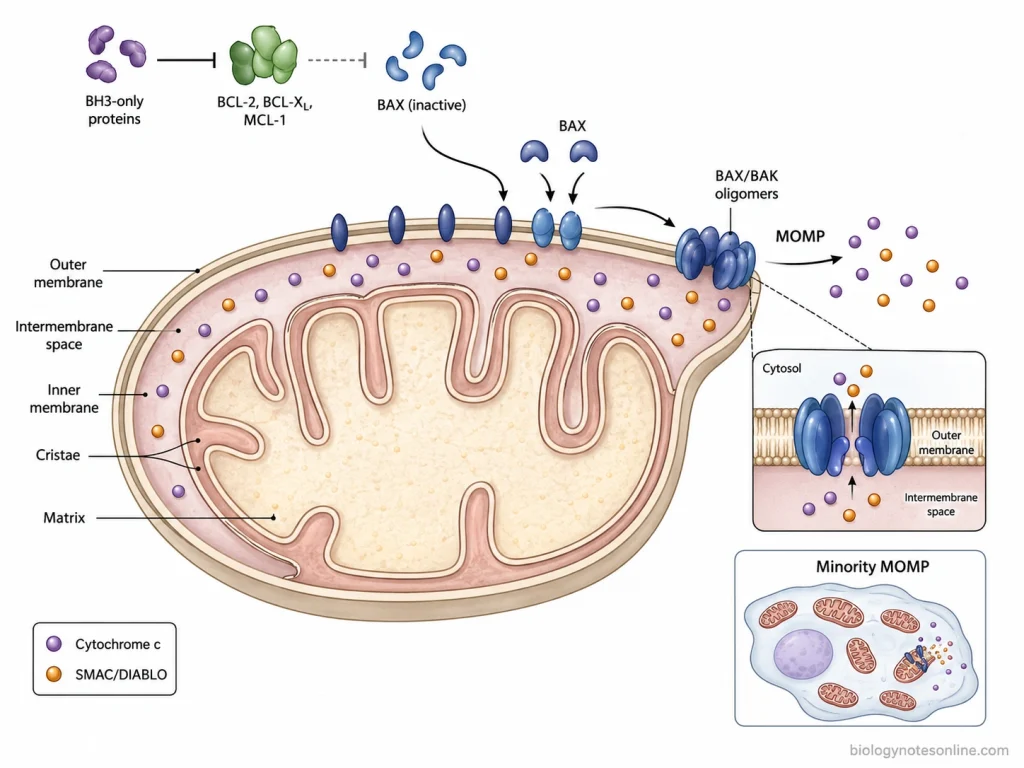
Mitochondrial outer membrane permeabilization (MOMP) is the process where permeability of the outer mitochondrial membrane is increased during intrinsic apoptosis. It is mainly controlled by the proteins of BCL-2 family. This step is important because after extensive MOMP, the cell is generally committed to apoptotic death.
During cellular stress, the BH3-only proteins become active. These proteins affect the balance between pro-apoptotic and anti-apoptotic members of BCL-2 family, where BCL-2, BCL-XL and MCL-1 normally act to prevent the mitochondrial apoptotic process. Their activity is inhibited. BAX and BAK are now allowed to become active.
The activated BAX and BAK accumulate in the outer mitochondrial membrane. Oligomerization takes place and openings are formed in the membrane. The mitochondrial outer membrane therefore becomes permeable. This is referred to as MOMP.
After this process, proteins of mitochondrial intermembrane space are released into the cytoplasm. The major one is cytochrome c. SMAC/DIABLO is also released. Cytochrome c is used for formation of apoptosome and activation of caspase-9, while SMAC/DIABLO inhibits IAP proteins which normally restrict the activity of caspases.
MOMP is considered as the commitment step or point of no return of intrinsic apoptosis. Once a large number of mitochondria become permeabilized, cytochrome c has already entered into cytoplasm and the caspase process can proceed. Normal mitochondrial functions are also affected severely. Because of this, even blocking of caspase activity may not always save the cell after extensive MOMP.
However, MOMP does not necessarily occur in every mitochondrion of the cell. Sometimes only a small population of mitochondria becomes permeabilized, whereas the remaining mitochondria stay functional. This is called incomplete or minority MOMP. In such condition, recovery of the cell may still be possible.
How Is the Intrinsic Apoptosis Pathway Regulated?
The intrinsic apoptosis pathway is regulated mainly by the proteins of BCL-2 family and their action on mitochondrial outer membrane. Some of these proteins prevent apoptosis. Some promote the process. Regulation also takes place after release of mitochondrial proteins, during activation of caspases. The following are some of the important regulatory mechanisms-
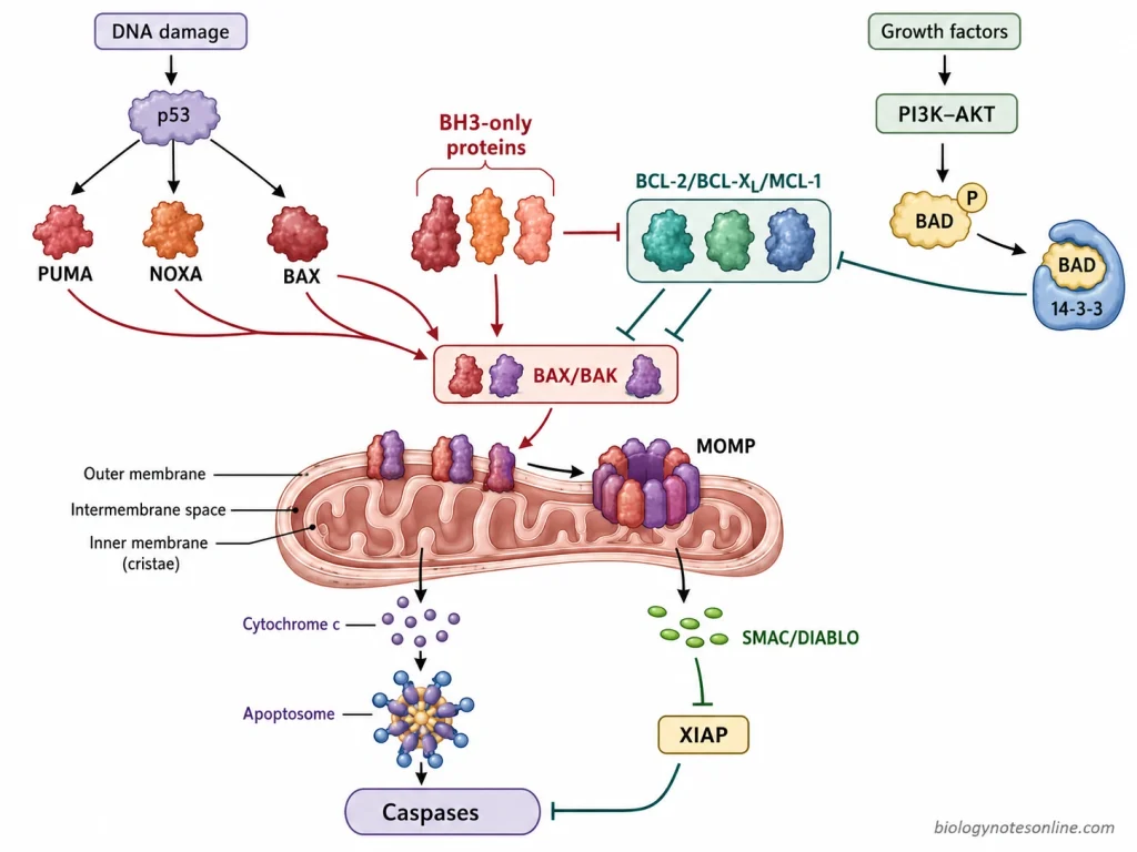
- BCL-2 family proteins- The major regulation before mitochondrial permeabilization is carried out by BCL-2 family proteins. BCL-2, BCL-XL and MCL-1 are anti-apoptotic members. They keep the activity of BAX and BAK under control and thus prevent mitochondrial apoptosis. BAX and BAK, on the other hand, are pro-apoptotic proteins required for MOMP. The relative activity of these two groups therefore controls whether the mitochondrial membrane will remain intact or become permeable.
- BH3-only proteins- BIM, BID, PUMA, NOXA and BAD are some of the important BH3-only proteins. These proteins are activated by different cellular stresses. Their major action is on the anti-apoptotic BCL-2 proteins. Some BH3-only proteins can also help in activation of BAX and BAK. During this process, the balance is shifted towards mitochondrial permeabilization.
- p53 regulation- p53 has an important role particularly during DNA damage. It can increase formation of pro-apoptotic proteins such as PUMA, NOXA and BAX. PUMA and NOXA then act through BCL-2 family proteins. When damage cannot be repaired properly, this regulation favors activation of intrinsic apoptosis.
- Growth and survival signals- Growth factors normally produce signals that help in survival of the cell. One important pathway involved is the PI3K-AKT pathway. Activated AKT phosphorylates BAD, a pro-apoptotic BH3-only protein. Phosphorylated BAD is retained by 14-3-3 proteins and its apoptotic activity becomes reduced. BCL-2 and BCL-XL can therefore continue their anti-apoptotic action.
- Regulation of BAX and BAK- In normal cells, pore-forming activity of BAX and BAK remains restricted. After sufficient apoptotic signals, this restriction is removed. The proteins become activated and oligomerization takes place at the outer mitochondrial membrane. MOMP is then produced, followed by release of cytochrome c.
- IAP proteins and SMAC/DIABLO- Regulation is also present after mitochondrial permeabilization. XIAP is an inhibitor of apoptosis protein (IAP) which inhibits caspase-9 and also the executioner caspases, caspase-3 and caspase-7. SMAC/DIABLO has opposite action. It is released from mitochondria during MOMP and inhibits the IAP proteins. Caspase activity can now continue.
Apoptosome Formation and the Caspase Cascade
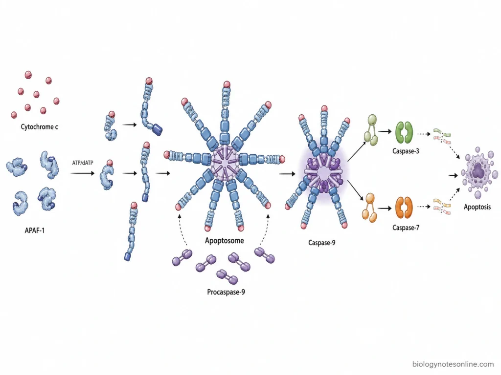
The apoptosome is a large protein complex which is formed in the cytoplasm during intrinsic apoptosis. Its major function is activation of caspase-9. After mitochondrial outer membrane permeabilization, cytochrome c comes out from mitochondrial intermembrane space into the cytoplasm.
Apoptosome formation
Cytochrome c binds with APAF-1 (apoptotic protease-activating factor-1) in the presence of ATP or dATP. APAF-1 before this remains in an inactive form. Binding of cytochrome c causes structural change in APAF-1 and nucleotide exchange takes place. Several APAF-1 molecules now join together.
A wheel-like heptameric complex is formed. This is referred to as the apoptosome.
The CARD regions of APAF-1 are located towards the central region of apoptosome. Procaspase-9 binds at this region.
Activation of caspase-9
In this step, procaspase-9 molecules are brought close together on the apoptosome. Caspase-9 is then activated mainly by their association and dimerization, rather than by cleavage alone. It is the initiator caspase of mitochondrial pathway.
Caspase cascade
The activated caspase-9 acts on the executioner procaspases. The major ones are caspase-3 and caspase-7. These procaspases are cleaved and converted into their active forms.
During this process, a series of proteolytic reactions starts inside the cell. Caspase-3 and caspase-7 act on different cellular proteins which are involved in cell structure and other normal functions of the cell. This stepwise activation of caspases is referred to as the caspase cascade.
Cellular Changes During the Execution Phase of Apoptosis
During the execution phase, activated executioner caspases act on different proteins of the cell and several structural changes are produced. The following are some of the important cellular changes-
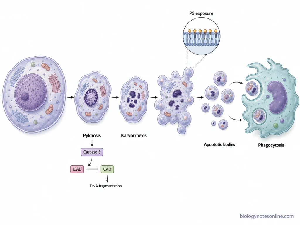
- Cell shrinkage- The apoptotic cell becomes smaller in size and loses its normal volume. Cytoplasm becomes dense, with the cellular organelles more closely packed together.
- Chromatin condensation- It is one of the characteristic changes of apoptosis. The nuclear chromatin becomes highly condensed and is commonly collected towards the inner side of nuclear membrane. This shrinkage and condensation of nucleus is referred to as pyknosis.
- DNA fragmentation- During this process, caspase-3 cleaves ICAD, which normally keeps caspase-activated DNase (CAD) inactive. CAD is now released and acts on nuclear DNA. The DNA is broken mainly at the regions between nucleosomes, producing the characteristic internucleosomal DNA fragments.
- Nuclear fragmentation- The condensed nucleus does not remain intact. It breaks into smaller nuclear fragments. This process is called karyorrhexis.
- Cytoskeletal breakdown and membrane blebbing- Executioner caspases cleave different cytoskeletal and associated proteins, which changes the normal shape and attachment of the cell. The cell becomes rounded. Its plasma membrane then forms repeated bulges or blebs, while membrane integrity is still largely maintained.
- Phosphatidylserine exposure- Phosphatidylserine (PS) is normally present mainly on the inner side of plasma membrane. During apoptosis, its distribution is changed and PS becomes exposed on the outer surface. Caspase-dependent activation of phospholipid scramblases and inhibition of flippases are involved in this change. The exposed PS acts as a recognition signal for removal of apoptotic cell.
- Formation of apoptotic bodies- With further membrane blebbing and cellular fragmentation, the cell breaks into small membrane-bound fragments called apoptotic bodies. These contain portions of cytoplasm, organelles and nuclear fragments.
- Removal of apoptotic cell- Apoptotic bodies are recognized and taken up by macrophages or neighboring cells. The cellular contents remain enclosed by membrane during their removal and therefore widespread leakage of intracellular materials usually does not occur. This is why apoptosis is generally removed without producing the marked inflammatory response seen with cell rupture.
Biological Significance of the Intrinsic Apoptotic Pathway
The intrinsic apoptotic pathway has different biological roles in maintaining normal cells and tissues. It removes the cells which are damaged, unwanted or no longer required. Some of the important functions are-
- Development of tissues and organs- During development, many cells are produced and some of them are not required in the mature tissue. These cells are removed by apoptosis. It helps in proper formation and shaping of different tissues and organs.
- Maintenance of tissue homeostasis- New cells are continuously formed in many tissues, whereas old and unwanted cells need to be removed. The apoptotic process maintains this cell number. Excess cells are eliminated without much disturbance of the surrounding tissue.
- Removal of damaged cells- Cells having severe DNA damage, oxidative stress or loss of survival signals may not be suitable for further survival. Such cells can be removed through the mitochondrial apoptotic pathway. This also prevents damaged genetic material from being passed to the daughter cells.
- Prevention of cancer- It is one of the important functions of intrinsic apoptosis. Cells having irreparable DNA damage or oncogenic stress are eliminated before their further multiplication. If this apoptotic control is lost, abnormal cells continue to survive and accumulate, which can support development of tumors.
- Regulation of immune cells- Intrinsic apoptosis also takes part during development and maintenance of lymphocytes. Unwanted or self-reactive lymphocytes are removed. After an immune response, excess immune cells which are no longer required can also undergo apoptosis. Disturbance of this process may result in abnormal accumulation of lymphocytes or autoimmune conditions.
- Balance between cell survival and death- The activity of BCL-2 family proteins helps in deciding whether a stressed cell will survive or undergo apoptosis. This balance is required for normal functioning of tissues. Less apoptosis can favor cancer and autoimmune conditions, while excessive loss of cells by apoptosis is associated with degenerative tissue damage.
Dysregulation of Intrinsic Apoptosis in Disease
The intrinsic apoptotic pathway needs proper balance between survival and death of the cell. Too little apoptosis allows abnormal cells to remain alive, whereas excess activation causes unnecessary loss of cells. Some of the important diseases associated with its dysregulation are-
- Cancer- Reduced intrinsic apoptosis is commonly seen in cancer. Tumor cells may have increased anti-apoptotic proteins such as BCL-2, BCL-XL and MCL-1, loss of pro-apoptotic signals, or defective p53 activity. The damaged cells which normally should undergo apoptosis can now survive and continue their multiplication. Resistance to anticancer treatment can also develop due to this increased apoptotic threshold.
- Autoimmune diseases- Intrinsic apoptosis is important for removal of unwanted and self-reactive lymphocytes. If these cells escape the normal death process, they can remain for longer period and accumulate. Abnormal expression of BCL-2 or loss of proteins such as BIM, BAX and BAK has been associated with failure of lymphocyte deletion and autoimmune conditions.
- Neurodegenerative diseases- In these conditions, the problem is generally excessive loss of cells rather than their survival. Mitochondrial dysfunction and different cellular stresses can activate the intrinsic pathway in neurons. Release of cytochrome c and activation of the caspase process causes neuronal cell loss, which has been implicated in different neurodegenerative disorders.
- Ischemia-reperfusion injury- During ischemia, cells are deprived of oxygen and nutrients. Reperfusion can further produce oxidative stress and mitochondrial damage. Intrinsic apoptosis is one of the cell death mechanisms activated during this process, particularly in cardiac and nervous tissues.
- Abnormal lymphocyte accumulation- The BCL-2-regulated pathway has an important role in controlling survival of lymphocytes. When this pathway is impaired, cells which have completed their function or are otherwise unwanted may fail to die. Excess lymphocyte survival can contribute to lymphoid malignancy as well as disorders of immune tolerance.
- Excessive tissue cell loss- Increased activation of mitochondrial apoptosis can also be harmful. Severe oxidative stress, mitochondrial damage and other intracellular stresses can produce activation of BAX and BAK followed by release of mitochondrial apoptotic proteins. When this occurs excessively, loss of functional cells contributes to tissue injury rather than normal homeostasis.
Intrinsic vs. Extrinsic Pathway of Apoptosis
| Features | Intrinsic Pathway | Extrinsic Pathway |
|---|---|---|
| Definition | It is a mitochondrial pathway of apoptosis which is activated mainly by stress arising inside the cell. | It is a receptor-mediated pathway which is activated by extracellular death signals. |
| Major trigger | DNA damage, oxidative stress, growth factor withdrawal, ER stress and other severe cellular damages. | Binding of extracellular death ligands with their corresponding death receptors. |
| Major site involved | Mitochondria play the major role in this pathway. | The process starts at the plasma membrane through death receptors. |
| Main regulatory proteins | Proteins of the BCL-2 family, including BCL-2, BCL-XL, MCL-1, BAX, BAK and BH3-only proteins. | Death receptors and their adaptor proteins are mainly involved, such as Fas, TNFR1 and FADD. |
| Membrane event | BAX and BAK are activated and mitochondrial outer membrane permeabilization (MOMP) takes place. | Death ligand binds with its receptor and receptor molecules assemble at the cell membrane. |
| Major protein complex formed | Apoptosome is formed from APAF-1, cytochrome c and procaspase-9. | Death-inducing signaling complex (DISC) is formed after receptor activation. |
| Initiator caspase | The major initiator caspase is caspase-9. | Mainly caspase-8, and caspase-10 in some cells. |
| Executioner caspases | Caspase-9 activates caspase-3 and caspase-7. | Caspase-8 activates executioner caspases, mainly caspase-3 and caspase-7. |
| Cytochrome c release | It is an important event. Cytochrome c is released from mitochondrial intermembrane space after MOMP. | Cytochrome c release is not required for direct receptor-mediated activation, although mitochondrial amplification can occur in some cells. |
| Role of mitochondria | Mitochondria have a central role in initiation of the pathway. | Mitochondria are not required for the initial signaling step. In some cells, the pathway is amplified through mitochondria. |
| Connection between pathways | The pathway can receive signals from the extrinsic pathway through BID. | Activated caspase-8 can cleave BID to form tBID, which then activates the mitochondrial pathway. |
| Final result | Activation of executioner caspases causes cleavage of cellular proteins and apoptotic changes. | It also activates executioner caspases and produces the characteristic changes of apoptosis. |
How the Two Pathways Connect Through BID/tBID
The extrinsic and intrinsic pathways of apoptosis are connected through the BH3-only protein BID. BID is normally present in the cytoplasm in an inactive form. During extrinsic apoptosis, activation of death receptors leads to formation of DISC and activation of caspase-8. The activated caspase-8 then cleaves BID. Its truncated and active form is called tBID (truncated BID).
tBID moves towards the outer mitochondrial membrane. Here, it acts on the pro-apoptotic proteins BAX and BAK, promoting their activation and oligomerization. Mitochondrial outer membrane permeabilization (MOMP) now takes place, followed by release of cytochrome c and other mitochondrial apoptotic proteins. Thus, a signal which initially started through the death receptor can also activate the mitochondrial pathway.
This connection is especially important in some cells where activation of death receptors alone is not sufficient for strong executioner caspase activity. These are referred to as type II cells. In such cells, cleavage of BID to tBID provides mitochondrial amplification of the apoptotic signal, whereas type I cells can carry out extrinsic apoptosis without this mitochondrial amplification.
References
- Adams, J. M., & Cory, S. (2007). Bcl-2-regulated apoptosis: Mechanism and therapeutic potential. Current Opinion in Immunology, 19(5), 488–496. https://doi.org/10.1016/j.coi.2007.05.004
- Aubrey, B. J., Kelly, G. L., Janic, A., Herold, M. J., & Strasser, A. (2018). How does p53 induce apoptosis and how does this relate to p53-mediated tumour suppression? Cell Death & Differentiation, 25(1), 104–113. https://doi.org/10.1038/cdd.2017.169
- Brown, K., Awan, N. A., Le, P. H., & Wilson, A. M. (2023). Histology, cell death. In StatPearls. StatPearls Publishing. https://www.ncbi.nlm.nih.gov/books/NBK526045/
- Cavalcante, G. C., Schaan, A. P., Cabral, G. F., Santana-da-Silva, M. N., Pinto, P., Vidal, A. F., & Ribeiro-Dos-Santos, Â. (2019). A cell’s fate: An overview of the molecular biology and genetics of apoptosis. International Journal of Molecular Sciences, 20(17), 4133. https://doi.org/10.3390/ijms20174133
- Cooper, G. M. (2000). Regulation of programmed cell death. In The cell: A molecular approach (2nd ed.). Sinauer Associates. https://www.ncbi.nlm.nih.gov/books/NBK9922/
- Croce, C. M., Vaux, D., Strasser, A., Opferman, J. T., Czabotar, P. E., & Fesik, S. W. (2025). The BCL-2 protein family: From discovery to drug development. Cell Death & Differentiation, 32, 1369–1381. https://doi.org/10.1038/s41418-025-01481-z
- Cullen, S. P., & Martin, S. J. (2009). Caspase activation pathways: Some recent progress. Cell Death & Differentiation, 16, 935–938. https://doi.org/10.1038/cdd.2009.59
- Datta, S. R., Dudek, H., Tao, X., Masters, S., Fu, H., Gotoh, Y., & Greenberg, M. E. (1997). Akt phosphorylation of BAD couples survival signals to the cell-intrinsic death machinery. Cell, 91(2), 231–241. https://doi.org/10.1016/S0092-8674(00)80405-5
- Fogg, V. C., Lanning, N. J., & MacKeigan, J. P. (2011). Mitochondria in cancer: At the crossroads of life and death. Chinese Journal of Cancer, 30(8), 526–539. https://doi.org/10.5732/cjc.011.10018
- Franklin, J. L. (2011). Redox regulation of the intrinsic pathway in neuronal apoptosis. Antioxidants & Redox Signaling, 14(8), 1437–1448. https://doi.org/10.1089/ars.2010.3596
- Galluzzi, L., Vitale, I., Aaronson, S. A., Abrams, J. M., Adam, D., Agostinis, P., Alnemri, E. S., Altucci, L., Amelio, I., Andrews, D. W., Annicchiarico-Petruzzelli, M., Antonov, A. V., Arama, E., Baehrecke, E. H., Barlev, N. A., Bazan, N. G., Bernassola, F., Bertrand, M. J. M., Bianchi, K., … Kroemer, G. (2018). Molecular mechanisms of cell death: Recommendations of the Nomenclature Committee on Cell Death 2018. Cell Death & Differentiation, 25(3), 486–541. https://doi.org/10.1038/s41418-017-0012-4
- Gong, Y.-N., Crawford, J. C., Heckmann, B. L., & Green, D. R. (2019). To the edge of cell death and back. The FEBS Journal, 286(3), 430–440. https://doi.org/10.1111/febs.14714
- Green, D. R., & Llambi, F. (2015). Cell death signaling. Cold Spring Harbor Perspectives in Biology, 7(12), a006080. https://doi.org/10.1101/cshperspect.a006080
- Guo, D., Liu, Z., Zhou, J., Ke, C., & Li, D. (2024). Significance of programmed cell death pathways in neurodegenerative diseases. International Journal of Molecular Sciences, 25(18), 9947. https://doi.org/10.3390/ijms25189947
- Hardwick, J. M., & Soane, L. (2013). Multiple functions of BCL-2 family proteins. Cold Spring Harbor Perspectives in Biology, 5(2), a008722. https://doi.org/10.1101/cshperspect.a008722
- He, B., Lu, N., & Zhou, Z. (2009). Cellular and nuclear degradation during apoptosis. Current Opinion in Cell Biology, 21(6), 900–912. https://doi.org/10.1016/j.ceb.2009.08.008
- He, J., Liu, D., Zhao, L., Zhou, D., Rong, J., Zhang, L., & Xia, Z. (2022). Myocardial ischemia/reperfusion injury: Mechanisms of injury and implications for management. Experimental and Therapeutic Medicine, 23(6), 430. https://doi.org/10.3892/etm.2022.11357
- Koff, J. L., Ramachandiran, S., & Bernal-Mizrachi, L. (2015). A time to kill: Targeting apoptosis in cancer. International Journal of Molecular Sciences, 16(2), 2942–2955. https://doi.org/10.3390/ijms16022942
- Lavrik, I. N., Golks, A., & Krammer, P. H. (2005). Caspases: Pharmacological manipulation of cell death. The Journal of Clinical Investigation, 115(10), 2665–2672. https://doi.org/10.1172/JCI26252
- Lees, A., Sessler, T., & McDade, S. (2021). Dying to survive: The p53 paradox. Cancers, 13(13), 3257. https://doi.org/10.3390/cancers13133257
- McIlwain, D. R., Berger, T., & Mak, T. W. (2013). Caspase functions in cell death and disease. Cold Spring Harbor Perspectives in Biology, 5(4), a008656. https://doi.org/10.1101/cshperspect.a008656
- Merheb, E., Zubair, M., & Bokhari, S. R. A. (2026). Apoptosis and cell death: Signaling in health and diseases. In StatPearls. StatPearls Publishing. https://www.ncbi.nlm.nih.gov/books/NBK499821/
- Pfeffer, C. M., & Singh, A. T. K. (2018). Apoptosis: A target for anticancer therapy. International Journal of Molecular Sciences, 19(2), 448. https://doi.org/10.3390/ijms19020448
- Pistritto, G., Trisciuoglio, D., Ceci, C., Garufi, A., & D’Orazi, G. (2016). Apoptosis as anticancer mechanism: Function and dysfunction of its modulators and targeted therapeutic strategies. Aging, 8(4), 603–619. https://doi.org/10.18632/aging.100934
- Reed, J. C. (2000). Mechanisms of apoptosis. The American Journal of Pathology, 157(5), 1415–1430. https://doi.org/10.1016/S0002-9440(10)64779-7
- Ricci, M. S., & Zong, W.-X. (2006). Chemotherapeutic approaches for targeting cell death pathways. The Oncologist, 11(4), 342–357. https://doi.org/10.1634/theoncologist.11-4-342
- Riedl, S. J., & Salvesen, G. S. (2007). The apoptosome: Signalling platform of cell death. Nature Reviews Molecular Cell Biology, 8(5), 405–413. https://doi.org/10.1038/nrm2153
- Sano, R., & Reed, J. C. (2013). ER stress-induced cell death mechanisms. Biochimica et Biophysica Acta (BBA) – Molecular Cell Research, 1833(12), 3460–3470. https://doi.org/10.1016/j.bbamcr.2013.06.028
- Sayers, T. J. (2011). Targeting the extrinsic apoptosis signaling pathway for cancer therapy. Cancer Immunology, Immunotherapy, 60(8), 1173–1180. https://doi.org/10.1007/s00262-011-1008-4
- Singh, R., Letai, A., & Sarosiek, K. (2019). Regulation of apoptosis in health and disease: The balancing act of BCL-2 family proteins. Nature Reviews Molecular Cell Biology, 20(3), 175–193. https://doi.org/10.1038/s41580-018-0089-8
- Tian, X., Srinivasan, P. R., Tajiknia, V., Sanchez Sevilla Uruchurtu, A. F., Seyhan, A. A., Carneiro, B. A., De La Cruz, A., Pinho-Schwermann, M., George, A., Zhao, S., Strandberg, J., Di Cristofano, F., Zhang, S., Zhou, L., Raufi, A. G., Navaraj, A., Zhang, Y., Verovkina, N., Ghandali, M., … El-Deiry, W. S. (2024). Targeting apoptotic pathways for cancer therapy. The Journal of Clinical Investigation, 134(14), e179570. https://doi.org/10.1172/JCI179570
- Van Cruchten, S., & Van Den Broeck, W. (2002). Morphological and biochemical aspects of apoptosis, oncosis and necrosis. Anatomia, Histologia, Embryologia, 31(4), 214–223. https://doi.org/10.1046/j.1439-0264.2002.00398.x
- Vitale, I., Pietrocola, F., Guilbaud, E., Aaronson, S. A., Abrams, J. M., Adam, D., Agostini, M., Agostinis, P., Alnemri, E. S., Altucci, L., Amelio, I., Andrews, D. W., Aqeilan, R. I., Arama, E., Baehrecke, E. H., Balachandran, S., Bano, D., Barlev, N. A., Bartek, J., … Galluzzi, L. (2023). Apoptotic cell death in disease—Current understanding of the NCCD 2023. Cell Death & Differentiation, 30(5), 1097–1154. https://doi.org/10.1038/s41418-023-01153-w
- Vogler, M., Braun, Y., Smith, V. M., Westhoff, M.-A., Pereira, R. S., Pieper, N. M., Anders, M., Callens, M., Vervliet, T., Abbas, M., Macip, S., Schmid, R., Bultynck, G., & Dyer, M. J. S. (2025). The BCL2 family: From apoptosis mechanisms to new advances in targeted therapy. Signal Transduction and Targeted Therapy, 10, 91. https://doi.org/10.1038/s41392-025-02176-0
- Youle, R. J., & Strasser, A. (2008). The BCL-2 protein family: Opposing activities that mediate cell death. Nature Reviews Molecular Cell Biology, 9(1), 47–59. https://doi.org/10.1038/nrm2308
- Zaman, S., Wang, R., & Gandhi, V. (2014). Targeting the apoptosis pathway in hematologic malignancies. Leukemia & Lymphoma, 55(9), 1980–1992. https://doi.org/10.3109/10428194.2013.855307