

Platelets are small anucleate cell fragments present in circulating blood and are formed from the cytoplasm of large bone marrow cells called megakaryocytes. In resting condition, they have a discoid shape and are usually about 2–3 μm in diameter. They circulate for about 10 days, and normal human blood contains approximately 150,000–400,000 platelets per μL. The major function of platelets is in hemostasis. When a blood vessel is injured, platelets adhere to the damaged area, become activated, release substances from their granules and aggregate with other platelets. This forms the platelet plug and also helps in the process of blood coagulation. Platelets do not contain a nucleus, but they contain different granules, membrane receptors and other cellular components which help in their activity. They are also involved in inflammation, vascular functions and other processes apart from blood clotting.
Key Characteristics of Platelets
- Platelets are small blood elements without nucleus and are formed by cytoplasmic fragmentation of large cells called megakaryocytes, mainly present in the bone marrow.
- The size of platelets is about 2–4 μm in diameter. In normal circulating condition, they are discoid in shape but this shape changes after activation.
- The normal platelet count in healthy individuals is approximately 150–450 × 10⁹/L of blood. Around 10¹¹ platelets are produced and removed every day for maintaining their number in the blood circulation.
- Platelets have a short life span of about 7–10 days. After this period, old platelets are removed from the circulation.
- Platelets do not contain nucleus, but different cellular components required for their activity are present. The cytoplasm contains different granules mainly alpha granules and dense granules. These granules contain proteins, nucleotides, calcium and several other active substances.
- Different glycoprotein receptors are present on the platelet surface. These receptors help in attachment of platelets to the injured blood vessel and also help in binding of one platelet with another.
- During activation, platelets show rapid change in their normal discoid shape and cytoplasmic projections are formed. The substances stored inside the granules are also released during this process. This helps the platelets in adhesion and aggregation at the damaged site.
- The major function of platelets is in hemostasis and thrombus formation. They adhere to the damaged blood vessel, become activated and aggregate with one another. This results in the formation of platelet plug and prevents excessive loss of blood.
- Platelets are also involved in different processes other than blood clotting. These include inflammation, immune response, tissue growth and wound healing, where different biologically active substances are released by the platelets.
Where Are Platelets Found in the Body?
- Platelets are mainly present in the circulating blood and move continuously through arteries, veins and smaller blood vessels. Nearly two-thirds of the total platelets are present in blood circulation during normal condition.
- The spleen also contains a considerable number of platelets. Approximately one-third of total platelet population is normally stored in spleen, which can again return into the blood circulation.
- Platelets are formed from large cells called megakaryocytes, which are mainly present in the bone marrow. During platelet formation, mature megakaryocytes release platelets into the blood vessels present in bone marrow. From here, the platelets enter into circulating blood.
- During injury of a blood vessel, the circulating platelets are accumulated at the injured region. They attach to the exposed vessel surface and become activated. Other platelets are then recruited at the same region, resulting in accumulation of large number of platelets during formation of platelet plug.
- After completing their normal period in circulation, old platelets are mainly removed through the spleen and liver. These organs are therefore associated with removal and normal turnover of platelets from the blood.
How Are Platelets Formed?
The formation of platelets is referred to as thrombopoiesis. It is a process that mainly takes place in the bone marrow, where the blood forming stem cells finally develop into large megakaryocytes. The cytoplasm of these mature cells is then used for formation of platelets which are released into blood circulation. The following are the steps involved in platelet formation-
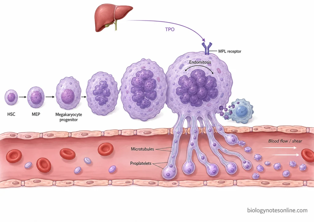
- The process starts from hematopoietic stem cells (HSCs) present in the bone marrow. These cells first form megakaryocyte-erythrocyte progenitors, followed by megakaryocyte progenitor cells. The cells then develop towards mature megakaryocytes.
- The major hormone involved in this process is thrombopoietin (TPO). It is produced mostly by the liver and acts through a receptor called MPL. TPO is involved in development of megakaryocyte cells and also helps to maintain the normal production of platelets.
- During development, megakaryocyte undergoes a special process known as endomitosis. In this process, replication of DNA takes place repeatedly but the cell does not divide into daughter cells. Thus, the cell becomes very large and polyploid, containing a multilobed nucleus.
- The cytoplasm also increases considerably with the maturation of megakaryocyte. Different granules and organelles are developed inside the cell. An extensive internal membrane system is also formed which provides the membrane required for formation of platelets.
- Mature megakaryocytes are generally present near the vascular sinusoids of bone marrow. In this step, the cytoplasm forms long and branched extensions into these blood vessels. These extensions are referred to as proplatelets.
- During this process, rearrangement of the cytoskeleton takes place. Microtubules are involved in elongation of the proplatelets and also help in movement of granules and other cellular components towards their ends. Platelet-sized structures are then formed mainly at the terminal region of these extensions.
- Finally, the proplatelets are released into blood where they separate to form individual platelets. Blood flow and shear forces also take part in this final process of platelet release. A single mature megakaryocyte can produce approximately 1,000–3,000 platelets. The remaining nuclear material of megakaryocyte is removed by macrophages.
Structure of Platelets
Platelets are small anucleated cell fragments having a complex internal structure. In resting condition, they are discoid in shape and contain different membrane systems, granules, cytoskeletal proteins and organelles. Nucleus is absent, but different cellular components required for platelet activity are present. The following are the major structural components of platelets-
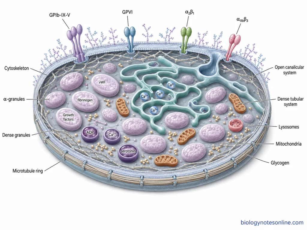
- The outer part of platelet is covered by a plasma membrane. It is made up of phospholipid bilayer containing cholesterol, glycolipids and different glycoproteins. A carbohydrate-rich layer called glycocalyx is present over the surface. Different receptors required for adhesion, activation and aggregation are also present in this membrane.
- The plasma membrane extends inside the platelet and forms a system of channels known as open canalicular system (OCS). It is connected with the outer surface of the platelet. This system increases the membrane surface and also provides a passage for release of substances from platelet granules.
- Another membrane system is the dense tubular system (DTS). These channels are not directly connected with the external surface. It is a remnant of smooth endoplasmic reticulum of megakaryocyte and acts as a storage site for calcium. Different enzymes involved in platelet activation are also associated with it.
- A well-developed cytoskeleton is present below the platelet membrane. It consists mainly of actin, myosin, spectrin and microtubules. A peripheral ring of microtubules helps to maintain the normal discoid shape of resting platelet. During activation, rearrangement of these components takes place and cytoplasmic projections are formed.
- Alpha granules (α-granules) are the largest and most abundant secretory granules present in platelets. About 50–80 alpha granules can be present in a platelet. They contain fibrinogen, von Willebrand factor (vWF), adhesion molecules, growth factors and several other proteins which are released after activation.
- Smaller dense granules (δ-granules) are also present in the platelet cytoplasm. These granules contain ADP, ATP, calcium and serotonin. During platelet activation, the stored substances are released and take part in further platelet responses.
- Lysosomes form another type of granule present inside platelets. They contain acid hydrolases and their number is less as compared to alpha granules.
- Mitochondria, glycogen particles and different small vesicular structures are also present in the cytoplasm. Mitochondria are involved in energy production, while glycogen is present as a stored source of energy.
Functions of Platelets
Platelets perform different functions in the blood and vascular system. The major function is associated with hemostasis, but they also take part in blood coagulation, maintenance of blood vessels, inflammatory reactions and repair of damaged tissues. The following are some of the important functions of platelets-
- The major function of platelets is to prevent excessive loss of blood after injury of a blood vessel. Platelets attach to the damaged region, become activated and other platelets are then aggregated at the same site. This forms a platelet plug, which acts as the initial barrier against bleeding.
- Platelets also take part in the process of blood coagulation. After strong activation, the platelet membrane provides a surface where different coagulation reactions can take place and formation of thrombin is increased. Fibrin is then formed around the platelet plug, making the developing clot more stable.
- After formation of the fibrin clot, platelets help in clot retraction. In this process, the contractile components present inside activated platelets pull the fibrin strands. The clot becomes smaller and more compact, and the edges of injured tissue are also brought closer.
- Another important function is the maintenance of vascular integrity. Platelets can accumulate at small gaps or damaged regions of blood vessels and prevent leakage of blood. This function is particularly important during inflammation, where the vessel wall may become more permeable without a large mechanical injury.
- During inflammatory reactions, activated platelets interact with leukocytes and endothelial cells. Different cytokines and chemokines are also released from platelet granules, which help in recruitment of neutrophils, monocytes and other immune cells towards the affected region.
- They also have a role in immune response and host defence. Platelets can recognize and interact with different microorganisms and release substances that influence immune cells. In some conditions, platelet activity also helps in limiting the spread of microorganisms through formation of localized clots.
- Platelets are involved in wound healing and tissue repair. At the injured site, different growth factors, cytokines and chemokines are released after platelet activation. These substances affect migration and proliferation of fibroblasts and other cells which are involved during repair of damaged tissue.
- Platelets also regulate formation of new blood vessels during angiogenesis. Their alpha granules contain different pro-angiogenic and anti-angiogenic factors. These factors act on endothelial cells and take part in growth and development of new vessels during tissue repair.
How Do Platelets Stop Bleeding?
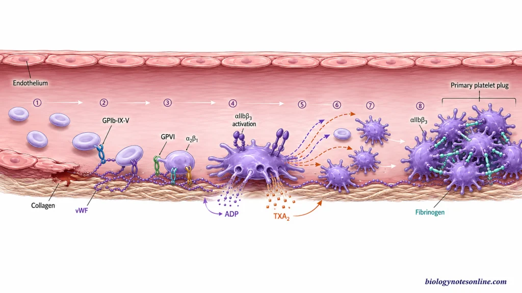
When a blood vessel is injured, platelets rapidly collect at the damaged region and form a temporary platelet plug. This process is referred to as primary hemostasis. It takes place through platelet adhesion, activation, release of different activating substances and finally aggregation of platelets.
Platelet Adhesion at the Injured Blood Vessel
In normal blood vessels, collagen present below the endothelial layer is not exposed to circulating platelets. After injury, the endothelial layer is damaged and subendothelial collagen becomes exposed. von Willebrand factor (vWF) present in blood binds to this exposed collagen.
The platelets are then captured at the injured surface. The GPIb-IX-V complex present on platelet membrane binds with collagen-bound vWF, which is particularly important during high blood flow. After this initial attachment, GPVI and integrin α2β1 bind to collagen. These interactions strengthen platelet adhesion and also start signals inside the platelet.
Platelet Activation and Shape Change
The attachment of platelet receptors with vWF and collagen does not only hold the platelet at the injured site. It also activates the platelet. Different signaling reactions are started inside the cell, especially after activation of GPVI and other platelet receptors.
During this process, rearrangement of platelet cytoskeleton takes place. The normal discoid platelet becomes more spherical and cytoplasmic projections are formed. This increases contact of platelet with the damaged surface and other platelets. At the same time, the platelet becomes more responsive and the αIIbβ3 integrin changes from a low-affinity to an active high-affinity form.
Granule Release and Recruitment of More Platelets
Activated platelets now release the substances stored inside their granules. ADP is released mainly from dense granules and acts on nearby platelets, resulting in further platelet activation. Thromboxane A₂ (TXA₂) is also formed by activated platelets and acts as another important signal for activation and recruitment of platelets.
In this step, the first activated platelets help to bring more circulating platelets towards the injured area. These newly recruited platelets are also activated and release more activating substances. Thus, platelet response at the damaged vessel increases rapidly.
Platelet Aggregation
After activation, integrin αIIbβ3 (GPIIb/IIIa) present on the platelet surface develops high affinity for fibrinogen. Fibrinogen can bind with αIIbβ3 receptors present on two adjacent activated platelets. It therefore acts as a bridge between the platelets.
More platelets are connected in the same way and a mass of aggregated platelets is formed at the site of injury. This forms the primary platelet plug, which closes the damaged region and reduces the loss of blood.
From the Platelet Plug to a Fibrin-Stabilized Clot
The formation of platelet plug and formation of fibrin clot are two closely related processes of hemostasis, but they are not the same process. Primary hemostasis mainly forms the platelet plug by adhesion, activation and aggregation of platelets. Coagulation (secondary hemostasis) produces fibrin which strengthens this platelet plug. Both processes start very early after injury and continue together rather than one process completely finishing before the other starts.
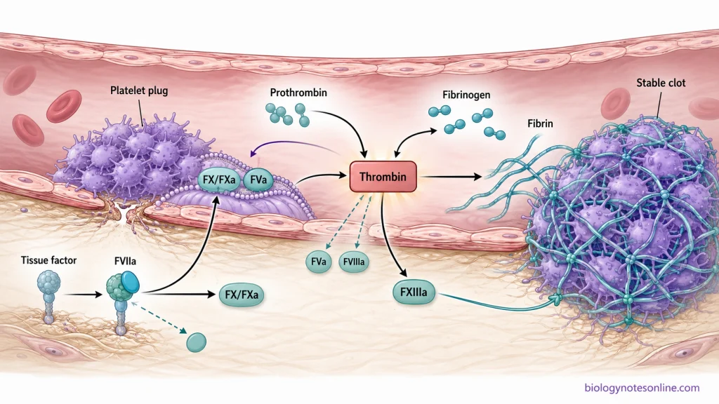
- After injury of the blood vessel, platelets adhere and aggregate at the damaged region and a temporary platelet plug is formed. This is referred to as primary hemostasis. At the same time, blood also comes in contact with tissue factor (TF) present in the injured extravascular tissue, which starts the coagulation reactions.
- Tissue factor binds with factor VIIa (FVIIa). The TF-FVIIa complex then activates factor X (FX) and also factor IX. Factor Xa together with its cofactor factor Va converts a small amount of prothrombin into thrombin. Thus, thrombin formation has already started while the platelet plug is being formed.
- Activated platelets now provide a suitable membrane surface for further coagulation reactions. In this step, coagulation factor complexes are assembled on the platelet surface and formation of thrombin is greatly increased. Thrombin itself further activates platelets and coagulation factors such as factors V and VIII. This produces amplification and propagation of the coagulation process around the injured region.
- The major action of this increased thrombin is conversion of soluble fibrinogen into insoluble fibrin. Fibrin molecules polymerize and form strands around and through the aggregated platelets. The initially formed platelet plug is therefore reinforced by a fibrin network.
- Thrombin also activates factor XIII to factor XIIIa. Factor XIIIa cross-links the fibrin strands, making the fibrin network stronger and more resistant to mechanical disruption. The temporary platelet-rich plug is now converted into a fibrin-stabilized hemostatic clot.
Platelet Count and Normal Reference Range
Platelet count is the number of platelets present in a particular volume of blood. It is generally measured during Complete Blood Count (CBC) and the value is expressed as ×10⁹/L or platelets/µL. In many laboratories, the normal reference range is taken as 150–400 ×10⁹/L or 150–450 ×10⁹/L. However, this value is not same for every individual and population. The platelet count can show variation depending upon age, sex, genetic background and population, and different laboratories may also use somewhat different reference limits. Thus, the reference range mentioned by the particular laboratory is generally considered during interpretation.
| Platelet count | Commonly used value | Meaning |
|---|---|---|
| Reference interval | About 150–400 or 150–450 ×10⁹/L | It is the commonly used adult range. The actual reference value can vary between different laboratories. |
| Low platelet count | Commonly below 150 ×10⁹/L | A decrease in platelet count below the reference limit is referred to as thrombocytopenia. Slightly lower values may also be present in some healthy individuals and populations. |
| High platelet count | Commonly 450 ×10⁹/L or more | An increased platelet count is referred to as thrombocytosis. |
How Is Platelet Count Measured?
Platelet count is generally measured from an anticoagulated blood sample with the help of an automated hematology analyzer. It is measured as one of the parameters during CBC. Different analyzers can use electrical impedance, optical light scatter or fluorescence-based methods for counting the platelets.
In impedance method, the platelets pass through a small counting aperture and produce electrical signals according to their volume. Optical methods are based on the light-scattering properties of platelets. Fluorescence-based platelet counting is also used in some hematology analyzers.
Sometimes, the platelet value obtained from the analyzer may not represent the actual platelet count. Platelets can form clumps in an EDTA-containing blood sample, which results in a falsely decreased platelet count. This is referred to as pseudothrombocytopenia. In such condition, peripheral blood smear examination or counting by another suitable method may be required.
What Does a Low Platelet Count Mean?
A decrease in the number of platelets below the lower reference limit is called thrombocytopenia. A platelet count below 150 ×10⁹/L is commonly considered as low in adults. This lower limit, however, is not universal for every population and some healthy individuals, particularly older individuals and certain populations, may have slightly lower platelet counts.
The low platelet count can occur due to decreased production of platelets, increased destruction or consumption of platelets, or increased sequestration of platelets in the spleen. Mild decrease may remain without any symptoms. When the count decreases further, bruising and bleeding can occur. At very low platelet count, the possibility of serious spontaneous bleeding is increased.
What Does a High Platelet Count Mean?
An increase in the number of platelets above the upper reference limit is known as thrombocytosis. A platelet count of 450 ×10⁹/L or more is generally used for this condition, although the upper reference value can be different between laboratories.
The increased platelet count may occur as a secondary or reactive response during infection, inflammation, tissue damage, iron deficiency and some other conditions. It can also occur due to a primary bone marrow disorder such as essential thrombocythemia. Sometimes a high platelet value is present only temporarily. Therefore, persistence of the increased count is generally checked during its evaluation.
Major Platelet Disorders
Platelet disorders are conditions associated with abnormal number or functioning of platelets. The number may be decreased or increased, while in some conditions the platelet count remains nearly normal but normal adhesion, secretion or aggregation is affected. These disorders can be acquired or inherited. The following are some of the major platelet disorders-
- Thrombocytopenia- When the number of circulating platelets is decreased, the condition is referred to as thrombocytopenia. It can develop because of decreased formation of platelets in bone marrow, increased destruction or consumption of platelets, or abnormal sequestration. The tendency of bleeding generally increases with a considerably low platelet count. However, platelet number alone does not always determine the clinical effect.
- Immune thrombocytopenia (ITP)- It is an acquired autoimmune platelet disorder in which circulating platelets are removed at an increased rate. During this condition, platelet production from megakaryocytes is also affected. The platelet count becomes low. Petechiae, bruising and other mucocutaneous bleeding can be present, and the severity varies between individuals.
- Thrombocytosis- An abnormal increase in circulating platelet number is known as thrombocytosis. It can be present as a reactive response during infection, inflammation, tissue injury, malignancy and some other conditions. In these cases, increase in platelet count occurs secondary to another condition and is not a primary disorder of platelet production.
- Essential thrombocythemia (ET)- It is a clonal myeloproliferative neoplasm with persistent increase in platelet production. Increased and abnormal proliferation of megakaryocytes takes place in the bone marrow. Mutations in JAK2, CALR or MPL are found in most cases. The condition can be associated with thrombosis as well as bleeding.
- Glanzmann thrombasthenia- It is an inherited disorder where platelet αIIbβ3 (GPIIb/IIIa) receptor is absent or does not function normally. This receptor is required for fibrinogen-mediated bridging between platelets. Hence, platelet aggregation becomes defective, while the platelet count is usually normal.
- Bernard-Soulier syndrome- In this disorder, the major defect is associated with platelet adhesion. It is an inherited platelet disorder caused by abnormalities of the GPIb-IX-V complex, which is involved in binding of platelet with von Willebrand factor. Large platelets are commonly present and the platelet count is also reduced.
- Storage pool disorders- These disorders affect the substances stored or released from platelet granules, which disturbs the normal amplification of platelet activation. Gray platelet syndrome is one of the inherited granule disorders. It is associated with deficiency of platelet alpha-granule contents.
- Acquired platelet function defects- Platelet function defects are not always inherited. Different drugs, particularly aspirin and other agents affecting platelet pathways, can interfere with normal platelet aggregation or secretion. Qualitative platelet abnormalities can also occur in chronic renal failure, some hematological disorders and other systemic conditions, even when the platelet count is not markedly decreased.
Biological Significance of Platelets
Platelets have important biological roles in maintenance of blood vessels and response of the body towards tissue injury. Their activity is not limited only to formation of platelet plug. They also interact with endothelial cells and immune cells and release different substances from their granules. Some of the important biological significance of platelets are-
- Hemostasis- The major function of platelets is prevention of blood loss after vascular injury. They adhere to the damaged vessel surface and aggregate together to form platelet plug. During this process, activated platelets also provide a surface for different coagulation reactions, which helps in formation of a stable hemostatic clot.
- Maintenance of vascular integrity- Platelets continuously circulate close to the vascular wall and respond when integrity of endothelium is disturbed. They help to seal small vascular injuries and prevent leakage of blood from the vessels. This function can also take place during inflammatory damage of blood vessels where there may not be a large external injury.
- Wound healing and tissue repair- Platelets are among the early blood elements accumulated at an injured region. After activation, different growth factors, cytokines and other biologically active substances are released from their granules. These substances take part in migration and growth of cells which are required during repair of the damaged tissue.
- Inflammatory response- Platelets also participate in inflammation. They interact with leukocytes and endothelial cells and release different inflammatory mediators. In this process, platelet activity can influence recruitment and functioning of neutrophils, monocytes and other inflammatory cells at the affected region.
- Immune response- Platelets can recognize and respond to different microbial and inflammatory signals. They contain different immune receptors and interact with several cells of innate as well as adaptive immune system. Platelets can also release antimicrobial and immunomodulatory substances, therefore they have a role in host defence in addition to hemostasis.
- Angiogenesis- Platelet alpha granules contain different factors which can regulate growth of new blood vessels. These include both angiogenesis-promoting and angiogenesis-inhibiting substances. Their release at the injured tissue helps in regulation of vascular growth during the process of tissue repair.
- Tissue remodeling- Platelets interact with different resident and circulating cells after tissue injury. The substances released from platelets can affect extracellular matrix, cell proliferation and other events taking place during restoration of injured tissue. Thus, platelet activity remains important beyond the initial stage of bleeding control.
References
- Agbani, E. O., & Poole, A. W. (2017). Procoagulant platelets: Generation, function, and therapeutic targeting in thrombosis. Blood, 130(20), 2171–2179. https://doi.org/10.1182/blood-2017-05-787259
- Ali, R. A., Wuescher, L. M., & Worth, R. G. (2015). Platelets: Essential components of the immune system. Current Trends in Immunology, 16, 65–78. https://pmc.ncbi.nlm.nih.gov/articles/PMC5096834/
- Almanaseer, A., Chin-Yee, B., Ho, J., Lazo-Langner, A., Schenkel, L., Bhai, P., Sadikovic, B., Chin-Yee, I. H., & Hsia, C. C. (2024). An approach to the investigation of thrombocytosis: Differentiating between essential thrombocythemia and secondary thrombocytosis. Advances in Hematology, 2024, 3056216. https://doi.org/10.1155/2024/3056216
- Andrews, R. K., & Berndt, M. C. (2004). Platelet physiology and thrombosis. Thrombosis Research, 114(5–6), 447–453. https://doi.org/10.1016/j.thromres.2004.07.020
- Balduini, C. L., & Noris, P. (2014). Platelet count and aging. Haematologica, 99(6), 953–955. https://doi.org/10.3324/haematol.2014.106260
- Bianchi, S., Torge, D., Rinaldi, F., Piattelli, M., Bernardi, S., & Varvara, G. (2022). Platelets’ role in dentistry: From oral pathology to regenerative potential. Biomedicines, 10(2), 218. https://doi.org/10.3390/biomedicines10020218
- Blair, P., & Flaumenhaft, R. (2009). Platelet α-granules: Basic biology and clinical correlates. Blood Reviews, 23(4), 177–189. https://doi.org/10.1016/j.blre.2009.04.001
- Bleeker, J. S., & Hogan, W. J. (2011). Thrombocytosis: Diagnostic evaluation, thrombotic risk stratification, and risk-based management strategies. Thrombosis, 2011, 536062. https://doi.org/10.1155/2011/536062
- Briggs, C., Harrison, P., & Machin, S. J. (2007). Continuing developments with the automated platelet count. International Journal of Laboratory Hematology, 29(2), 77–91. https://doi.org/10.1111/j.1751-553X.2007.00909.x
- Bryckaert, M., Rosa, J.-P., Denis, C. V., & Lenting, P. J. (2015). Of von Willebrand factor and platelets. Cellular and Molecular Life Sciences, 72(2), 307–326. https://doi.org/10.1007/s00018-014-1743-8
- Cattaneo, M. (2025). Pseudothrombocytopenia and other conditions associated with spuriously low platelet counts. Haematologica, 110(8), 1677–1692. https://doi.org/10.3324/haematol.2024.286234
- Fountain, J. H., & Lappin, S. L. (2023). Physiology, platelet. In StatPearls. StatPearls Publishing. https://www.ncbi.nlm.nih.gov/books/NBK470328/
- Gale, A. J. (2011). Current understanding of hemostasis. Toxicologic Pathology, 39(1), 273–280. https://doi.org/10.1177/0192623310389474
- Gawaz, M. (2005). Platelets and inflammation. In M. Quinn & D. Fitzgerald (Eds.), Platelet function: Assessment, diagnosis, and treatment (pp. 115–147). Humana Press. https://doi.org/10.1007/978-1-59259-917-2_5
- Gawaz, M., & Vogel, S. (2013). Platelets in tissue repair: Control of apoptosis and interactions with regenerative cells. Blood, 122(15), 2550–2554. https://doi.org/10.1182/blood-2013-05-468694
- Geddis, A. E. (2009). The regulation of proplatelet production. Haematologica, 94(6), 756–759. https://doi.org/10.3324/haematol.2009.006577
- Ghoshal, K., & Bhattacharyya, M. (2014). Overview of platelet physiology: Its hemostatic and nonhemostatic role in disease pathogenesis. The Scientific World Journal, 2014, 781857. https://doi.org/10.1155/2014/781857
- Hitchcock, I. S., Hafer, M., Sangkhae, V., & Tucker, J. A. (2021). The thrombopoietin receptor: Revisiting the master regulator of platelet production. Platelets, 32(6), 770–778. https://doi.org/10.1080/09537104.2021.1925102
- Ho-Tin-Noé, B., Boulaftali, Y., & Camerer, E. (2018). Platelets and vascular integrity: How platelets prevent bleeding in inflammation. Blood, 131(3), 277–288. https://doi.org/10.1182/blood-2017-06-742676
- Jansen, E. E., & Hartmann, M. (2021). Clot retraction: Cellular mechanisms and inhibitors, measuring methods, and clinical implications. Biomedicines, 9(8), 1064. https://doi.org/10.3390/biomedicines9081064
- Jinna, S., Karra, S., Penney, S. W., & Khandhar, P. B. (2025). Thrombocytopenia. In StatPearls. StatPearls Publishing. https://www.ncbi.nlm.nih.gov/books/NBK542208/
- Josefsson, E. C., Vainchenker, W., & James, C. (2020). Regulation of platelet production and life span: Role of Bcl-xL and potential implications for human platelet diseases. International Journal of Molecular Sciences, 21(20), 7591. https://doi.org/10.3390/ijms21207591
- Kaushansky, K. (2008). Historical review: Megakaryopoiesis and thrombopoiesis. Blood, 111(3), 981–986. https://doi.org/10.1182/blood-2007-05-088500
- Lam, F. W., Vijayan, K. V., & Rumbaut, R. E. (2015). Platelets and their interactions with other immune cells. Comprehensive Physiology, 5(3), 1265–1280. https://doi.org/10.1002/cphy.c140074
- Locatelli, L., Colciago, A., Castiglioni, S., & Maier, J. A. (2021). Platelets in wound healing: What happens in space? Frontiers in Bioengineering and Biotechnology, 9, 716184. https://doi.org/10.3389/fbioe.2021.716184
- Machlus, K. R., Thon, J. N., & Italiano, J. E., Jr. (2014). Interpreting the developmental dance of the megakaryocyte: A review of the cellular and molecular processes mediating platelet formation. British Journal of Haematology, 165(2), 227–236. https://doi.org/10.1111/bjh.12758
- Martínez, C. E., Smith, P. C., & Palma Alvarado, V. A. (2015). The influence of platelet-derived products on angiogenesis and tissue repair: A concise update. Frontiers in Physiology, 6, 290. https://doi.org/10.3389/fphys.2015.00290
- Mititelu, A., Onisâi, M.-C., Roșca, A., & Vlădăreanu, A. M. (2024). Current understanding of immune thrombocytopenia: A review of pathogenesis and treatment options. International Journal of Molecular Sciences, 25(4), 2163. https://doi.org/10.3390/ijms25042163
- Mohan, G., Malayala, S. V., Mehta, P., & Balla, M. (2020). A comprehensive review of congenital platelet disorders, thrombocytopenias and thrombocytopathies. Cureus, 12(10), e11275. https://doi.org/10.7759/cureus.11275
- Nurden, A. T., & Nurden, P. (2020). Inherited thrombocytopenias: History, advances and perspectives. Haematologica, 105(8), 2004–2019. https://doi.org/10.3324/haematol.2019.233197
- Palta, S., Saroa, R., & Palta, A. (2014). Overview of the coagulation system. Indian Journal of Anaesthesia, 58(5), 515–523. https://doi.org/10.4103/0019-5049.144643
- Patel, S. R., Hartwig, J. H., & Italiano, J. E., Jr. (2005). The biogenesis of platelets from megakaryocyte proplatelets. The Journal of Clinical Investigation, 115(12), 3348–3354. https://doi.org/10.1172/JCI26891
- Perez Botero, J., Lee, K., Branchford, B. R., Bray, P. F., Freson, K., Lambert, M. P., Luo, M., Mohan, S., Ross, J. E., Bergmeier, W., Di Paola, J., & ClinGen Platelet Disorder Variant Curation Expert Panel. (2020). Glanzmann thrombasthenia: Genetic basis and clinical correlates. Haematologica, 105(4), 888–894. https://doi.org/10.3324/haematol.2018.214239
- Periayah, M. H., Halim, A. S., & Mat Saad, A. Z. (2017). Mechanism action of platelets and crucial blood coagulation pathways in hemostasis. International Journal of Hematology-Oncology and Stem Cell Research, 11(4), 319–327. https://pmc.ncbi.nlm.nih.gov/articles/PMC5767294/
- Rumbaut, R. E., & Thiagarajan, P. (2010). Platelet-vessel wall interactions in hemostasis and thrombosis. Morgan & Claypool Life Sciences. https://doi.org/10.4199/C00007ED1V01Y201002ISP004
- Sim, X., Poncz, M., Gadue, P., & French, D. L. (2016). Understanding platelet generation from megakaryocytes: Implications for in vitro-derived platelets. Blood, 127(10), 1227–1233. https://doi.org/10.1182/blood-2015-08-607929
- Sonmez, O., & Sonmez, M. (2017). Role of platelets in immune system and inflammation. Porto Biomedical Journal, 2(6), 311–314. https://doi.org/10.1016/j.pbj.2017.05.005
- Stiff, P. J. (1990). Platelets. In H. K. Walker, W. D. Hall, & J. W. Hurst (Eds.), Clinical methods: The history, physical, and laboratory examinations (3rd ed., Chapter 154). Butterworths. https://www.ncbi.nlm.nih.gov/books/NBK262/
- Tefferi, A., Vannucchi, A. M., & Barbui, T. (2024). Essential thrombocythemia: 2024 update on diagnosis, risk stratification, and management. American Journal of Hematology, 99(4), 697–718. https://doi.org/10.1002/ajh.27216
- Tomaiuolo, M., Brass, L. F., & Stalker, T. J. (2017). Regulation of platelet activation and coagulation and its role in vascular injury and arterial thrombosis. Interventional Cardiology Clinics, 6(1), 1–12. https://doi.org/10.1016/j.iccl.2016.08.001
- Tyagi, T., Jain, K., Gu, S. X., Qiu, M., Gu, V. W., Melchinger, H., Rinder, H., Martin, K. A., Gardiner, E. E., Lee, A. I., Tang, W. H., & Hwa, J. (2022). A guide to molecular and functional investigations of platelets to bridge basic and clinical sciences. Nature Cardiovascular Research, 1(3), 223–237. https://doi.org/10.1038/s44161-022-00021-z
- Vieira-de-Abreu, A., Campbell, R. A., Weyrich, A. S., & Zimmerman, G. A. (2012). Platelets: Versatile effector cells in hemostasis, inflammation, and the immune continuum. Seminars in Immunopathology, 34(1), 5–30. https://doi.org/10.1007/s00281-011-0286-4
- Xu, X. R., Carrim, N., Dias Neves, M. A., McKeown, T., Stratton, T. W., Matos Pinto Coelho, R., Lei, X., Chen, P., Xu, J., Dai, X., Li, B. X., & Ni, H. (2016). Platelets and platelet adhesion molecules: Novel mechanisms of thrombosis and anti-thrombotic therapies. Thrombosis Journal, 14(Suppl 1), 29. https://doi.org/10.1186/s12959-016-0100-6
- Yadav, S., & Storrie, B. (2017). The cellular basis of platelet secretion: Emerging structure/function relationships. Platelets, 28(2), 108–118. https://doi.org/10.1080/09537104.2016.1257786