

Ribosome is a ribonucleoprotein complex which acts as the main site for protein synthesis in both prokaryotic and eukaryotic cells. It is made up of ribosomal RNA (rRNA) and proteins. The ribosome consists of two unequal subunits, a small subunit and a large subunit.
During protein synthesis, the small subunit reads the codons present on messenger RNA (mRNA). Transfer RNA (tRNA) brings the corresponding amino acids. The large subunit helps in the formation of peptide bonds between these amino acids, resulting in formation of a growing polypeptide chain. This process is referred to as protein synthesis.
In bacteria, the complete ribosome is 70S, which is formed of 30S and 50S subunits. Whereas, the cytoplasmic ribosome of eukaryotic cells is 80S and consists of 40S and 60S subunits.
Ribosome Location in Cells
Ribosomes are present at different locations inside a cell. Their location also depends upon the type of cell and the protein being synthesized. The following are the main locations-
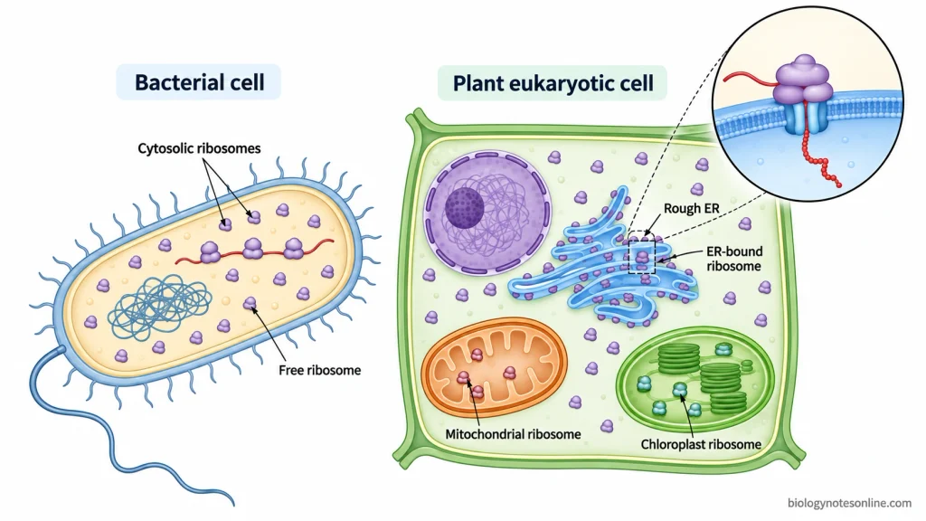
- In prokaryotic cells, ribosomes are present freely in the cytoplasm. These are the sites for synthesis of cellular proteins.
- In eukaryotic cells, many ribosomes remain free in the cytosol. Proteins which remain in cytosol, or later go into nucleus, mitochondria, chloroplast and peroxisomes are generally synthesized by these free ribosomes.
- Some ribosomes remain attached with the cytosolic surface of rough endoplasmic reticulum (RER). Because of these attached ribosomes the ER gets its rough appearance. They mainly synthesize proteins entering the secretory pathway, including secreted and membrane proteins.
- Ribosomes are also present inside mitochondria. These mitochondrial ribosomes occur in the matrix and synthesize some of the proteins encoded by mitochondrial genome.
- In plant and algal cells, another group of ribosomes occurs inside chloroplasts. They are located in the chloroplast stroma and take part in translation of chloroplast-encoded proteins.
Ribosome Structure
The structure of ribosome can be described as follows-
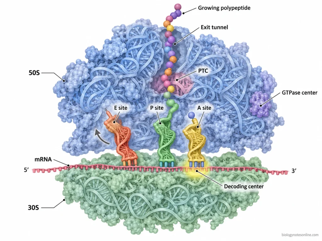
- Composition- Ribosome is a ribonucleoprotein complex made up of ribosomal RNA (rRNA) and several ribosomal proteins. It is not surrounded by any membrane. rRNA forms a major structural and functional part of the ribosome.
- Subunits- Each ribosome consists of two unequal parts, a small subunit and a large subunit. These subunits remain separate and come together to form a functional ribosome during protein synthesis.
- Small subunit- The smaller subunit contains one major rRNA along with ribosomal proteins. It holds the mRNA and contains the decoding center, where codon of mRNA is matched with anticodon of tRNA. An mRNA channel is also present in this subunit.
- Large subunit- It contains more rRNA and proteins and forms the catalytic part of ribosome. The peptidyl transferase center (PTC) is present here which is involved in formation of peptide bonds. Newly formed polypeptide passes out through a peptide exit tunnel.
- Binding sites- Ribosome contains three important tRNA-binding sites, called A, P and E sites. A site (aminoacyl site) receives incoming aminoacyl-tRNA. P site (peptidyl site) holds the tRNA carrying growing polypeptide chain. The discharged tRNA moves through the E site (exit site) before leaving the ribosome.
- 70S ribosome- It is formed of 30S small subunit and 50S large subunit. The 30S subunit contains 16S rRNA, whereas 50S contains 23S and 5S rRNAs. This type is characteristic of bacterial ribosomes.
- 80S ribosome- Cytoplasmic ribosome of eukaryotic cells consists of 40S and 60S subunits. The 40S subunit contains 18S rRNA. On the other hand, the 60S subunit contains 28S, 5.8S and 5S rRNAs, along with ribosomal proteins.
- Svedberg unit- The letter S represents the sedimentation coefficient or Svedberg unit. The values of two ribosomal subunits are not added mathematically. Therefore, 30S and 50S form a 70S ribosome, whereas 40S and 60S form an 80S ribosome.
Chemical Composition of Ribosomes
Ribosome is a ribonucleoprotein particle made up mainly of ribosomal RNA (rRNA) and ribosomal proteins. In bacterial ribosome, RNA forms nearly two-thirds of the total mass and the remaining part is mainly protein.
rRNA- It forms the major structural framework of ribosome. rRNA also takes part in catalytic activity. Different rRNA molecules are present in different ribosomal subunits.
Ribosomal proteins- These proteins remain associated with rRNA. They help in proper folding, stability and formation of ribosomal subunits. Their number and type differ between bacterial and eukaryotic ribosomes.
70S ribosome- The 30S subunit contains 16S rRNA together with ribosomal proteins. The 50S subunit contains 23S and 5S rRNAs with proteins.
80S ribosome- The 40S subunit contains 18S rRNA. Whereas, the 60S subunit contains 28S, 5.8S and 5S rRNAs along with ribosomal proteins.
Functional Centers and Binding Sites of Ribosome
The important functional centers and binding sites of ribosome are summarized below-
| Center/Site | Location | Function |
|---|---|---|
| mRNA binding site | Small ribosomal subunit | It binds the mRNA and keeps it in proper position during translation. The mRNA passes through the small subunit where its codons are exposed for reading. |
| Decoding center | Small subunit | This is the site where codon of mRNA is checked with the anticodon of incoming tRNA. It plays a major role in selection of the correct aminoacyl-tRNA. |
| A site | Between small and large subunits | A site (aminoacyl site) receives the incoming aminoacyl-tRNA carrying the next amino acid. Codon-anticodon pairing takes place here during elongation. |
| P site | Between both ribosomal subunits | P site (peptidyl site) holds the tRNA carrying the growing polypeptide chain. During initiation, initiator tRNA is also positioned in this site. |
| E site | Ribosomal subunit interface | E site (exit site) holds the deacylated tRNA for a short period. The tRNA then leaves the ribosome from this site. |
| PTC | Large ribosomal subunit | Peptidyl transferase center (PTC) is the catalytic center of ribosome. In this site, peptide bond is formed between amino acids attached to the A-site and P-site tRNAs. rRNA forms the major catalytic part. |
| GTPase center | Large subunit | GTPase-associated center (GAC) is the region where different translation GTPases interact with ribosome. It is involved during initiation and elongation, including the action of translation factors. |
| Exit tunnel | Large ribosomal subunit | The newly formed polypeptide enters the polypeptide exit tunnel from the PTC and passes through it toward outside of ribosome. The tunnel is made mainly of rRNA with some ribosomal proteins. |
How Ribosomes Make Proteins
Protein synthesis by ribosome is referred to as translation. In this process, the codons present in mRNA are read and amino acids are joined in a particular sequence. It mainly includes three stages, initiation, elongation and termination. The process is as follows-

Step 1- Binding of mRNA
The translation process starts with binding of small ribosomal subunit with mRNA. The starting region of mRNA is recognized. Start codon (AUG) is then placed at the proper position for beginning of protein synthesis.
Step 2- Binding of initiator tRNA
A special initiator tRNA carrying methionine binds with the AUG codon by codon-anticodon pairing. This tRNA is present at the P site (peptidyl site). The A site remains empty at this stage.
Step 3- Formation of complete ribosome
After this, the large ribosomal subunit joins with the small subunit. The complete functional ribosome is now formed. mRNA and initiator tRNA are kept in their proper position and elongation can start.
Step 4- Entry of aminoacyl-tRNA
The next aminoacyl-tRNA enters into the A site carrying its specific amino acid. Its anticodon pairs with the corresponding codon of mRNA.
In this way, correct tRNA is selected according to the sequence present in mRNA.
Step 5- Peptide bond formation
In this step, a peptide bond is formed between the amino acid present at P site and amino acid brought to the A site. This reaction takes place at the peptidyl transferase center (PTC) of large ribosomal subunit. rRNA performs the major catalytic role.
The growing polypeptide chain is now transferred to the tRNA of A site.
Step 6- Translocation
The ribosome then moves forward along mRNA by one codon or three nucleotides. The tRNA carrying growing polypeptide chain shifts from A site to P site. At the same time, empty tRNA moves toward the E site (exit site) and finally comes out from the ribosome.
The A site becomes free again. Another aminoacyl-tRNA enters this site and the same steps are repeated. Thus, amino acids are added one after another and the polypeptide chain becomes longer.
Step 7- Termination
Elongation continues until any one of the stop codons (UAA, UAG or UGA) reaches the A site. These codons are normally not recognized by any tRNA. Instead, a release factor binds with the ribosome.
This results in release of completed polypeptide chain. After this, mRNA and tRNA separate and the ribosome dissociates into large and small subunits. These subunits can again take part in another round of protein synthesis.
Ribosome Types Across Cells
Ribosomes are not of same type in all cells. Based on their size and cellular location, the common types are as follows-
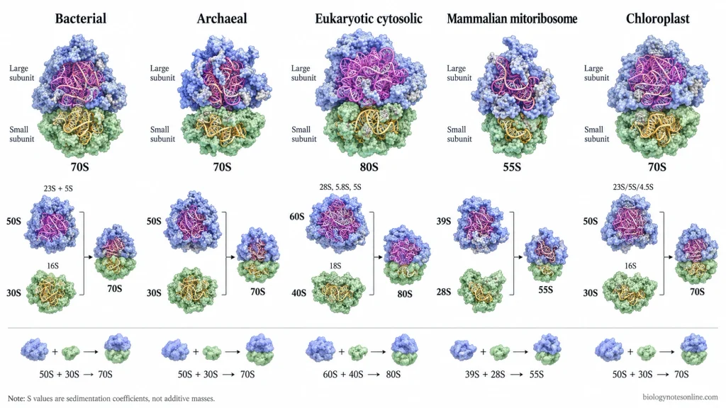
- Bacterial ribosome (70S)- These ribosomes are present in bacterial cytoplasm. The 70S ribosome is of two subunits, 30S and 50S. 30S contains 16S rRNA. Whereas, 50S contains 23S and 5S rRNAs.
- Archaeal ribosome (70S)- The ribosome of archaea is also 70S, consisting of 30S small and 50S large subunit. Its major rRNAs are 16S, 23S and 5S. However, it is not completely similar with bacterial ribosome. A greater set of ribosomal proteins are present in archaeal ribosomes.
- Eukaryotic ribosome (80S)- It is found in the cytoplasm of eukaryotic cells. The ribosome consists of 40S small subunit and 60S large subunit. 40S contains 18S rRNA, while 60S contains 28S, 5.8S and 5S rRNAs. These may occur freely in the cytoplasm or remain attached with rough endoplasmic reticulum.
- Mitochondrial ribosome- These are also called mitoribosomes and are present inside mitochondria. Their size differs among eukaryotic organisms. Mammalian mitoribosome is 55S (28S + 39S). In fungi, it is around 74S, while higher plant mitoribosomes have been reported around 77-78S.
- Chloroplast ribosome (70S)- Chloroplast of plants and algae contains bacterial-type 70S ribosome. It is made up of 30S and 50S subunits. The 30S contains 16S rRNA. The larger 50S subunit has 23S, 5S and 4.5S rRNAs.
Ribosome Biogenesis
Ribosome biogenesis is the formation of mature ribosomal subunits from rRNA and ribosomal proteins. In eukaryotes, most of this process starts in the nucleolus, followed by further processing in nucleoplasm and cytoplasm. The major steps are as follows-

Step 1- Pre-rRNA formation
The process starts with transcription of ribosomal DNA (rDNA). RNA polymerase I produces a large precursor rRNA (pre-rRNA) in nucleolus. In mammalian cells, it is formed as 47S pre-rRNA, which contains the sequences for 18S, 5.8S and 28S rRNAs.
5S rRNA is formed separately by RNA polymerase III.
Step 2- Processing of pre-rRNA
The large pre-rRNA is now cut and processed at different sites. Internal and external spacer sequences are removed. After processing, 18S, 5.8S and 28S rRNAs are produced.
Several nucleotides of rRNA are also modified during this process with the help of small nucleolar ribonucleoproteins (snoRNPs). Some processing starts even when pre-rRNA synthesis is going on.
Step 3- Ribosomal protein formation
Genes of ribosomal proteins are transcribed by RNA polymerase II. Their mRNAs pass into cytoplasm, where ribosomal proteins are synthesized.
These proteins are then transported back into nucleus and nucleolus. Here, they associate with newly formed rRNA.
Step 4- Pre-ribosome assembly
rRNA, ribosomal proteins and several ribosome biogenesis factors now come together. A large 90S pre-ribosomal particle is formed in the nucleolus.
Assembly and processing take place together. The complete pre-rRNA does not need to finish first.
Step 5- Formation of pre-40S and pre-60S
The 90S particle is further processed and separated into two developing subunits.
The part containing 18S rRNA develops into pre-40S subunit. Whereas, 5.8S and 28S rRNAs, 5S rRNA and large-subunit proteins form the pre-60S subunit.
Step 6- Nuclear maturation and export
Both pre-40S and pre-60S undergo further maturation while passing from nucleolus to nucleoplasm. More ribosomal proteins are added. Some temporary assembly factors are removed or exchanged.
The two pre-ribosomal subunits are then exported separately through the nuclear pore complex (NPC) into cytoplasm.
Step 7- Cytoplasmic maturation
Final maturation occurs after the particles reach cytoplasm. Remaining rRNA processing is completed and associated assembly factors are released. Immature particles are also prevented from entering translation during these quality-control steps.
Finally, mature 40S and 60S ribosomal subunits are formed. They remain separate and join on mRNA during translation to form the functional 80S ribosome.
Biological and Medical Significance
Some of the important biological and medical significance of ribosomes are-
- Protein synthesis- Ribosome is the main site for translation of mRNA into proteins. Thus, cellular proteins and enzymes are produced with the help of ribosomes.
- Cell growth- Continuous synthesis of proteins is required for cell growth, division and normal cellular activities. Ribosomes carry out this basic process.
- Antibiotic target- Bacterial ribosome is one of the major targets of antibiotics. Drugs can bind with different functional regions of ribosome and block bacterial protein synthesis.
- Ribosomal diseases- Defects in ribosomal proteins or ribosome biogenesis can cause a group of disorders called ribosomopathies. Diamond-Blackfan anemia and Treacher Collins syndrome are some examples.
- Cancer- Abnormal ribosome biogenesis and translation are found in many cancers. Increased formation of ribosomes helps in rapid protein production required by growing cancer cells.
- Drug development- Structure and functional sites of ribosome are studied for development of new antimicrobial drugs, particularly against antibiotic-resistant bacteria.
Dysfunctions of Ribosome and Disorders
Defects in ribosomal proteins, rRNA processing or assembly of ribosomal subunits can disturb normal ribosome formation. Such disorders are commonly referred to as ribosomopathies. They particularly affect tissues with high requirement for cell growth and protein synthesis, although the clinical effects are different in different disorders.
Some of the important ribosomal dysfunctions are-
- Defective ribosome biogenesis- Mutation in a ribosomal protein or its assembly factor can decrease formation of mature ribosomes. The amount of functional ribosome is reduced.
- Abnormal rRNA processing- Defects during rRNA cleavage, modification or maturation can prevent proper assembly of ribosomal subunits. Some ribosomopathies arise by this mechanism.
- Defective translation- An abnormal ribosome may change the synthesis of cellular proteins. Protein production may be reduced or particular mRNAs may not be translated normally.
- Nucleolar stress- Disturbance of ribosome biogenesis produces nucleolar stress. In many ribosomopathies, this can activate p53, resulting in cell-cycle arrest or apoptosis.
Disorders Associated with Ribosome Dysfunction
- Diamond-Blackfan anemia (DBA)- It is a congenital bone marrow failure disorder caused commonly by defects in ribosomal protein genes (RPS or RPL genes). Severe reduction of red blood cell production is characteristic. Congenital abnormalities can also occur.
- Shwachman-Diamond syndrome (SDS)- It is associated commonly with defects in SBDS, which is required for proper maturation and function of ribosomal subunits. Bone marrow abnormalities and exocrine pancreatic insufficiency are important features.
- Treacher Collins syndrome (TCS)- Defects involving TCOF1 and some RNA polymerase I related genes interfere with rRNA production and ribosome biogenesis. The disorder mainly produces craniofacial abnormalities.
- 5q- syndrome- It is an acquired myelodysplastic disorder in which loss of RPS14 contributes to defective ribosome function. A characteristic anemia is produced.
- Dyskeratosis congenita- Some forms involve DKC1 and related factors affecting rRNA modification, along with their other cellular functions. Bone marrow failure and increased cancer susceptibility can occur.
- Cartilage-hair hypoplasia- Mutation affecting RMRP can disturb rRNA processing. Short-limbed growth, sparse hair, immune defects and anemia are commonly associated with the disorder.
References
- Alberts, B., Johnson, A., Lewis, J., Raff, M., Roberts, K., & Walter, P. (2002). Molecular biology of the cell (4th ed.). Garland Science. https://www.ncbi.nlm.nih.gov/books/NBK21054/
- Bhavsar, R. B., Makley, L. N., & Tsonis, P. A. (2010). The other lives of ribosomal proteins. Human Genomics, 4(5), 327–344. https://doi.org/10.1186/1479-7364-4-5-327
- Blanchet, S., & Ranjan, N. (2022). Translation phases in eukaryotes. In K.-D. Entian (Ed.), Ribosome biogenesis: Methods and protocols (pp. 217–228). Humana. https://doi.org/10.1007/978-1-0716-2501-9_13
- Brown, T. A. (2002). Genomes (2nd ed.). Wiley-Liss. https://www.ncbi.nlm.nih.gov/books/NBK21128/
- Burwick, N., Shimamura, A., & Liu, J. M. (2011). Non-Diamond Blackfan anemia disorders of ribosome function: Shwachman Diamond syndrome and 5q- syndrome. Seminars in Hematology, 48(2), 136–143. https://doi.org/10.1053/j.seminhematol.2011.01.002
- Cooper, G. M. (2000). The cell: A molecular approach (2nd ed.). Sinauer Associates. https://www.ncbi.nlm.nih.gov/books/NBK9839/
- Dörner, K., Ruggeri, C., Zemp, I., & Kutay, U. (2023). Ribosome biogenesis factors—from names to functions. The EMBO Journal, 42(7), e112699. https://doi.org/10.15252/embj.2022112699
- Gitareja, K., Chelliah, S. S., Sanij, E., Sandhu, S., Kang, J., & Khot, A. (2025). Ribosome biogenesis and function in cancer: From mechanisms to therapy. Cancers, 17(15), 2534. https://doi.org/10.3390/cancers17152534
- Giuliano, M. G., & Engl, C. (2021). The lifecycle of ribosomal RNA in bacteria. In I. Kotta-Loizou (Ed.), RNA damage and repair (pp. 27–51). Springer. https://doi.org/10.1007/978-3-030-76571-2_2
- Kaczanowska, M., & Rydén-Aulin, M. (2007). Ribosome biogenesis and the translation process in Escherichia coli. Microbiology and Molecular Biology Reviews, 71(3), 477–494. https://doi.org/10.1128/MMBR.00013-07
- Kang, J., Brajanovski, N., Chan, K. T., Xuan, J., Pearson, R. B., & Sanij, E. (2021). Ribosomal proteins and human diseases: Molecular mechanisms and targeted therapy. Signal Transduction and Targeted Therapy, 6, 323. https://doi.org/10.1038/s41392-021-00728-8
- Klinge, S., & Woolford, J. L., Jr. (2019). Ribosome assembly coming into focus. Nature Reviews Molecular Cell Biology, 20(2), 116–131. https://doi.org/10.1038/s41580-018-0078-y
- Laursen, B. S., Sørensen, H. P., Mortensen, K. K., & Sperling-Petersen, H. U. (2005). Initiation of protein synthesis in bacteria. Microbiology and Molecular Biology Reviews, 69(1), 101–123. https://doi.org/10.1128/MMBR.69.1.101-123.2005
- Lin, J., Zhou, D., Steitz, T. A., Polikanov, Y. S., & Gagnon, M. G. (2018). Ribosome-targeting antibiotics: Modes of action, mechanisms of resistance, and implications for drug design. Annual Review of Biochemistry, 87, 451–478. https://doi.org/10.1146/annurev-biochem-062917-011942
- Liutkute, M., Samatova, E., & Rodnina, M. V. (2020). Cotranslational folding of proteins on the ribosome. Biomolecules, 10(1), 97. https://doi.org/10.3390/biom10010097
- Maehama, T., Nishio, M., Otani, J., Mak, T. W., & Suzuki, A. (2023). Nucleolar stress: Molecular mechanisms and related human diseases. Cancer Science, 114(5), 2078–2086. https://doi.org/10.1111/cas.15755
- Mattick, J., & Amaral, P. (2022). RNA, the epicenter of genetic information: A new understanding of molecular biology. CRC Press. https://doi.org/10.1201/9781003109242
- Moraleva, A. A., Deryabin, A. S., Rubtsov, Y. P., Rubtsova, M. P., & Dontsova, O. A. (2022). Eukaryotic ribosome biogenesis: The 40S subunit. Acta Naturae, 14(1), 14–30. https://doi.org/10.32607/actanaturae.11540
- Nakhoul, H., Ke, J., Zhou, X., Liao, W., Zeng, S. X., & Lu, H. (2014). Ribosomopathies: Mechanisms of disease. Clinical Medicine Insights: Blood Disorders, 7, 7–16. https://doi.org/10.4137/CMBD.S16952
- Narla, A., & Ebert, B. L. (2010). Ribosomopathies: Human disorders of ribosome dysfunction. Blood, 115(16), 3196–3205. https://doi.org/10.1182/blood-2009-10-178129
- Narla, A., Hurst, S. N., & Ebert, B. L. (2011). Ribosome defects in disorders of erythropoiesis. International Journal of Hematology, 93(2), 144–149. https://doi.org/10.1007/s12185-011-0776-0
- Oborská-Oplová, M., Fischer, U., Altvater, M., & Panse, V. G. (2022). Eukaryotic ribosome assembly and nucleocytoplasmic transport. In K.-D. Entian (Ed.), Ribosome biogenesis: Methods and protocols (pp. 99–126). Humana. https://doi.org/10.1007/978-1-0716-2501-9_7
- Panse, V. G., & Johnson, A. W. (2010). Maturation of eukaryotic ribosomes: Acquisition of functionality. Trends in Biochemical Sciences, 35(5), 260–266. https://doi.org/10.1016/j.tibs.2010.01.001
- Rye, C., Wise, R., Jurukovski, V., DeSaix, J., Choi, J., & Avissar, Y. (2016). Biology. OpenStax. https://openstax.org/books/biology/pages/1-introduction
- Tomal, A., Kwasniak-Owczarek, M., & Janska, H. (2019). An update on mitochondrial ribosome biology: The plant mitoribosome in the spotlight. Cells, 8(12), 1562. https://doi.org/10.3390/cells8121562
- Venturi, G., & Montanaro, L. (2020). How altered ribosome production can cause or contribute to human disease: The spectrum of ribosomopathies. Cells, 9(10), 2300. https://doi.org/10.3390/cells9102300
- Wilson, D. N., & Cate, J. H. D. (2012). The structure and function of the eukaryotic ribosome. Cold Spring Harbor Perspectives in Biology, 4(5), a011536. https://doi.org/10.1101/cshperspect.a011536
- Zoschke, R., & Bock, R. (2018). Chloroplast translation: Structural and functional organization, operational control, and regulation. The Plant Cell, 30(4), 745–770. https://doi.org/10.1105/tpc.18.00016