What is Yeast?
Yeast are tiny, single-celled fungi that play a crucial role in creating everyday delights like bread, beer, and wine through a process called fermentation. These minuscule organisms have an impressive ability to multiply and adapt, making them essential for various culinary and scientific applications.
These unicellular organisms, belonging to the Fungus Kingdom, are classified under the subkingdom Dikarya. They come in a variety of species, with the majority falling into the Ascomycota phylum, while a few are categorized as Basidiomycota.
Yeast cells primarily reproduce through budding or binary fission, both of which are methods of asexual reproduction. In the budding process, a new yeast cell emerges as a bud from the parent cell, eventually breaking away to become an independent identical cell. In binary fission, the cell’s genome replicates and divides, leading to the formation of two new cells from the parent cell.
Some yeast species, known as dimorphic fungi, can switch between a yeast phase and a hyphal phase, resembling thread-like structures. This versatility enables them to thrive in various environments, particularly those rich in sugars. Yeast can be found on flowers, plant leaves, fruits, soil, deep-sea environments, skin surfaces, and even the intestinal tracts of warm-blooded animals.
For yeast to flourish, they require moist surroundings with an ample supply of simple and soluble nutrients. In environments with these conditions, yeast can grow and multiply.
Although certain yeast strains, such as Candida, can cause infections like Candidiasis, many yeast types are incredibly useful. Some of the notable ones include:
- Baker’s Yeast: Essential for leavening bread and causing dough to rise.
- Nutritional Yeast: Packed with nutrients and often used as a flavor-enhancing ingredient.
- Brewer’s Yeast: Vital in the production of beer, aiding in the fermentation process.
- Distiller’s and Wine Yeast: Crucial for the production of alcoholic beverages like wine and spirits.
In addition to their role in food and beverage production, yeast, particularly the bread yeast Saccharomyces cerevisiae, has been extensively employed by scientists to study important cellular processes. This remarkable microorganism continues to leave its mark on both culinary delights and scientific exploration.
Microscopy Techniques
- Yeast, those tiny organisms with big impacts, can be brought into focus using two distinct microscopy methods: bright field microscopy and fluorescence microscopy. These techniques allow us to unveil the hidden world of yeast in ways that both fascinate and inform.
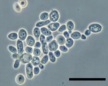
- With a high-powered bright field microscope, like the compound microscope, yeast and their budding counterparts become visible at an impressive magnification of 1000 times. This reveals oval-shaped microstructures, the essential building blocks of yeast cells. Bright field microscopy also unveils the mesmerizing dance of yeast during fermentation in a sugary solution and offers insights into their reproduction process through budding.
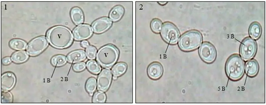
- Yet, there’s more to yeast than meets the eye. A fluorescent microscope takes us deeper, enabling the identification of cell organelles residing within yeast cells and showing us how they’re distributed within. Among these microscopic components are nuclei, mitochondria, vacuoles, endoplasmic reticulum, and the protective cell wall.
- Step into a world of microscopic wonder as you explore a collection of magnified images showcasing budding yeast. While they might appear similar at first glance, these yeast cells come in a variety of sizes and shapes, each holding its own intriguing story.
- In the realm of microscopy, yeast becomes a captivating subject, revealing its intricate details and processes, and inspiring us to delve further into the mysteries of the microscopic universe.
Materials Required
- Yeast Source: Opt for yeast cake or active yeast, and if using the latter, a smidgen of sugar.
- Warm Water: A cup of comfortably warm water serves as the liquid foundation.
- Mixing Bowl: A small bowl will facilitate the blending process.
- Stirring Tools: Utilize mixing sticks or a spoon for thorough mixing.
- Fluid Transfer Device: Employ a medicine dropper or pipette for precise fluid transfer.
- Slide Ensemble: You will require glass slides and cover slips to create the slide structure.
- Staining Agents: Consider artificial dyes designed for staining*, if aiming to enhance visibility.
Sample Preparation
The process of preparing a sample slide of yeast for microscopic analysis is a meticulous endeavor that enables us to uncover the intricacies of these microscopic organisms. By following a systematic procedure and utilizing accessible materials, a window into the world of yeast becomes accessible for scrutiny.
- Source Selection: The initial step involves selecting a suitable yeast source. Among the options, a cultivated yeast variety like yeast cake, obtainable from baking supply stores, stands as a convenient choice. This specialized yeast harbors a sugar-consuming fungus, thereby offering an ideal medium for observation. Alternatively, active yeast paired with a small quantity of sugar may be employed to serve as the source material.
- Gathering Materials: To embark on the sample preparation journey, several essential materials are required:
- Yeast source (yeast cake or active yeast)
- Sugar (if not using yeast cake)
- Microscope slides
- Cover slips
- Microscope
- Microscopy-friendly staining agents (if desired)
- Water or suitable liquid medium (such as saline solution)
- Creating the Yeast Solution: The subsequent phase necessitates the formulation of a viable yeast solution. This can be accomplished by following these steps:
- Dissolve a small amount of yeast cake in water or a liquid medium to create a suspension. Alternatively, combine active yeast and a spoonful of sugar with the liquid medium to encourage yeast activity.
- Gently mix the solution to ensure even distribution of yeast particles.
- Preparation of the Slide: Constructing the slide for microscopy entails careful execution:
- Place a droplet of the yeast solution onto a clean microscope slide.
- Gently position a cover slip over the droplet, ensuring minimal air bubbles are trapped.
- Microscopic Analysis: With the sample slide ready, it’s time to venture into the microscopic realm:
- Place the prepared slide under the microscope’s lens and adjust the focus to achieve a clear view.
- Observe the yeast cells and their various components at different magnifications, employing techniques such as bright field or fluorescence microscopy.
- Optional Staining: If deeper insights are sought, employing staining agents designed for microscopy can enhance the visibility of specific cellular structures or organelles. This step may involve carefully introducing the staining agent to the yeast solution before slide preparation.
In essence, the preparation of a yeast sample for microscopic investigation involves meticulous attention to detail, from source selection to slide creation. This methodical approach empowers scientists and enthusiasts alike to delve into the captivating world of yeast, unraveling its mysteries and unveiling its hidden features.
Procedures
The process of preparing and examining a yeast sample under a microscope involves a series of simple yet crucial steps. By following these procedures, we can unravel the secrets of yeast at a microscopic level:
Preparation
- Slice and Mix: Take a small piece, about a quarter of the yeast cake, and combine it with water. Blend until it transforms into a paste-like texture. Add around a pint of water to dilute the mixture. Next, introduce a tablespoon of sugar and stir until it fully dissolves.Alternatively, if using active yeast, mix one packet of it with one tablespoon of sugar and one cup of warm water. Stir the mixture thoroughly, ensuring there are no lumps of yeast or sugar. Allow the solution to rest for nearly an hour.
- Observe Bubbling: If you wish to witness the bread yeast’s bubbling process, use active dry bread yeast. Mix the yeast solution in a glass bottle and cover the bottle’s top with a balloon. After approximately 10 minutes or when the balloon inflates due to carbon dioxide production, you can proceed.
Staining
- Consider Staining: For improved visibility of distinct yeast cell components, staining techniques may be necessary. Popular dyes include calcofluor white, DAPI, DASPMI, FM4-64, and DIOC6. Different dyes might demand specific procedures for effective staining. Extensive research and collaboration with an experienced professional can ensure accurate staining of yeast cells and other specimens.
Viewing
- Transfer and Prepare Slide: Using a medicine dropper or a pipette, place a drop of the prepared yeast solution onto a glass slide. Gently position a cover slip over the drop, ensuring proper alignment with the slide’s edges. Remove any excess solution.
- Microscopic Examination: Position the prepared slide on the microscope’s stage. Select the highest power objective lens, typically 60x or 100x, which, combined with the ocular lens, results in a total magnification of 600x to 1000x.
By following these systematic steps, the microscopic realm opens up, granting us access to the hidden wonders of yeast cells. This process offers a rewarding opportunity to explore and appreciate the intricate details of these remarkable microorganisms.
Observation
With Brightfield Microscopy
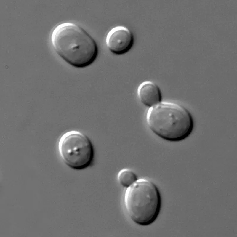
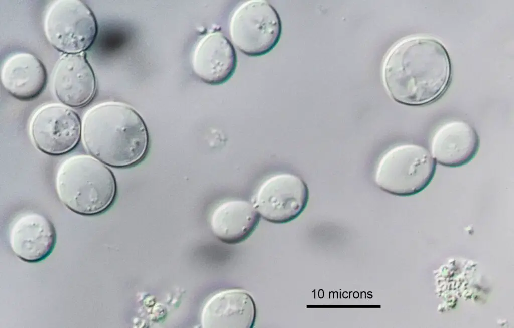
- Oval Organisms: At higher magnifications, such as 1000x and beyond, yeast cells reveal themselves as oval, egg-shaped entities. These distinctive shapes serve as identifiers of these remarkable organisms.
- Budding Structures: The microscope also grants us the privilege of witnessing budding, a form of reproduction in yeast. Some cells exhibit small buds emerging from their surfaces.
- Fermentation Clues: When the specimen contains sugar, bubbles become evident in the view. These bubbles, a byproduct of the fermentation process, offer visual evidence of the microorganisms’ metabolic activity.
With Fluorescence Microscopy
- Unveiling Organelles: Fluorescence microscopy becomes a powerful tool for unveiling the inner world of yeast cells. This technique allows us to peer into the intricate realm of cell organelles.
- Challenges and Solutions: While yeast cells’ small size and protective cell wall pose challenges, fluorescence microscopy conquers these barriers. By employing various dyes, we enhance contrast and distinguish different organelles.
Specific Dyes for Clarity
- Nuclei – DAPI: The DAPI dye sheds light on nuclei within the yeast cells, enabling us to pinpoint their locations.
- Vacuoles – FM4-64: The FM4-64 dye highlights vacuoles, offering insights into these vital storage compartments.
- Endoplasmic Reticulum and Mitochondria – DIOC6: DIOC6 dye aids in the visualization of both endoplasmic reticulum and mitochondria.
- Mitochondria – DASPMI: DASPMI dye further aids in highlighting mitochondria, crucial energy producers within cells.
- Cell Wall – Calcofluor White: The Calcofluor White dye assists in staining the cell wall, enhancing visibility of this essential structure.
Unlocking Minutiae Through Optimal Magnification
- Yeast Size: Yeast cells are notably small, with diameters ranging from 5 to 10 micrometers. Given their size, high magnification is essential for accurate observation.
- Magnification Recommendations: For optimal visual clarity, employ 60x or 100x objectives with a numerical aperture of 1.4. These settings ensure both bright images and maximum resolution.
- Digital Recording: When using a digital camera, a 60x magnification with a numerical aperture of 1.4 is suggested. This captures intricate details, maintaining a balance between visual fidelity and image size.
In essence, the act of observing yeast cells under different microscopy techniques opens up realms of understanding. Through these methods, we unravel the mysteries of yeast cells’ inner workings, furthering our comprehension of these remarkable microorganisms.
Yeast Under the Microscope Images


Yeast Under the Microscope Video
FAQ
What is yeast, and why is it interesting to study under a microscope?
Yeast is a single-celled fungus used in various applications, from baking to brewing. Studying yeast under a microscope allows us to explore its cellular structures and processes, providing insights into its vital functions.
How can I prepare a yeast sample for microscopy?
You can prepare a yeast sample by creating a solution using yeast cake or active yeast, along with water and, optionally, sugar. The yeast solution is then placed on a glass slide with a cover slip for observation.
What are the typical shapes of yeast cells?
Yeast cells often appear oval or egg-shaped when observed under the microscope. Some cells may exhibit budding, which indicates their reproductive process.
Can I observe the fermentation process of yeast under the microscope?
Yes, if you include sugar in the yeast solution, you can witness the fermentation process through the appearance of bubbles in the specimen. These bubbles are a result of the carbon dioxide produced during fermentation.
How can fluorescence microscopy help in studying yeast?
Fluorescence microscopy is used to observe the organelles within yeast cells. It employs specific dyes to highlight structures like nuclei, mitochondria, vacuoles, endoplasmic reticulum, and the cell wall.
Why is staining necessary when observing yeast cells?
Yeast cells are small and can be challenging to differentiate. Staining enhances visibility by coloring specific cellular components, making it easier to identify and study them.
What are some common dyes used for staining yeast cells?
Common dyes include DAPI for nuclei, FM4-64 for vacuoles, DIOC6 for endoplasmic reticulum and mitochondria, DASPMI for mitochondria, and Calcofluor White for the cell wall.
What is the recommended magnification for observing yeast cells?
To capture fine details, a magnification of 60x or 100x, combined with a numerical aperture of 1.4, is suggested. This provides clarity and resolution in the microscopic view.
Can I see yeast cells using a digital camera?
Yes, a digital camera can be used to capture images of yeast cells under the microscope. For optimal results, a magnification of 60x with a numerical aperture of 1.4 is recommended.
Why is studying yeast under the microscope important?
Observing yeast cells under the microscope allows us to delve into their intricate structures, behaviors, and functions. This knowledge contributes to various fields, including biotechnology, food production, and scientific research.
References
- https://www.microscopeclub.com/yeast-under-microscope/
- https://www.microscopemaster.com/yeast-cells-under-the-microscope.html
- https://www2.mrc-lmb.cam.ac.uk/microscopes4schools/yeast.php
- https://biokimicroki.com/microscopic-observation-of-yeasts/
- https://moticmicroscopes.com/blogs/articles/microscopy-of-yeast
- https://naomedical.com/info/yeast-under-microscope.html
- https://microscopeclarity.com/yeast-an-amazing-microorganism/