What is Paramecium?
- Paramecium is a remarkable genus of unicellular microorganisms that falls within the phylum Ciliophora, a diverse group of eukaryotes characterized by their use of hair-like structures called cilia for movement and feeding. Paramecia are abundant in various aquatic habitats, including freshwater bodies such as ponds, lakes, and streams. Their presence in these environments is vital to the balance of ecosystems, as they serve both as predators, consuming bacteria and other small organisms, and as prey for larger organisms in the microbial food web.
- Distinctive in appearance, Paramecia showcase an elongated shape, typically ranging from 50 to 350 micrometers in length. This elongation facilitates their efficient movement through water, driven by the rhythmic beating of their cilia. The coordinated motion of these cilia not only propels the Paramecium forward but also generates currents that sweep food particles into the organism’s oral groove. This feeding process involves engulfing the captured particles into a specialized feeding organelle called the oral groove, which then leads to the formation of food vacuoles where digestion occurs.
- As protists, Paramecia belong to a group of diverse eukaryotic microorganisms that don’t fit neatly into the classification of plants, animals, or fungi. This complexity has made them invaluable subjects of scientific research, particularly in the early exploration of cellular processes, genetics, and cellular regeneration. Due to their relatively large size and ease of cultivation, Paramecia have served as model organisms to unravel fundamental biological concepts and mechanisms.
- One of the intriguing aspects of Paramecia is their capacity for intricate behaviors. These microorganisms exhibit responsiveness to environmental stimuli, allowing them to tactically navigate their surroundings and avoid obstacles in their path. Additionally, their ability to display negative geotaxis, moving against gravity, demonstrates their adaptation to diverse ecological niches within aquatic habitats.
- A notable feature of Paramecia’s reproductive biology is conjugation, a form of sexual reproduction. During conjugation, two individual Paramecia come together and exchange genetic material, enhancing genetic diversity within the population. This process contributes to the resilience and adaptability of Paramecia in fluctuating environments.
- In summary, Paramecium exemplifies the captivating intricacies of the microscopic world, offering profound insights into cellular biology and ecological dynamics. As an integral part of aquatic ecosystems and a subject of ongoing scientific exploration, these remarkable microorganisms continue to broaden our understanding of life’s complexity and evolution.
Requirement for Observation of Paramecium Under Microscope
The observation and study of Paramecium, a type of microscopic organism, require specific tools and materials to ensure accurate and detailed examination. These components play a crucial role in microscopy, allowing scientists and researchers to gain insights into the biology and behavior of Paramecium. Here are the essential requirements for conducting microscopy of Paramecium:
- Paramecium Culture: Before microscopy, it is necessary to have a healthy and thriving population of Paramecium. These microorganisms are typically cultured in a controlled environment to provide them with the necessary nutrients and conditions for growth. A suitable Paramecium culture serves as the source of specimens for microscopic analysis.
- Protoslo Solution: Protoslo solution is a specialized substance used to temporarily slow down the movement of Paramecium. This is important because Paramecia are known for their rapid and agile motion, which can make detailed observation challenging. Adding a small amount of Protoslo solution to the microscope slide helps immobilize the Paramecia, making it easier to focus on specific features and behaviors.
- Microscope Slides: Microscope slides are flat, transparent surfaces where the Paramecia samples are placed for examination under the microscope. These slides provide a stable platform for observing the organisms and allow for precise focusing and magnification. Properly prepared slides are essential to ensure clear and accurate observations.
- Transfer Pipettes: Transfer pipettes are slender tools used to carefully transfer a small sample of Paramecia from the culture to the microscope slide. The pipettes allow for controlled and accurate placement of the organisms on the slide, minimizing disturbance to their natural state.
- Coverslips: Coverslips are thin, transparent pieces of glass that are gently placed over the Paramecia sample on the microscope slide. They serve to protect the sample, prevent drying, and provide a flat surface for the microscope objective to focus on. Coverslips also help maintain a consistent distance between the microscope lens and the specimen.
In conclusion, the process of microscopy for studying Paramecium involves several important requirements. These include having a healthy Paramecium culture, using Protoslo solution to slow down movement, utilizing microscope slides for sample placement, employing transfer pipettes for precise transfer, and using coverslips to protect and enhance visibility. These tools and materials collectively enable researchers to delve into the intricate details of Paramecium biology and behavior under the microscope.
Culture Preparation
The process of preparing a culture of Paramecium is a critical step to ensure their well-being and growth. Here’s how to do it:
- Unpacking and Aeration: When you receive your Paramecium culture, begin by gently loosening the lid of the container. To provide the culture with air, use a plastic pipette. Dip the tip of the pipette into the culture water, and then gently squeeze the bulb to release a bit of air into the water. Release the bulb, allowing it to fill with air again. Repeat this action four more times. This process helps replenish the oxygen that might have been depleted during shipping.
- Lid Replacement: After aerating the culture, put the lid back on the container. It’s important not to screw the lid on too tightly. A loose lid allows for proper airflow while preventing any excess moisture buildup.
- Storage: Find a suitable place to store the Paramecium culture. Choose a dark cupboard or storage area that is away from direct sunlight. This dark environment helps create the optimal conditions for the Paramecia to thrive and avoids unnecessary exposure to light.
By following these steps, you’re taking the necessary measures to ensure the health and success of your Paramecium culture. This initial preparation sets the stage for observing and studying these fascinating microorganisms.
Paramecium culture procedure
Creating a Paramecium culture is a systematic process that involves several steps to ensure the proper growth and health of these microorganisms. Here is a simplified explanation of the Paramecium culture procedure:
Materials Needed:
- 2L flask
- 2.5g cerophyl powder (wheat grass powder)
- 3/4 g dibase sodium phosphate powder
- Klebsiella starter culture
- Large jar (<2L)
- Hotplate
Procedure:
- Start with a clean 2L flask and fill it with 1L of distilled water.
- Measure out 2.5g of wheat grass powder and 3/4 of a gram of sodium phosphate. Add these powders to the water in the flask.
- Heat the mixture on a hotplate until it comes to a boil.
- Allow the mixture to cool down to room temperature.
- Optionally, you can filter the cooled mixture through cotton or a special nitex filter to remove any wheat grass particles.
- Another optional step is to autoclave the cooled and filtered mixture. Autoclaving helps ensure that the culture is free from unwanted contaminants.
- Inoculate the mixture with klebsiella medium. Take a piece of the agar with bacteria and add it directly to the mixture. The agar piece should be about the size of your thumbnail.
- Let the mixture incubate overnight (8-12 hours) at a temperature of 37 degrees Celsius. This higher temperature is necessary for the bacteria to grow well.
- Transfer the mixture into a large jar. You can also add the mix directly to an existing Paramecium culture for faster growth.
- Allow the Paramecium population to expand over 1-3 days.
- A mature culture should show a cloud-like appearance in the jar, indicating a visible Paramecium population. You can confirm this using a dissection microscope where the presence of Paramecia should be clearly noticeable.
- Every 1-4 weeks, depending on the need, replenish the culture with new food by repeating steps 1-8.
By following these steps, you can create and maintain a healthy Paramecium culture, providing an environment for their growth and observation.
Slide Preparation of Paramecium
- Begin by placing your Paramecium culture on your workspace. Take a moment to observe the culture with your naked eye and make note of what you see.
- To collect a sample from the culture, hold the pipette bulb between your thumb and forefinger. Lower the pipette tip to the bottom of the culture jar. Gently release the pressure on the bulb, allowing the sample to be drawn into the pipette. It’s a good idea to take the sample from the bottom of the jar, where the Paramecium concentration is likely to be highest.
- On a microscope slide, dispense 2-3 drops of your collected sample. Add a drop of Protoslo solution as well. Thoroughly mix the sample and Protoslo together on the slide.
- Carefully place a coverslip over the mixture on the slide. This coverslip helps create a flat and clear viewing surface.
- To locate the Paramecia on the slide, start by using the low-power objective on your microscope. Position the Paramecium in the center of the field of view. Once you’ve found it, switch to the high-power objective and refocus the microscope to see the Paramecium in greater detail.
By following these steps, you can create a wet mount slide that allows you to observe Paramecia under a microscope. This process helps you study these microscopic organisms and gain insights into their biology and behavior.
Observation and Result
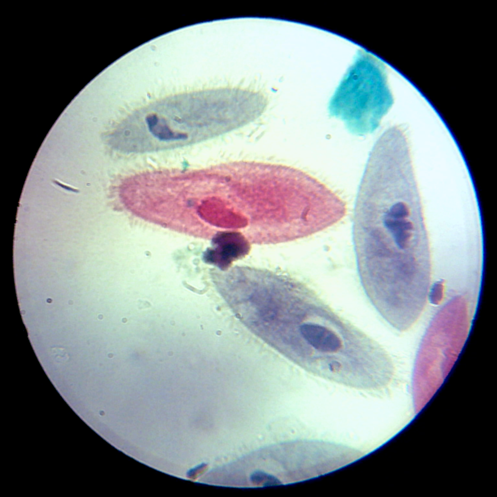
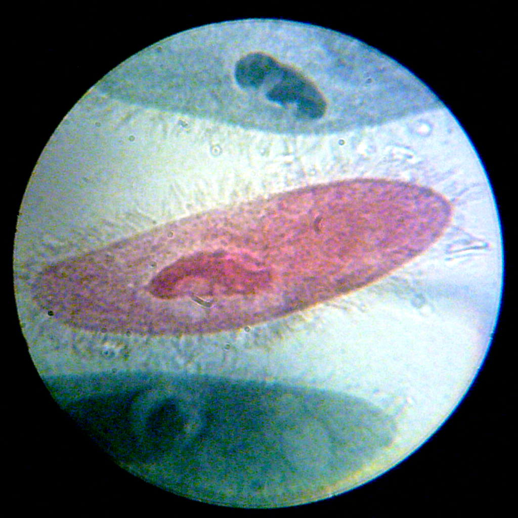
When observing Paramecium under a microscope, you can notice several distinct features and structures that reveal important aspects of their biology. Here are the observations you can make and the results you can infer from these observations:
- Oral Groove: On one side of the Paramecium’s body, you can see a depression known as the oral groove. This groove extends from the front of the Paramecium to a point beyond the middle. The purpose of the oral groove is to guide food particles, particularly bacteria, into the pharynx or gullet. As food particles collect in the bottom of this groove, they form new food vacuoles.
- Food Vacuoles: Within the cytoplasm, you can observe spherical structures of varying sizes known as food vacuoles. These food vacuoles serve as storage units for food during the digestion process.
- Contractile Vacuole: Another noticeable structure is the contractile vacuole. This clear, spherical structure often expands to a larger size and then suddenly collapses. It contracts by releasing its contents into the surrounding environment. Interestingly, the contractile vacuole is located externally to the cell itself. Take note of the number of contractile vacuoles you can observe in the Paramecium.
- Radiating Canals: Connected to the contractile vacuoles, you might see channels in the cytoplasm that give a star-like pattern to the contractile vacuoles. These radiating canals play a role in the movement and function of the contractile vacuole.
- Cilia: Covering the surface of the Paramecium, you will see hair-like organelles called cilia. These cilia are responsible for locomotion, enabling the Paramecium to move. They also play a role in food retrieval. Cilia in the oral groove sweep particles towards the pharynx in a coordinated motion.
In conclusion, observing Paramecium under a microscope allows you to witness these fascinating structures and behaviors. By carefully studying these features, you can gain insights into how Paramecium interact with their environment, obtain and process food, and move in their aquatic habitats.
Paramecium Under Microscope Images


Paramecium Under Microscope Video

FAQ
What is Paramecium?
Paramecium is a single-celled, microscopic organism belonging to the phylum Ciliophora. It is known for its distinct shape and cilia-covered surface.
Why is Paramecium observed under a microscope?
Microscopic observation allows us to study Paramecium’s anatomy, behavior, and interactions with its environment in detail.
How do you prepare a Paramecium slide for microscopy?
A Paramecium slide is prepared by placing a sample of the culture on a slide, adding Protoslo for immobilization, and covering it with a coverslip.
What are cilia, and how are they important for Paramecium?
Cilia are hair-like structures on Paramecium’s surface that help with movement and food capture by creating currents in the water.
What is the function of the oral groove in Paramecium?
The oral groove guides food particles, mainly bacteria, into the pharynx for digestion and nutrient absorption.
What are food vacuoles in Paramecium?
Food vacuoles are structures in the cytoplasm that store and digest food particles, playing a crucial role in the organism’s nutrition.
What is a contractile vacuole, and what does it do?
The contractile vacuole helps regulate water balance by expelling excess water from the cell through sudden contractions.
How can I count contractile vacuoles under a microscope?
Observe the Paramecium’s movement and monitor the sudden contractions of the contractile vacuole; the number of visible contractions can indicate the health of the cell.
What are radiating canals in Paramecium?
Radiating canals are channels connected to the contractile vacuoles, assisting in the expulsion of excess water.
How can I focus on Paramecium using different microscope objectives?
Begin with the low-power objective to locate Paramecia. Once found, switch to the high-power objective for a detailed view and refocus to observe finer structures.