

Plasmolysis is a process in which the protoplast of a plant cell shrinks away from the cell wall because of loss of water from the cell. It occurs when a living plant cell is placed in a hypertonic solution having lower water potential than that of the cell. During this process, water moves out from the vacuole and cytoplasm by osmosis. The vacuole becomes smaller and the turgor pressure is lost.
As the loss of water continues, the plasma membrane along with the cytoplasm starts pulling away from the rigid cell wall. This condition is referred to as plasmolysis. The cell wall does not shrink in the same manner because it is rigid. When the plasmolysed living cell is again placed in a suitable hypotonic solution, water enters into the cell and the protoplast returns toward its original position. This reverse process is known as deplasmolysis.
Structures Involved in Plasmolysis
The major structures involved during plasmolysis are as follows-
- Cell wall- It is the rigid outer covering of the plant cell and retains the general shape of cell during plasmolysis. The protoplast moves away from this wall. Thus, a space becomes visible between the cell wall and the plasma membrane.
- Plasma membrane- The plasma membrane occurs just below the cell wall and surrounds the protoplast. During loss of water, it is pulled inward together with the shrinking protoplast. At some regions, thin connections between the membrane and cell wall remain present. These are referred to as Hechtian strands.
- Vacuole and Tonoplast- The large central vacuole contains the cell sap and occupies a major volume of a mature plant cell. In a hypertonic medium, water is largely lost from the vacuole and hence its volume decreases. Tonoplast (vacuolar membrane) also adjusts with the decreasing vacuolar volume. The turgor pressure of the cell goes down during this process.
- Cytoplasm and Protoplast- Cytoplasm along with the structures enclosed within the plasma membrane forms the living protoplast. It loses water during plasmolysis. As the volume is reduced, the protoplast shrinks and gets pulled away from the rigid cell wall, producing the characteristic plasmolysed condition.
Why Does Plasmolysis Occur?
Plasmolysis occurs due to the movement of water from a plant cell when it is kept in a hypertonic solution. The major reasons are as follows-
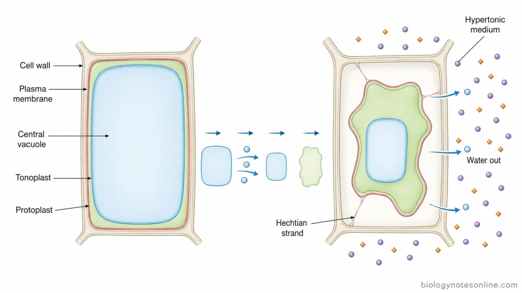
- Hypertonic external solution- When the surrounding solution contains more dissolved solutes as compared to the cell sap, it has a lower water potential. This creates a difference between the water potential inside and outside of the cell.
- Movement of water by osmosis- Due to this difference, water starts moving out through the selectively permeable plasma membrane. The movement takes place from higher water potential to lower water potential. Thus, water is lost from the plant cell into its surrounding solution.
- Loss of water from vacuole- The large vacuole contains most of the water of a mature plant cell. During this process, the vacuole loses water and becomes smaller. Hence, the turgor pressure which normally keeps the protoplast pressed against the cell wall also decreases.
- Decrease in protoplast volume- As more water goes out, the cytoplasm along with vacuole starts shrinking. The rigid cell wall does not shrink along with it. Therefore, the protoplast gradually pulls away from the cell wall and the cell becomes plasmolysed.
- Loss of turgidity- A normal plant cell remains turgid when sufficient water is present. During plasmolysis this condition is lost because the cell loses water and turgor pressure goes down. The cell becomes flaccid, and with further water loss, separation of the protoplast from cell wall takes place.
What Happens During Plasmolysis?
The process of plasmolysis occurs in a sequence of changes in the vacuole, protoplast and plasma membrane. The following are the major changes-
- Water moves out of the cell- When the plant cell is exposed to a hypertonic solution, water begins to move out through the selectively permeable membranes by osmosis. The outward movement is due to the lower water potential of the external solution.
- Vacuole decreases in volume- The central vacuole is the major compartment from which water is lost during plasmolysis. Thus, the vacuole starts becoming smaller. The tonoplast also adjusts with this decrease in vacuolar volume.
- Turgor pressure falls- Loss of water from the vacuole results in decrease of turgor pressure. At first, the cell loses its turgid condition. Further loss of water allows the protoplast to withdraw from the wall.
- Protoplast contracts- During this process, the volume of the living protoplast becomes smaller. The rigid cell wall, however, generally retains its position and shape. The shrinking takes place mainly in the membrane-covered protoplast.
- Plasma membrane separates from the cell wall- As the protoplast contracts further, the plasma membrane starts pulling away from some regions of the cell wall. The separation may first appear as concave pockets. At some attachment regions the membrane remains connected with the wall through fine Hechtian strands.
- Plasmolysis becomes more extensive- With continued water loss, a greater part of the protoplast is separated from the cell wall and it can become more rounded or convex. This is a more advanced plasmolysed condition. The cell wall itself does not normally collapse during ordinary plasmolysis. Collapse of both the wall and protoplast is a different response, referred to as cytorrhysis.
Stages of Plasmolysis
Based on the extent of separation of the protoplast from the cell wall, plasmolysis can be observed through different stages. The major stages are as follows-
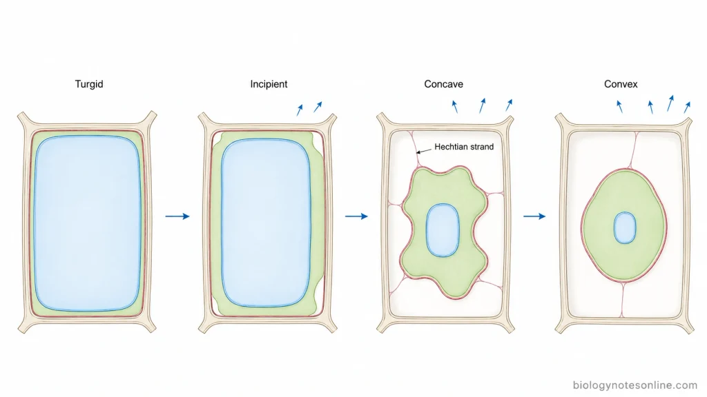
1. Incipient Plasmolysis
Incipient plasmolysis is the initial stage of plasmolysis. In this stage, turgor pressure becomes almost zero and the plasma membrane just starts separating from the cell wall at some regions. The protoplast has only started to withdraw. This point is also used for determination of osmotic properties of plant cells.
2. Concave Plasmolysis
With further loss of water, greater separation of the protoplast takes place. The plasma membrane pulls away unevenly from the cell wall and inward curved regions are produced. This condition is called concave plasmolysis. At several places the protoplast may still remain connected to the wall by Hechtian strands, therefore the withdrawn surface does not become completely rounded.
3. Convex Plasmolysis
When water loss continues, the protoplast becomes much more contracted and separation from the cell wall is extensive. It takes a rounded or convex form inside the cell. This is referred to as convex plasmolysis. The extent of protoplast retraction is greater at this stage, although some wall-plasma membrane connections may still remain present.
Types of Plasmolysis
Plasmolysis is mainly classified into two types based on the shape taken by the protoplast after separation from the cell wall. These include concave plasmolysis and convex plasmolysis.
1. Concave Plasmolysis
Concave plasmolysis- In this type, the protoplast does not separate uniformly from the cell wall. The plasma membrane pulls inward at different regions and several concave pockets are formed. Some portions still remain attached with the cell wall, hence giving an irregular appearance to the plasmolysed protoplast. Hechtian strands may be present between the withdrawing plasma membrane and cell wall. Concave plasmolysis can change into convex form with further withdrawal of the protoplast.
2. Convex Plasmolysis
Convex plasmolysis- It is the type where the protoplast separates more evenly from the cell wall and becomes rounded. The withdrawn protoplast shows convex surfaces and may appear almost spherical inside the rigid cell wall. This form has a greater and more uniform separation as compared to the concave type. However, connections with the wall through Hechtian structures can still remain at some places.
What Is Deplasmolysis?
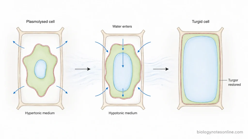
Deplasmolysis is the reverse process of plasmolysis in which a plasmolysed living plant cell again takes up water and returns toward its normal turgid condition. It occurs when the plasmolysed cell is transferred from a hypertonic medium to water or a suitable hypotonic solution.
During this process, water enters into the cell by osmosis. The vacuole starts increasing in volume and turgor pressure is gradually restored. As more water enters, the contracted protoplast expands and the plasma membrane moves back towards the cell wall. Thus, the separation produced during plasmolysis is reduced and the cell becomes turgid again. Deplasmolysis shows that plasmolysis can be reversible when the cell remains living and has not undergone irreversible injury.
How Is Plasmolysis Observed in the Laboratory?
Plasmolysis can be easily observed in plant cells under a light microscope. Onion epidermal cells or Elodea leaf cells are commonly used for this demonstration. The following steps are involved-
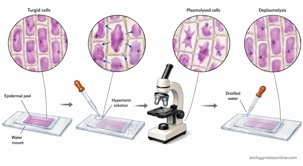
- Preparation of cells- A thin onion epidermal peel or an Elodea leaf is placed on a clean microscopic slide with water and covered with a coverslip. The cells are first observed under the microscope. In this condition, the protoplast remains pressed near the cell wall and the vacuole occupies a large portion of the cell.
- Addition of hypertonic solution- A concentrated salt or sugar solution is now brought in contact with the plant cells. Salt solutions and sucrose solutions are commonly used for this purpose in laboratory demonstrations.
- Water starts moving out- The hypertonic solution causes water to move out from the cells by osmosis. The vacuole becomes smaller and the turgid condition is gradually lost.
- Observation of plasmolysis- After some time, the protoplast is seen withdrawing from the cell wall. The plasma membrane and cytoplasmic contents are no longer closely pressed against the wall. This visible separation indicates plasmolysis.
- Comparison of cells- The cells before and after addition of the hypertonic solution can be compared under the microscope. Normal cells show an expanded protoplast, while plasmolysed cells show a contracted protoplast separated from the wall. If the plasmolysed cells are again supplied with distilled water, recovery of the cells may also be observed by deplasmolysis.
Examples of Plasmolysis
Plasmolysis can be observed in many living plant cells when they are exposed to concentrated salt or sugar solutions. Some of the common examples are as follows-
- Onion epidermal cells- Red onion epidermis is commonly used for showing plasmolysis under microscope. When these cells are placed in a strong salt solution such as KCl or NaCl, water comes out from the cells and the protoplast gets separated from the cell wall. The coloured vacuole also makes this change easier to observe.
- Elodea leaf cells- Plasmolysis can also be seen in the cells of Elodea. A concentrated NaCl or sucrose solution causes the protoplast to shrink and pull away from the wall. Chloroplasts which were present along the cell periphery become collected with the contracted protoplast.
- Arabidopsis cells- In experimental studies, cells of Arabidopsis thaliana are plasmolysed using hyperosmotic compounds such as sucrose, mannitol or sorbitol. During this condition, the vacuole loses water first, followed by shrinking and withdrawal of the protoplast.
- Moss protonemal cells- Plasmolysis is also produced in living moss protonemata by using concentrated salt or sugar solutions. It is used to make the plasma membrane, cell wall and membrane-wall connections more clearly distinguishable. Fine Hechtian strands can also remain attached during this process.
- Large algal cells- Cells of genera such as Nitella and Chara have also been used for studying plasmolysis. Their large cells make changes of the protoplast and its surface layers suitable for experimental observation.
Biological Significance of Plasmolysis
Some of the important biological significance of plasmolysis are as follows-
- Indicates osmotic stress- Plasmolysis shows excessive loss of water from a plant cell under hypertonic condition.
- Study of water relations- It is used to study turgor pressure, osmotic behaviour and water relations of plant cells.
- Shows membrane-wall connection- During plasmolysis, some parts of plasma membrane remain connected with the cell wall through Hechtian strands.
- Study of cell wall integrity- Plasmolysis helps in studying the mechanical association between the plasma membrane and cell wall.
- Study of dehydration response- It is useful to observe changes in vacuole, protoplast and plasma membrane during water loss and recovery.
Quick Summary Table of Plasmolysis
| Feature | Plasmolysis |
|---|---|
| Definition | Shrinking of protoplast away from the cell wall due to loss of water. |
| Condition required | Cell is placed in a hypertonic solution. |
| Movement of water | Water moves out from the cell by osmosis. |
| Vacuole | Becomes smaller due to loss of water. |
| Turgor pressure | Decreases and finally becomes very low. |
| Protoplast | Shrinks and gets pulled away from the cell wall. |
| Cell wall | Remains rigid and maintains the general cell shape. |
| Main types | Concave plasmolysis and convex plasmolysis. |
| Stages | Incipient, concave and convex plasmolysis. |
| Reverse process | Deplasmolysis. |
| Common examples | Onion epidermal cells and Elodea leaf cells. |
| Significance | Used to study osmosis, turgor, water relations and membrane-wall connection. |
References
- Arico, D. S., Dickmann, J. E. M., Hamant, O., & Canut, H. (2023). The plasma membrane–cell wall nexus in plant cells: Focus on the Hechtian structure. The Cell Surface, 10, 100115. https://doi.org/10.1016/j.tcsw.2023.100115
- Canut, H., Carrasco, A., Galaud, J. P., Cassan, C., Bouyssou, H., Vita, N., Ferrara, P., & Pont-Lezica, R. (1998). High affinity RGD-binding sites at the plasma membrane of Arabidopsis thaliana links the cell wall. The Plant Journal, 16(1), 63–71. https://doi.org/10.1046/j.1365-313x.1998.00276.x
- Cheng, X., Lang, I., Adeniji, O. S., & Griffing, L. (2017). Plasmolysis-deplasmolysis causes changes in endoplasmic reticulum form, movement, flow, and cytoskeletal association. Journal of Experimental Botany, 68(15), 4075–4087. https://doi.org/10.1093/jxb/erx243
- Harant, D., & Lang, I. (2021). 3D dissection of structural membrane-wall contacts in filamentous moss protonemata. International Journal of Molecular Sciences, 22(1), 158. https://doi.org/10.3390/ijms22010158
- Haswell, E. S., & Verslues, P. E. (2015). The ongoing search for the molecular basis of plant osmosensing. Journal of General Physiology, 145(5), 389–394. https://doi.org/10.1085/jgp.201411295
- Kaplan, F., Lewis, L. A., Herburger, K., & Holzinger, A. (2013). Osmotic stress in Arctic and Antarctic strains of the green alga Zygnema (Zygnematales, Streptophyta): Effects on photosynthesis and ultrastructure. Micron, 44, 317–330. https://doi.org/10.1016/j.micron.2012.08.004
- Lang, I., Barton, D. A., & Overall, R. L. (2004). Membrane-wall attachments in plasmolysed plant cells. Protoplasma, 224(3–4), 231–243. https://doi.org/10.1007/s00709-004-0062-6
- Lang, I., Sassmann, S., Schmidt, B., & Komis, G. (2014). Plasmolysis: Loss of turgor and beyond. Plants, 3(4), 583–593. https://doi.org/10.3390/plants3040583
- Osterhout, W. J. V. (1943). Studies of the inner and outer protoplasmic surfaces of large plant cells: I. Plasmolysis due to salts. Journal of General Physiology, 27(2), 139–142. https://doi.org/10.1085/jgp.27.2.139
- Reisen, D., Marty, F., & Leborgne-Castel, N. (2005). New insights into the tonoplast architecture of plant vacuoles and vacuolar dynamics during osmotic stress. BMC Plant Biology, 5, 13. https://doi.org/10.1186/1471-2229-5-13
- Thompson, D. S., & Islam, A. (2021). Plant cell wall hydration and plant physiology: An exploration of the consequences of direct effects of water deficit on the plant cell wall. Plants, 10(7), 1263. https://doi.org/10.3390/plants10071263
- Weber, A., Braybrook, S., Huflejt, M., Mosca, G., Routier-Kierzkowska, A.-L., & Smith, R. S. (2015). Measuring the mechanical properties of plant cells by combining micro-indentation with osmotic treatments. Journal of Experimental Botany, 66(11), 3229–3241. https://doi.org/10.1093/jxb/erv135
- Yoneda, A., Ohtani, M., Katagiri, D., Hosokawa, Y., & Demura, T. (2020). Hechtian strands transmit cell wall integrity signals in plant cells. Plants, 9(5), 604. https://doi.org/10.3390/plants9050604