

Developmental biology is a branch of biology that studies how an organism develops and changes through its life, including the embryonic and other developmental processes. Development is not only an increase in size.
It includes formation of different cell types, their arrangement in correct places, and the changes in form, size, and function of the developing organism with time through cell differentiation, morphogenesis, and growth.
The field covers both animal and plant development. In plants, development extends much beyond the embryo, as meristems continue to produce new cells and structures during post-embryonic growth.
Most cells of an organism are genetically related and generally carry the same DNA, still they can become structurally and functionally different by expressing different sets of genes. The major question in developmental biology is how such cells become different from one another and then get organized into tissues and organs to make a functional organism.
Levels of Developmental Organization
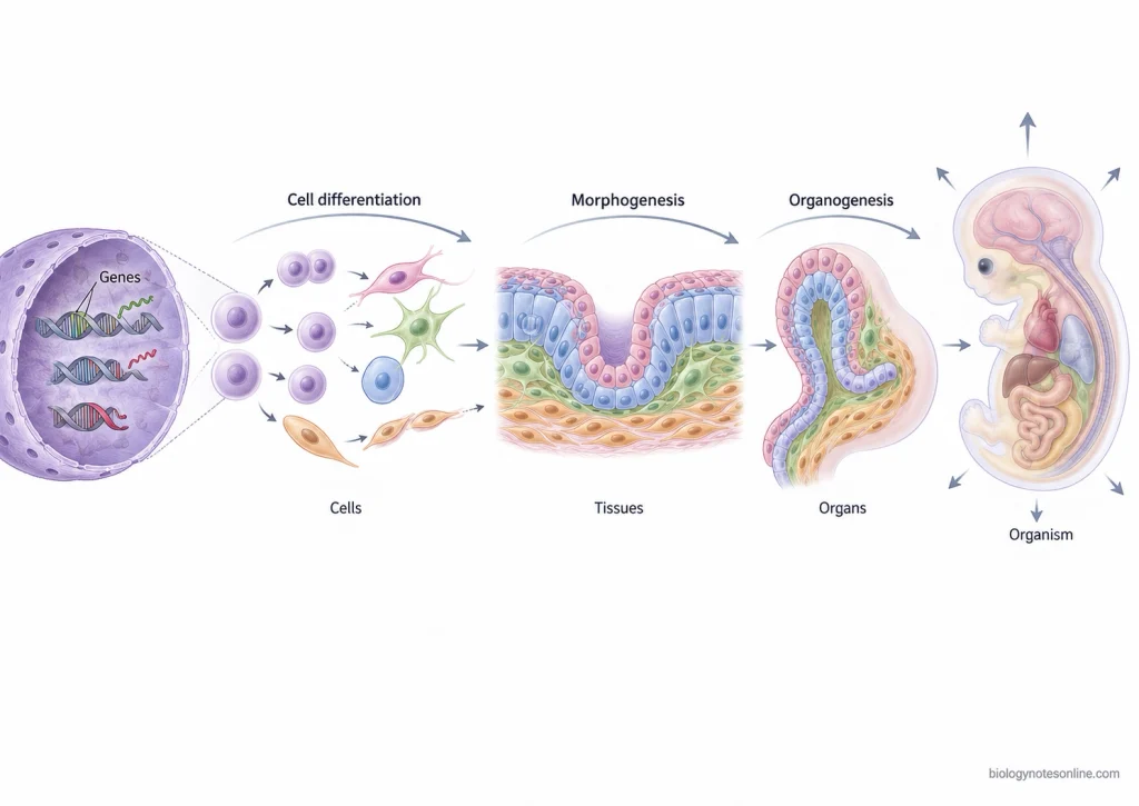
- Molecular or genetic level- Development at the molecular level is based largely on the controlled expression of genes. Although most cells contain essentially the same genome, different sets of genes are expressed in different cells. Cell signaling and gene regulation take part in deciding cell identity and developmental responses.
- Cellular level- Cells divide, grow and become different from one another by cell differentiation. During development, cells can also move from one place to another, change their shape, attach with other cells or undergo programmed cell death. These cellular activities are used to build up the developing body.
- Tissue level- Different cells are not distributed randomly. They are arranged into organized groups and tissues, with a particular position and structural pattern. This formation of ordered form is referred to as morphogenesis, and involves cell movement, adhesion, division, growth and changes in cell shape.
- Organ level- Tissues are further brought together in a definite arrangement to produce organs. The formation of organs during development is known as organogenesis. In animal embryos, tissues derived from the germ layers differentiate and interact during formation of the different organs.
- Organism level- At this level, development involves coordination of the different tissues and organs within the complete organism. Growth of different body parts has to be regulated with their position, size and function while the organism is developing.
Developmental Biology vs Embryology
Embryology and developmental biology overlap closely, but they are not exactly the same. Embryology is mainly concerned with formation, structure and development of the embryo, while developmental biology includes embryonic development and other developmental processes also. The field can cover development from fertilization through later life, including post-embryonic growth, metamorphosis, regeneration and aging.
| Feature | Developmental Biology | Embryology |
|---|---|---|
| Scope | It is a broader field studying the mechanisms by which organisms develop. Gene expression, cell differentiation, morphogenesis, tissue and organ formation and other developmental changes are studied. | It is the study of formation and development of the embryo. Descriptive, comparative and experimental study of embryos are major parts of embryology. |
| Life stages covered | Development is studied during embryonic as well as other stages. Depending on the organism and question, it extends to post-embryonic development, metamorphosis, regeneration and aging. Plant development also continues extensively after embryogenesis. | Mainly covers the embryonic stages, from fertilization and formation of the embryo through the developmental events occurring before later post-embryonic stages. |
| Typical questions | How do cells having related genetic information become different cell types? How are body patterns produced, and how are cells arranged into tissues and organs? Developmental biology also asks how developmental mechanisms continue or change during later stages of life. | How is an embryo formed? What happens during cleavage, gastrulation and organ formation, and which embryonic cells or tissues give rise to particular structures? Experimental embryology also studies how embryonic cells respond when their normal development is altered. |
| Examples | Cell differentiation, gene regulation during development, body-axis formation, morphogenesis, organ development, plant post-embryonic growth, metamorphosis and regeneration. | Fertilization, cleavage and gastrulation, germ-layer formation, early vertebrate development and development of embryonic organs. |
Core Principles of Developmental Biology
Developmental biology is not organized around one single theory. The following are some of the important recurring principles used to understand how development takes place.
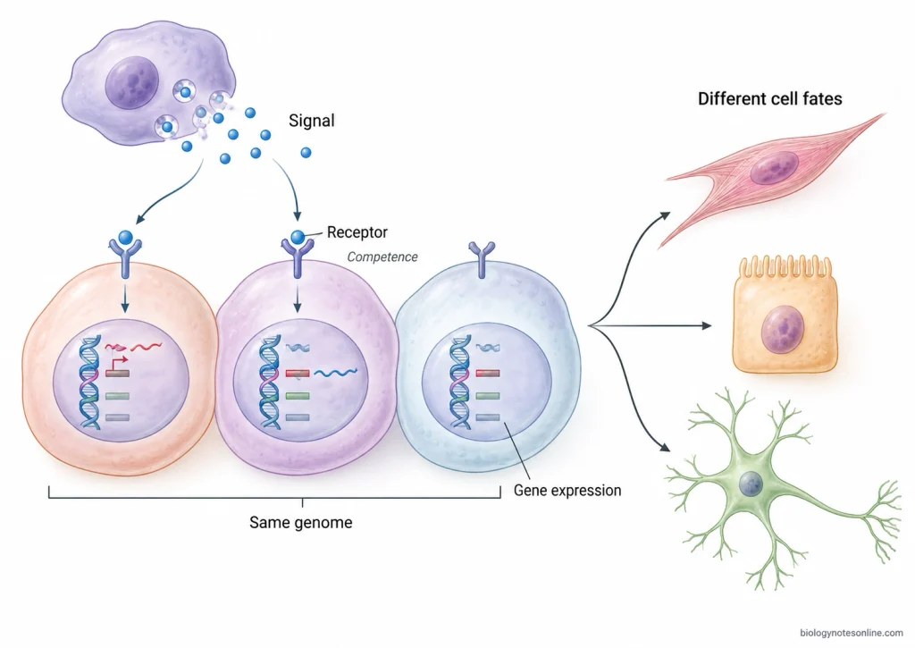
- Genomic equivalence and differential gene expression- Most somatic cells of an organism contain essentially the same genetic information, but all genes are not expressed in every cell. Different sets of genes are switched on or kept inactive at particular places and times. This differential gene expression produces different cell types from genetically related cells.
- Cell fate specification and determination- A developing cell gradually becomes committed to a particular fate. Specification is an earlier and more reversible state, whereas determination represents a more stable commitment before final differentiation. Cell fate may depend on factors already present inside the cell or on information received from surrounding cells.
- Cell-cell signaling and induction- Cells do not develop independently. One group of cells can send signals that change the developmental pathway of another group, which is referred to as induction. The responding cells must also have the proper “competence” to respond. Wnt, Hedgehog, BMP, FGF and Notch are among the signaling systems repeatedly used during development.
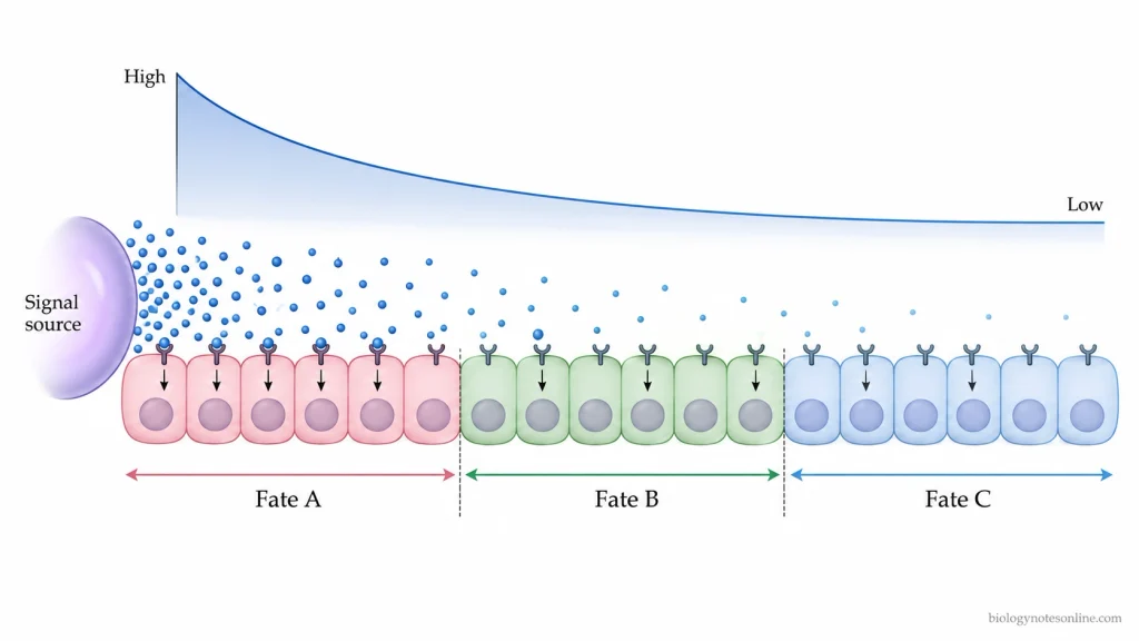
- Positional information and morphogens- The fate of a cell can depend on where it is located in a developing tissue. Some signaling molecules form concentration gradients called morphogen gradients, and cells at different positions can respond differently to the amount or duration of the signal. It is one important mechanism of producing spatial differences within an initially similar group of cells.
- Pattern formation- Development requires cells of different types to be placed in particular spatial arrangements. Body axes, tissue boundaries and regions of future organs are established during this process. Pattern formation is produced by interactions between gene regulation, signaling, positional information and cell behaviour rather than by a single mechanism.
- Morphogenesis- Formation of a body involves more than making different types of cells. Cells change shape, move, divide, become polarized and attach to or separate from neighbouring cells. Such cellular activities, together with mechanical forces, build tissues and organs having a definite three-dimensional form.
- Growth and programmed cell death- Cell proliferation and growth increase the size of developing tissues. At the same time, selected cells can be removed by programmed cell death, including apoptosis. Cell death is used during development for controlling cell number and in the formation or sculpting of some structures.
- Conservation and change of developmental mechanisms- Many developmental genes and signaling pathways are shared between widely different organisms and are repeatedly used at different stages or places. Changes in these developmental gene-regulatory systems, their timing and spatial use can produce differences in organismal form. This relationship between development and evolutionary change is studied in evolutionary developmental biology (evo-devo).
Major Stages of Animal Development
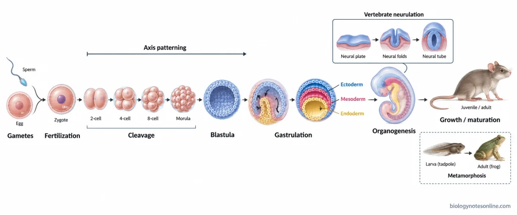
Developmental biology itself does not occur in a fixed series of stages. The following are the major stages of animal development commonly used to study it. Their timing and terminology differ among animals. Some processes also overlap. For example, axis formation can begin during oocyte formation, cleavage or gastrulation depending on the organism, while neurulation is characteristic of vertebrate development and is not a universal stage of all animals.
| Developmental stage | Major event |
|---|---|
| Gametogenesis and fertilization | Formation of gametes followed by sperm-egg fusion and formation of the zygote |
| Cleavage and blastulation | Rapid cell divisions produce numerous blastomeres and an early multicellular embryo |
| Gastrulation | Extensive cell movements and organization of the germ layers |
| Axis formation and patterning | Establishment of body coordinates and spatial identities |
| Neurulation | Formation of the neural tube in vertebrates |
| Organogenesis | Germ-layer derivatives interact and form tissues and organs |
| Growth and postembryonic development | Increase in body size, maturation and later developmental changes, including metamorphosis in animals having a larval stage |
Gametogenesis and Fertilization
Gametogenesis is the formation of gametes, such as sperm and eggs. Germ cells undergo meiosis during this process, producing the haploid condition required for sexual reproduction. Oogenesis and spermatogenesis differ considerably in their cellular details.
During fertilization, the sperm and egg interact and their genetic material is brought together. The egg is also activated to start development, producing the zygote from which embryonic development proceeds.
Cleavage and Blastulation
After fertilization, the zygote undergoes a series of mitotic divisions called cleavage. The large egg cytoplasm is divided among many smaller cells known as blastomeres. There is little increase in the overall amount of embryonic material during these early divisions.
As cleavage proceeds, an early multicellular embryo is produced. In many animals it forms a blastula, although its structure and the pattern of cleavage can be very different between animal groups.
Gastrulation and Germ-Layer Formation
Gastrulation involves large-scale movement and rearrangement of embryonic cells. Cells acquire new positions and new neighbours, changing the relatively simple early embryo into a multilayered body plan.
In triploblastic animals, three primary germ layers are organized during this period, the ectoderm, mesoderm and endoderm. These layers later give rise to different tissues and organs. The form of gastrulation itself varies greatly across animal groups.
Axis Formation and Patterning
The developing embryo must establish positional information. Major body axes include the anterior-posterior, dorsal-ventral and left-right axes, but these are not necessarily established as one separate stage after gastrulation.
Axis specification may start very early. In some animals it is linked with oocyte organization or cleavage, while in others important parts of axis formation continue through gastrulation. Cells are then patterned according to their position along these developing axes.
Neurulation in Vertebrates
In vertebrates, neurulation is the developmental process by which the early nervous system is formed. Neural ectoderm gives rise to the neural plate and this tissue is reorganized to produce the neural tube, the precursor of the brain and spinal cord.
Two modes are recognized, primary neurulation and secondary neurulation. Their contribution differs between vertebrate groups and also along different regions of the same embryo.
Organogenesis
After establishment of the major body plan, cells from the germ layers interact, differentiate and become arranged into tissues and organs. This process is referred to as organogenesis. An individual organ can contain tissues derived from more than one germ layer.
Cell migration, differentiation and tissue interactions occur extensively during this stage. Different organs also start their development at overlapping times rather than appearing one after another in a simple sequence.
Growth, Maturation, and Postembryonic Development
Development continues after the major embryonic organs have been formed. Body size increases, tissues mature and organs acquire their later functional state. Postembryonic development can differ greatly depending on the animal life cycle.
In animals having a distinct larval form, further development may include metamorphosis. Larval tissues can be remodeled, removed or replaced while adult structures develop. This is seen prominently in amphibians and insects, while metamorphic changes also occur in many other animal groups.
Model Systems in Developmental Biology
Only a limited number of organisms are used very extensively as model systems in developmental biology. They can be grown or bred under laboratory condition, their development can be followed experimentally, and genetic or embryological manipulation is possible. Many basic developmental mechanisms are also conserved between widely separated animals, allowing a gene or developmental pathway discovered in one model to be studied in another. Different models, however, are useful for different developmental problems.
| Model system | Major experimental advantage | Typical developmental question |
|---|---|---|
| Drosophila melanogaster | Short generation time, easy breeding and powerful genetic analysis | How genes establish body axes, segments and the body pattern. |
| Caenorhabditis elegans | Small transparent body and highly reproducible cell lineage | How individual cells acquire their fate, divide, differentiate or undergo programmed cell death. |
| Zebrafish (Danio rerio) | Transparent externally developing embryos, large broods and rapid development | How vertebrate organs, tissues and cell populations form in a living embryo. |
| Xenopus | Large externally developing embryos which can be injected, cut and transplanted easily | How embryonic induction, axis formation and early vertebrate organogenesis are controlled. |
| Chick | Embryo can be directly observed, grafted and experimentally manipulated inside or outside the egg | How tissues interact during neural, limb and other vertebrate development. |
| Mouse (Mus musculus) | Mammalian development combined with extensive targeted genetic manipulation | How genes regulate mammalian embryogenesis, organ formation and developmental abnormalities. |
| Arabidopsis thaliana | Small plant, short life cycle, easy cultivation and strong molecular-genetic tools | How meristems, roots, shoots, leaves and flowers are produced during plant development. |
| Sea urchins | Large numbers of transparent, synchronously developing embryos can be fertilized and manipulated in vitro | How fertilization, cell specification, gastrulation and developmental gene regulatory networks operate. |
| Ascidians | Simple chordate embryos with rapid development and highly stereotyped cell lineages | How cell fate, morphogenesis and the basic chordate body plan are established. |
| Axolotls and planarians | Extensive ability to regenerate lost structures | How stem cells, positional information and tissue patterning rebuild damaged body parts. |
Drosophila and Caenorhabditis elegans
Drosophila melanogaster is one of the major genetic models of animal development. It can be bred rapidly and large numbers of mutant animals can be produced and examined. Genetic studies in Drosophila have been especially important for finding genes involved in axis formation, segmentation, positional information and body-plan formation.
Caenorhabditis elegans- It is a small transparent nematode with a highly reproducible developmental lineage. The adult hermaphrodite has 959 somatic cells, and the origin of these cells has been traced from the early embryo. This makes it especially useful for studying cell lineage, asymmetric cell division, cell-fate specification, neuronal differentiation and programmed cell death. Individual cells can be watched in the living animal.
Zebrafish, Xenopus, Chick, and Mouse
Zebrafish- The embryos of zebrafish develop outside the female and remain largely transparent during early development. Large numbers can be obtained. Development is rapid. Living cells, blood vessels and developing organs can be followed directly by microscopy, while genetic screens are used to identify genes controlling vertebrate development.
Xenopus- Frogs of the genus Xenopus produce large eggs and embryos that develop externally. The early blastomeres can be injected with RNA, DNA or other materials, and embryonic tissues are suitable for cutting, grafting and explant experiments. It is widely used to work out embryonic induction, germ-layer specification, axis formation and organogenesis.
Chick embryos are quite different experimentally. The embryo can be reached through the egg and its tissues can be removed, transplanted or labelled while development continues. Such manipulation has been used extensively for questions involving neural induction, neural crest migration and limb patterning.
Mouse- Mouse is the principal model for studying development in a mammalian system. Genes can be removed, altered or expressed in selected tissues, allowing developmental effects to be examined in the whole animal. Mammalian organogenesis, embryonic signaling and genetic causes of developmental defects are major areas studied with mouse models.
Arabidopsis and Plant Development
Arabidopsis thaliana is a small flowering plant used extensively for genetic and molecular study of plant development. It has a compact, well-characterized genome, produces many seeds and is comparatively easy to grow and manipulate.
Plant development continues from active meristems, rather than being largely completed during embryogenesis as in many animals. Arabidopsis is therefore used to study maintenance of stem-cell populations in meristems, root and shoot patterning, organ initiation and flower development. Mutant plants can survive quite major alterations of organ number or arrangement, making developmental changes readily visible.
Other Specialized Developmental Models
Sea urchins- A single spawning can provide very large numbers of eggs. Fertilization takes place experimentally in seawater, embryos develop synchronously and they are optically clear. Sea urchins have been especially useful for studies of fertilization, early cell specification, gastrulation and developmental gene regulatory networks (GRNs).
Ascidians- These marine chordates have simple embryos with stereotyped cleavage and highly reproducible cell lineages. Their embryos contain relatively few cells, so changes in cell fate and morphogenetic movements can be followed at cellular level. They are used for studying early chordate development, notochord formation and the relation between cell fate and embryo shape.
Axolotls are used where regeneration is the main developmental question. After limb loss, a regenerative program forms new tissues and restores the missing structure, providing a vertebrate system for studying pattern formation and the signals required for successful regeneration.
Planarians- These flatworms contain adult stem cells called neoblasts and can regenerate extensive portions of the body after injury. They are used to study pluripotent stem cells, tissue renewal, re-establishment of body polarity and how positional information is put back during regeneration.
How Developmental Biology Is Studied
The study of developmental biology combines observation with experimental changes to the developing organism. Anatomical, experimental and genetic approaches are used together, while molecular and imaging methods allow the same questions to be studied at cell and gene level.
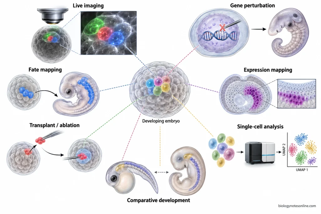
- Normal development is first followed through different developmental stages. Embryos can be examined by microscopy, histological sections and live imaging to see when cells divide, move, change shape or form tissues. Fluorescent reporters make it possible to follow selected cells and proteins in a living embryo over time.
- Cells can be labelled and then followed to find out what they become later. This is used to prepare fate maps and trace cell lineages. A labelled embryonic region may later be found in a particular tissue or organ, linking the early cell position with its developmental fate.
- Parts of an embryo may be removed, transplanted or placed beside another tissue and their development is then observed. Such experiments help to find whether a tissue can develop by itself, whether another tissue provides an inductive signal, and at what time its developmental fate becomes restricted. These experimental embryology approaches test cell and tissue interactions directly.
- Developmental mutants are compared with normal organisms. If disruption of a gene causes the eye, limb, body axis or another structure to develop abnormally, the gene can be investigated for its role in that process. Large genetic screens have been particularly useful for finding genes whose developmental functions were previously unknown.
- A selected gene can also be deliberately changed. Gene knockout, transgenic experiments, RNA interference and newer genome-editing methods such as CRISPR-Cas are used to reduce, remove or modify gene activity and observe what happens during development. Genes may also be altered in selected cells or at a particular developmental time where suitable systems are available.
- Where and when a gene is active is studied along with what happens when that gene is disturbed. RNA localization, reporter genes and protein-detection methods can reveal spatial and temporal patterns of gene expression. An expression pattern by itself does not establish the function of the gene, so it is commonly compared with genetic or experimental perturbation.
- Modern developmental studies can examine gene expression in individual cells rather than averaging many different cells together. Single-cell transcriptomics is used to identify cell states, intermediate populations and changes occurring while cells differentiate. Combined with lineage information, it can help to work out which progenitor cells give rise to particular descendants and how cell-fate decisions develop.
- Development is also compared between organisms. Similar developmental genes, structures or cell behaviours can be examined in different species, while differences are used to study how developmental processes have changed during evolution. Experiments in several model organisms are useful because all developmental questions cannot be tested equally well in one species.
Applications of Developmental Biology
- Congenital disorders- Developmental biology is used to understand how genetic changes or abnormal developmental processes produce congenital malformations. Animal models are also used to study similar developmental defects in humans.
- Teratology and developmental toxicity- It helps to study how drugs, chemicals, infections and other environmental factors interfere with normal embryo development. The developmental stage at which exposure occurs can strongly affect the type of abnormality produced.
- Assisted reproduction- Knowledge of fertilization and early embryo development is applied in in vitro fertilization (IVF) and embryo culture. Culture conditions are studied and improved for supporting preimplantation development before embryo transfer.
- Stem cells and regenerative medicine- Principles of cell differentiation are used to direct stem cells into particular cell and tissue types. Such studies are important for repair or replacement of damaged tissues and for regenerative medicine.
- Organoids and tissue engineering- Developmental signals can be reproduced in culture to make three-dimensional organoids from stem cells. These models are used to study human organ development, diseases and for drug screening.
- Cancer biology- Several signaling pathways controlling normal development, including Wnt, Notch and Hedgehog, are also altered or reactivated in different cancers. Their study helps in understanding tumor growth, cell migration and possible therapeutic targets.
- Crop improvement- Plant developmental studies are used to modify flowering, branching, meristem activity and plant architecture. Developmental regulators are important targets for improving crop yield and other agronomic characters.
History of Developmental Biology
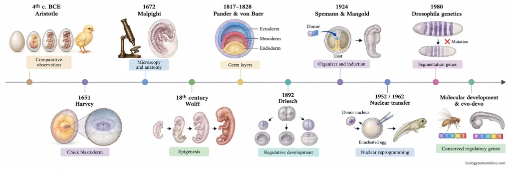
- Developmental studies can be traced to Aristotle in the fourth century BCE, who compared animal embryos and followed chick development by opening eggs at different stages. Early work was mainly descriptive, asking how visible structures appear during development.
- In 1651, William Harvey argued that animals originate from eggs. After microscopes became available, Marcello Malpighi published a microscopic description of chick development in 1672. These observations also became part of the long debate between “preformation” and epigenesis.
- During the eighteenth century, Kaspar Friedrich Wolff supported epigenesis from observations showing embryonic structures forming progressively rather than simply enlarging from preformed structures. In the early nineteenth century, Christian Pander and Karl Ernst von Baer helped establish germ layers and comparative embryology as a specialized field.
- By the late nineteenth century, embryology moved from description to experiment. Wilhelm Roux altered early embryos to test developmental mechanisms, while Hans Driesch in 1892 separated sea urchin blastomeres and found that isolated early cells could produce complete larvae. This gave experimental evidence for regulative development.
- Hans Spemann and Hilde Mangold reported their transplantation experiments in 1924. A region of the amphibian gastrula could induce formation of a second body axis in another embryo, providing an experimental basis for embryonic induction and the “organizer” concept.
- Genetics and embryology became increasingly separated during the early twentieth century. Genetics concentrated mainly on transmission of hereditary information, while embryologists studied how traits and structures appeared during development. Molecular biology later brought these questions back together.
- Nuclear-transfer experiments changed another major idea. Briggs and King in 1952 produced tadpoles using nuclei transferred from early frog embryos, and later Gurdon’s experiments showed that nuclei from differentiated Xenopus cells still retained genetic information required for extensive development. Cell differentiation could therefore be studied mainly as a change in gene activity rather than simple loss of genes.
- During the 1970s and 1980s, developmental biology became strongly genetic and molecular. Large-scale mutant studies by Christiane Nüsslein-Volhard and Eric Wieschaus identified genes controlling segmentation in Drosophila, with their major screen reported in 1980.
- Studies of homeobox and Hox genes then showed that related developmental regulatory genes operate in widely different animals. Molecular genetics, embryology and evolutionary studies increasingly came together, forming much of modern developmental genetics and evolutionary developmental biology (evo-devo).
Current Research Directions in Developmental Biology
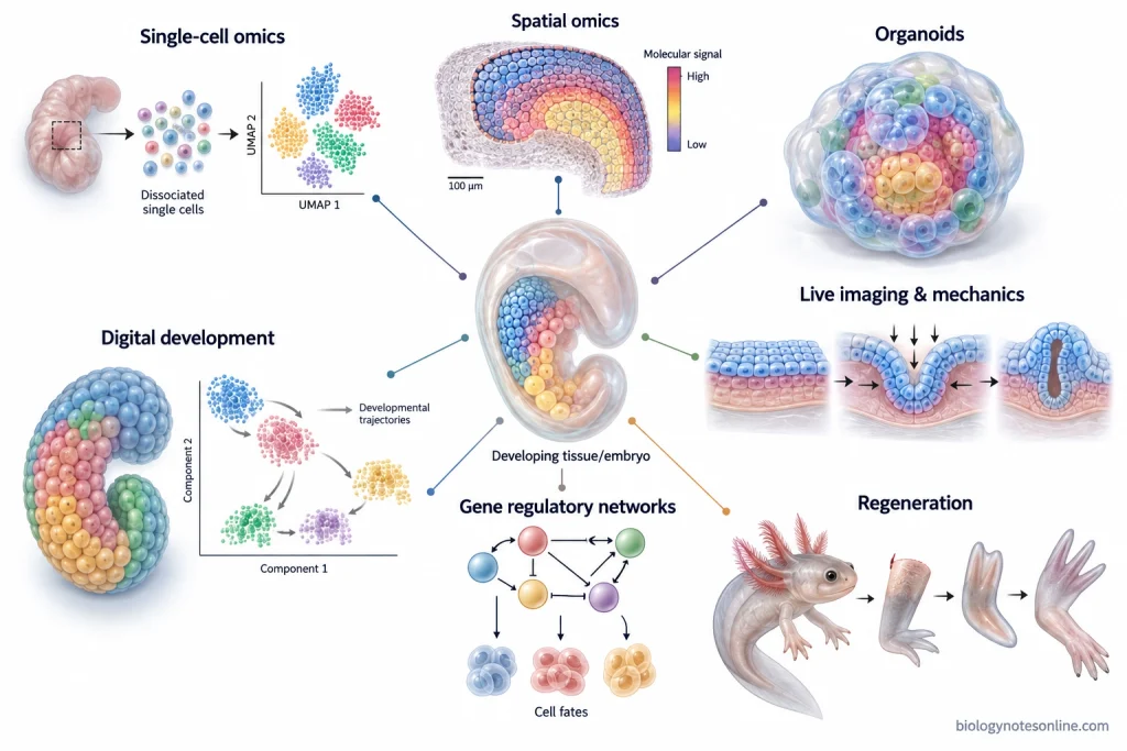
- Single-cell and spatial biology- Single-cell sequencing is being used to identify developmental cell states and differentiation pathways, while spatial transcriptomics keeps this molecular information connected with the position of cells inside tissues. Developmental atlases can now follow cellular diversity with very high resolution.
- Embryo models- Stem cells can self-organize into embryo-like models that reproduce selected events of early development. Human models are increasingly used for studying lineage formation, gastrulation and early organ development where direct experimental study of natural embryos is limited. Fidelity and standardization of these models are still major research problems.
- Organoids- Three-dimensional organoids are being developed to reproduce parts of tissue organization and organ development in vitro. Brain, intestinal and other organoid systems are used to work out human developmental mechanisms, genetic disorders and responses to experimental perturbations.
- Developmental mechanics- Development is studied not only through genes and chemical signals. Mechanical force, tissue tension, cell adhesion and material properties can change cell behaviour and shape a growing tissue. New imaging and force-measurement methods are being used to follow these events during morphogenesis.
- Gene regulatory networks- A major direction is to work out how transcription factors, regulatory DNA and signaling pathways control cell-fate decisions over time. Single-cell data together with genetic perturbations are increasingly used for reconstructing these gene regulatory networks (GRNs) and testing their functions.
- Regeneration and repair- Axolotls, planarians and other highly regenerative animals are studied to understand how cells recover positional information and rebuild missing structures. Stem-cell activation, pattern formation and developmental signaling after injury are important questions in this area.
- Computational developmental biology- Large imaging, single-cell and spatial datasets require computational methods for reconstructing cell trajectories and three-dimensional development. “Digital embryo” and predictive models are also being developed to connect molecular changes with later developmental events.
- Evolutionary developmental biology- Current evo-devo research compares developmental genes and regulatory systems between species to find how changes in development produce different body forms. Increasing genomic and molecular data allow developmental mechanisms to be connected more directly with phenotypic evolution.
- Plant developmental atlases- Single-cell and spatial approaches are now being applied across plant life cycles also. A recent Arabidopsis atlas mapped hundreds of thousands of nuclei from different organs and developmental stages, providing new ways to study changing cell identities in roots, shoots, flowers and reproductive tissues.
Developmental Biology at a Glance
| Topic | Quick summary for exams |
|---|---|
| Definition | Developmental biology is the study of how an organism develops, grows and changes during its life. |
| Central question | It studies how genetically related cells become different cell types and organize into tissues, organs and a functional organism. |
| Scope | Includes animal and plant development. It extends beyond embryology to postembryonic growth, maturation, metamorphosis, regeneration and other developmental changes. |
| Major levels | Molecular/genetic → cellular → tissue → organ → whole organism. |
| Main principles | Differential gene expression, cell-fate specification, induction, positional information, pattern formation, morphogenesis, growth and programmed cell death. |
| Embryology vs developmental biology | Embryology mainly deals with embryonic development. Developmental biology has a broader scope and includes embryonic as well as later developmental processes. |
| Animal developmental sequence | Gametogenesis and fertilization → cleavage and blastulation → gastrulation → patterning → organogenesis → growth and maturation. |
| Gastrulation | Cells are reorganized and the primary germ layers are established. |
| Germ layers | Ectoderm, mesoderm and endoderm are the three primary germ layers of triploblastic animals. |
| Neurulation | Formation of the neural tube during vertebrate development. It is not a universal stage of all animals. |
| Morphogenesis | Development of the form and three-dimensional organization of tissues and organs through cell movement, shape change, division and other cellular activities. |
| Cell differentiation | Process by which cells acquire different structural and functional characteristics through differences in gene expression. |
| Pattern formation | Establishment of spatial organization, body axes and positional identities during development. |
| Important animal models | Drosophila, C. elegans, zebrafish, Xenopus, chick and mouse. |
| Plant model | Arabidopsis thaliana is widely used for studying meristems, roots, shoots, flowers and plant developmental genetics. |
| Specialized models | Sea urchins and ascidians are useful for early development. Axolotls and planarians are important for regeneration studies. |
| How it is studied | Microscopy, fate mapping, transplantation, mutant analysis, gene manipulation, lineage tracing, gene-expression studies and single-cell methods are commonly used. |
| Major applications | Congenital-disorder research, developmental toxicology, IVF, stem-cell biology, regenerative medicine, organoids, cancer research and crop improvement. |
| Historical shift | The field moved from descriptive embryology to experimental embryology, genetics, molecular developmental biology and modern single-cell studies. |
| Current research | Single-cell and spatial biology, embryo models, organoids, developmental mechanics, gene regulatory networks, regeneration and evo-devo. |
References
- A look back at embryo models. (2025). Nature Methods, 22, 449–450. https://doi.org/10.1038/s41592-025-02642-1
- Alata-Jimenez, N., & Simoes-Costa, M. (2026). The chicken embryo as a model for developmental genomics. Developmental Biology, 536, 41–55. https://doi.org/10.1016/j.ydbio.2026.04.016
- Alberts, B., Johnson, A., Lewis, J., Raff, M., Roberts, K., & Walter, P. (2002). Molecular biology of the cell (4th ed.). Garland Science. https://www.ncbi.nlm.nih.gov/books/NBK21054/
- Arya, R., & White, K. (2015). Cell death in development: Signaling pathways and core mechanisms. Seminars in Cell & Developmental Biology, 39, 12–19. https://doi.org/10.1016/j.semcdb.2015.02.001
- Christian, J. L. (2012). Morphogen gradients in development: From form to function. Wiley Interdisciplinary Reviews: Developmental Biology, 1(1), 3–15. https://doi.org/10.1002/wdev.2
- Forsthoefel, D. J., & Newmark, P. A. (2009). Emerging patterns in planarian regeneration. Current Opinion in Genetics & Development, 19(4), 412–420. https://doi.org/10.1016/j.gde.2009.05.003
- Gaarslev, N., Swinnen, G., & Soyk, S. (2021). Meristem transitions and plant architecture—Learning from domestication for crop breeding. Plant Physiology, 187(3), 1045–1056. https://doi.org/10.1093/plphys/kiab388
- Gilbert, S. F. (2000). Developmental biology (6th ed.). Sinauer Associates. https://www.ncbi.nlm.nih.gov/books/NBK9983/
- Griffiths, J. A., Scialdone, A., & Marioni, J. C. (2018). Using single-cell genomics to understand developmental processes and cell fate decisions. Molecular Systems Biology, 14(4), e8046. https://doi.org/10.15252/msb.20178046
- Gualtieri, R., De Gregorio, V., Candela, A., Travaglione, A., Genovese, V., Barbato, V., & Talevi, R. (2024). In vitro culture of mammalian embryos: Is there room for improvement? Cells, 13(12), 996. https://doi.org/10.3390/cells13120996
- Gulati, G. S., D’Silva, J. P., Liu, Y., Wang, L., & Newman, A. M. (2025). Profiling cell identity and tissue architecture with single-cell and spatial transcriptomics. Nature Reviews Molecular Cell Biology, 26(1), 11–31. https://doi.org/10.1038/s41580-024-00768-2
- He, J., Zhang, X., Xia, X., Han, M., Li, F., Li, C., Li, Y., & Gao, D. (2020). Organoid technology for tissue engineering. Journal of Molecular Cell Biology, 12(8), 569–579. https://doi.org/10.1093/jmcb/mjaa012
- Herrera-Álvarez, S., & Thornton, J. W. (2026). Mechanisms of phenotypic evolution from molecules to organisms: Integrating evolutionary biochemistry and evolutionary developmental biology. Annual Review of Ecology, Evolution, and Systematics, 57. Advance online publication. https://doi.org/10.1146/annurev-ecolsys-102924-043757
- Hubert, K. A., & Wellik, D. M. (2023). Hox genes in development and beyond. Development, 150(1), dev192476. https://doi.org/10.1242/dev.192476
- Irion, U., & Nüsslein-Volhard, C. (2022). Developmental genetics with model organisms. Proceedings of the National Academy of Sciences of the United States of America, 119(30), e2122148119. https://doi.org/10.1073/pnas.2122148119
- Irish, V. F., & Benfey, P. N. (2004). Beyond Arabidopsis: Translational biology meets evolutionary developmental biology. Plant Physiology, 135(2), 611–614. https://doi.org/10.1104/pp.104.041632
- Kessler, D. S. (Ed.). (2009–2011). Colloquium series on developmental biology. Morgan & Claypool Life Sciences. https://www.ncbi.nlm.nih.gov/books/NBK53200/
- Lee, T. A., Illouz-Eliaz, N., Nobori, T., Xu, J., Jow, B., Nery, J. R., & Ecker, J. R. (2025). A single-cell, spatial transcriptomic atlas of the Arabidopsis life cycle. Nature Plants, 11(9), 1960–1975. https://doi.org/10.1038/s41477-025-02072-z
- Lemaire, P., Smith, W. C., & Nishida, H. (2008). Ascidians and the plasticity of the chordate developmental program. Current Biology, 18(14), R620–R631. https://doi.org/10.1016/j.cub.2008.05.039
- López, S. L., Cirio, M. C., & Garagna, S. (2023). Editorial: Early animal development: From fertilization to gastrulation. Frontiers in Cell and Developmental Biology, 11, 1146778. https://doi.org/10.3389/fcell.2023.1146778
- Mahla, R. S. (2016). Stem cells applications in regenerative medicine and disease therapeutics. International Journal of Cell Biology, 2016, 6940283. https://doi.org/10.1155/2016/6940283
- Martik, M. L., Lyons, D. C., & McClay, D. R. (2016). Developmental gene regulatory networks in sea urchins and what we can learn from them. F1000Research, 5, 203. https://doi.org/10.12688/f1000research.7381.1
- National Research Council, Committee on Developmental Toxicology. (2000). Recent advances in developmental biology. In Scientific frontiers in developmental toxicology and risk assessment. National Academies Press. https://www.ncbi.nlm.nih.gov/books/NBK225682/
- National Research Council, Committee on Research Opportunities in Biology. (1989). Development. In Opportunities in biology. National Academies Press. https://www.ncbi.nlm.nih.gov/books/NBK217800/
- Nowotschin, S., & Hadjantonakis, A.-K. (2014). Live imaging mouse embryonic development: Seeing is believing and revealing. Methods in Molecular Biology, 1092, 405–420. https://doi.org/10.1007/978-1-60327-292-6_24
- Nüsslein-Volhard, C., & Wieschaus, E. (1980). Mutations affecting segment number and polarity in Drosophila. Nature, 287(5785), 795–801. https://doi.org/10.1038/287795a0
- Nwabo Kamdje, A. H., Kamga, P. T., Simo, R. T., Vecchio, L., Seke Etet, P. F., Muller, J. M., Bassi, G., Lukong, E., Goel, R. K., Amvene, J. M., & Krampera, M. (2017). Developmental pathways associated with cancer metastasis: Notch, Wnt, and Hedgehog. Cancer Biology & Medicine, 14(2), 109–120. https://doi.org/10.20892/j.issn.2095-3941.2016.0032
- Ribatti, D. (2014). The chemical nature of the factor responsible for embryonic induction: An historical overview. Organogenesis, 10(1), 38–43. https://doi.org/10.4161/org.28026
- Seyedhassantehrani, N., Otsuka, T., Singh, S., & Gardiner, D. M. (2017). The axolotl limb regeneration model as a discovery tool for engineering the stem cell niche. Current Stem Cell Reports, 3(3), 156–163. https://doi.org/10.1007/s40778-017-0085-5
- Shin, J., Halberg-Spencer, S., Liu, Y., Hazra, S., Lee, E. D.-I., & Roy, S. (2026). Modeling dynamics, cell type specificity, and perturbations in gene regulatory networks. Annual Review of Genomics and Human Genetics, 27, 103–129. https://doi.org/10.1146/annurev-genom-120922-103729
- Tandon, P., Conlon, F., Furlow, J. D., & Horb, M. E. (2017). Expanding the genetic toolkit in Xenopus: Approaches and opportunities for human disease modeling. Developmental Biology, 426(2), 325–335. https://doi.org/10.1016/j.ydbio.2016.04.009
- Villeneuve, C., McCreery, K. P., & Wickström, S. A. (2025). Measuring and manipulating mechanical forces during development. Nature Cell Biology, 27, 575–590. https://doi.org/10.1038/s41556-025-01632-x
- Wang, D., Villenave, R., Stokar-Regenscheit, N., & Clevers, H. (2026). Human organoids as 3D in vitro platforms for drug discovery: Opportunities and challenges. Nature Reviews Drug Discovery, 25(3), 204–226. https://doi.org/10.1038/s41573-025-01317-y
- Wu, H., & Wang, H. (2026). Progress in stem cell-based embryo models and their applications in developmental biology and biomedicine. Nature Reviews Molecular Cell Biology, 27(3), 178–193. https://doi.org/10.1038/s41580-025-00942-0
i need to study so i downloaded this aap and it is good
this is very good