

What is Embryology?
Embryology is a branch of biology which deals with the formation and development of an embryo. It studies different changes taking place from fertilization and formation of zygote to development of tissues, organs and basic body structure.
Embryology is not restricted only to humans. Development of animals, plants and other multicellular organisms can also be studied under embryology. However, human embryology and animal embryology are commonly studied in medical and biomedical fields.
The scope of embryology includes fertilization, cleavage, early embryonic development, formation of germ layers, cell differentiation, morphogenesis and formation of organs. Developmental patterns between different organisms can also be compared. This is referred to as comparative embryology.
Embryology and Embryogenesis
Embryology and embryogenesis are two different terms. Embryology is the field which studies embryo development, while embryogenesis is the actual process of formation and development of an embryo.
During embryogenesis, the fertilized cell undergoes repeated cell divisions. The formed cells then become differentiated and get organized into different tissues and body structures. Thus, embryogenesis is one of the major processes studied under embryology.
Embryology and Developmental Biology
Developmental biology has a wider scope as compared to embryology. Embryology mainly deals with development occurring during the embryonic stages.
Developmental biology studies embryonic development together with the cellular, genetic and molecular mechanisms involved during development. It also includes different developmental processes occurring after the embryonic stage. Growth, differentiation, regeneration and metamorphosis are included where these processes occur.
Therefore, embryology is an important part of developmental biology. Both are related fields, but they are not exactly the same.
Embryo, Fetus and Conceptus
The terms embryo, fetus and conceptus are used during development. Their exact use depends upon the organism and the context.
An embryo is the developing organism during the early developmental stage when major tissues and basic body structures are being formed. In humans, the embryonic period generally continues up to the end of eighth week after fertilization.
After this stage, the developing organism is referred to as a fetus. During fetal period, further growth takes place. Differentiation of the already formed organs and body structures also continues.
The term conceptus has a broader meaning. It includes the embryo or fetus along with its associated extraembryonic structures, such as fetal membranes and placenta. Thus, the embryo or fetus forms only one part of the complete conceptus.
Embryonic Development
Embryonic development is a series of developmental changes which starts after the formation of zygote. The single-celled zygote undergoes repeated divisions and produces a large number of cells. These cells later move to different positions, become differentiated and are arranged into tissues and organs.
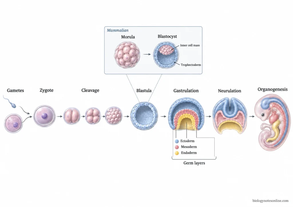
The following are the major processes involved in embryonic development-
Gametogenesis and Fertilization
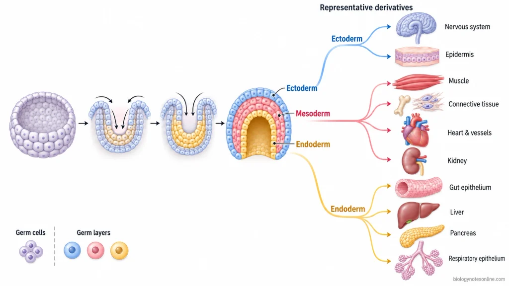
- Gametogenesis is the process by which mature male and female gametes are formed from germ-cell precursors. During this process, meiosis takes place and haploid gametes are produced having half number of chromosomes.
- During fertilization, the male and female gametes come together and fuse. Their genetic materials are brought into the same cell.
- This results in the formation of a diploid zygote. It is the first cell of the developing organism.
- Gametes are produced from the germ-cell lineage. Germ cells should not be confused with the germ layers, which are formed later during embryonic development.
Cleavage and Blastula Formation
After formation of zygote, it undergoes rapid mitotic divisions. This process is called cleavage.
- The cells produced during cleavage are called blastomeres. Their number increases after every division. However, the overall size of embryo does not increase greatly during the early cleavage period.
- Further divisions result in formation of an early multicellular embryo. A cavity may develop within it and the embryo forms a blastula. Its structure is not exactly same in all animals.
- In mammals, the dividing blastomeres initially form a compact ball-like structure called morula. Later, a cavity develops within it and the structure becomes a blastocyst.
- The mammalian blastocyst contains an outer cell layer called trophectoderm and a group of cells inside, known as the inner cell mass. The inner cell mass contributes to formation of the embryo proper.
Morula and blastocyst are therefore commonly used during mammalian development. All animals do not pass through identical structures.
Gastrulation and Germ Layers
Gastrulation is the process in which cells of the early embryo move from their previous positions and become rearranged. During this process, a relatively simple embryo is converted into a more organized structure.
The primary germ layers are established during gastrulation. In triploblastic animals, three germ layers are formed. These are ectoderm, mesoderm and endoderm.
Some of the important derivatives of these germ layers are-
| Germ layer | Representative derivatives |
|---|---|
| Ectoderm | Epidermis and related structures, nervous system |
| Mesoderm | Muscles, connective tissues, skeleton, circulatory system, kidneys and major parts of reproductive system |
| Endoderm | Epithelial lining of digestive and respiratory tracts, tissues of liver and pancreas |
These germ layers later develop into different tissues and organs of the body.
Germ layers and germ cells are different. Germ layers are embryonic tissue layers formed during gastrulation, whereas germ cells belong to the reproductive-cell lineage and later produce gametes.
Gastrulation also establishes the basic organization of the embryo. Cells are placed in different regions, and the body axes begin to become more clearly defined.
Neurulation and Body Patterning
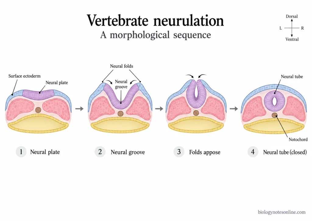
Neurulation is an important process during development of vertebrate embryos. In this process, a part of ectoderm becomes neural ectoderm and forms the neural plate.
The neural plate then changes its shape. Its lateral regions rise upward to form neural folds and a neural groove is present between them. These neural folds later come together and fuse.
As a result, the neural tube is formed. It later develops into major structures of the central nervous system.
The process of neurulation is not exactly identical throughout all vertebrates. Different regions can differ in the way by which neural tissue is converted into a tube.
During this developmental period, different cells and tissues are also arranged according to their position within the embryo. This establishes the basic body pattern, including organization along the major body axes.
Organogenesis and Differentiation
After formation of the germ layers, development of different organs begins. This process is referred to as organogenesis.
Cells derived from ectoderm, mesoderm and endoderm continue to divide and become specialized. This specialization of cells is called differentiation.
During organ formation, different tissues also interact with one another. These interactions help in determining the developmental fate and arrangement of the nearby cells.
In this process, cells become arranged into different structural forms such as sheets, tubes and compact tissue masses. These structures later develop into recognizable tissues and organs.
The formed organs continue to grow and become more organized. Further differentiation and tissue remodeling continue as embryonic development proceeds.
Control of Embryonic Development
Embryonic development is controlled by the interaction of genes, signals between cells and the position occupied by cells within the embryo. Generally, all embryonic cells contain the same genetic information, but different sets of genes become active in different cells. As a result, different cells acquire different developmental fates.
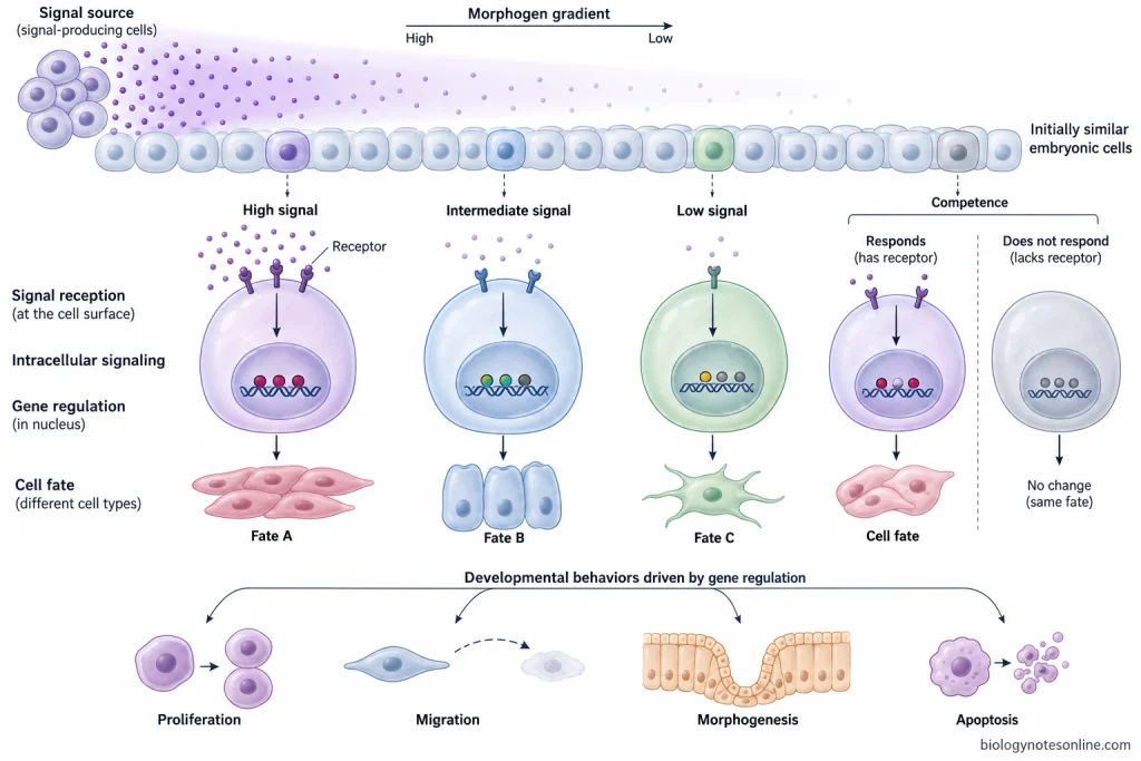
Some of the important mechanisms controlling embryonic development are-
1. Gene Regulation
Different genes are switched on or switched off at different periods of development. The proteins produced from these genes control different processes like cell division, differentiation, movement and formation of tissues.
Transcription factors have an important role in this process. They can activate or suppress other genes and establish a particular pattern of gene activity within a developing cell. Thus, cells having the same DNA can finally develop into very different types of cells.
2. Cell Signaling and Embryonic Induction
Cells present in an embryo do not develop independently. They communicate with nearby cells through different signaling molecules.
When one group of cells affects the developmental fate of another group, this process is called embryonic induction. The tissue producing the signal acts as an inducer. However, the responding tissue must have the ability to receive and respond to the particular signal.
This ability of tissue is referred to as competence. Inductive interactions can occur repeatedly, and several such interactions take place during formation of organs.
3. Morphogens and Positional Information
Some developmental signals can act over a distance. They form a concentration gradient across a group of cells. Such signaling molecules are called morphogens.
Cells located at different positions receive different amounts of the signal. Depending upon its concentration, and sometimes the period of exposure, different genes become activated. Different cell fates are then produced.
In this way, cells obtain information about their position within the developing embryo.
4. Cell-Cell and Tissue Interactions
Development also depends upon direct interactions between neighbouring cells and tissues. After receiving signals from surrounding cells, a cell may change its shape, rate of division or even its developmental fate.
These interactions are particularly important during tissue formation. One developing tissue can influence another tissue. Sometimes, the responding tissue also signals back.
Such repeated interactions help in formation and proper organization of different organs.
5. Cell Position and Developmental Fate
The position of a cell within an embryo can determine what the cell will become. Cells located in different regions are exposed to different signals and surrounding tissues.
During development, cells may also move from one region to another. Their new position can expose them to a different developmental environment. As a result, gene expression and developmental fate of these cells may change.
Therefore, position is an important factor in formation of the basic body pattern.
6. Timing of Developmental Signals
Developmental signals must act at a suitable stage. The same signal does not always produce the same response in a cell during every period of development.
The response depends upon the state of the particular cell, its previous developmental history and its ability to respond to that signal. This ability is related to competence.
Thus, both position and timing take part in the control of embryonic development.
7. Changes in Cell Behaviour
Control of development is finally expressed as different changes in cell behaviour. Cells may divide, stop dividing, migrate, change their shape, attach to other cells or undergo differentiation.
Mechanical forces produced inside the growing tissues also help in shaping the embryo. These forces act together with biochemical signals during morphogenesis. They do not act as a completely separate process.
8. Programmed Removal of Cells
Some cells are deliberately removed during normal development by programmed cell death (apoptosis). It is not always associated with damage.
Removal of selected cells can help in shaping the developing structures. It also removes cells which are no longer required.
Therefore, formation of an embryo does not depend only on production and differentiation of cells. Controlled removal of particular cells also takes part in normal embryonic development.
Major Approaches in Embryology
Embryology is studied by different approaches depending upon what part of development is being examined. Some are based on the structural observation of embryo, some compare the embryos of different organisms, while other approaches are used to find out the mechanisms controlling the developmental process. These are not considered as one fixed “types of embryology”. Some of the important approaches of embryology are as follows-
| Approach | Focus | Main question | Use |
|---|---|---|---|
| Descriptive Embryology | Structural changes during development | What changes occur in the developing embryo? | Observation and description of developmental stages |
| Comparative and Evolutionary Embryology | Development in different organisms | How developmental patterns differ or remain similar among species? | Comparison of development and evolutionary relationship |
| Experimental and Molecular Embryology | Mechanism of development | How cells and tissues are controlled during development? | Manipulation, lineage study, genetics and imaging |
| Medical Embryology and Teratology | Human development | How normal development takes place and how it may become abnormal? | Study of normal human development and developmental abnormalities |
Descriptive Embryology
Descriptive embryology is the study of embryo by observing and describing the changes which occur during development. The embryo is observed at different developmental periods and the structures formed during these periods are recorded.
It deals with the changes taking place during cleavage, gastrulation, formation of body structures and organ development. The position, shape and appearance of developing structures can be followed from one stage to another. Thus, it mainly tells what is happening in the embryo during development.
Comparative and Evolutionary Embryology
Comparative embryology is based on comparison of embryonic development between different organisms. The similarities and differences in their developmental pattern are studied.
For example, embryos of related animal groups may show some common developmental structures, although later these structures can develop differently. Such comparisons are useful for identifying common developmental patterns.
Evolutionary embryology relates these developmental patterns with evolutionary relationships. Similar development of homologous structures can provide information about common ancestry, whereas modification of these developmental processes produces differences between organisms.
Similarity between embryos, however, does not mean that embryo of one animal passes through the adult forms of another animal. An embryo develops as an embryo of its own species. The similarities represent shared developmental characters, not a repetition of adult evolutionary forms.
Experimental and Molecular Embryology
Unlike descriptive embryology which is mainly based on observation, experimental embryology studies development by changing some part of the developing embryo and observing its result.
A cell or group of cells may be removed, transplanted, marked or experimentally changed. What happens after this gives information about its developmental role. It is also used to determine whether a particular embryonic region forms a structure by itself or requires interaction with another tissue.
Lineage studies follow a selected cell and its descendants during development. By this, the structures produced from that cell can be worked out.
Molecular embryology deals with genes and molecules involved in the developmental process. Genetic methods, molecular techniques and imaging are used along with experimental approaches. Fluorescent labeling can also be used to follow cells in the living embryo. In this way, developmental events can be studied while they are taking place.
Medical Embryology and Teratology
Medical embryology deals mainly with normal development of the human embryo and fetus. It studies the formation and development of different tissues, organs and body structures from fertilization onwards.
It forms an important part of medical study because the arrangement of many adult structures can be understood from their embryonic development.
Teratology is concerned with abnormal development. It studies developmental abnormalities and the factors which can disturb the normal developmental process.
The effect of such factors depends partly upon when the disturbance occurs and which structure is developing during that period. Thus, medical embryology deals mainly with normal human development, while teratology deals with its abnormal developmental changes.
Human Embryology
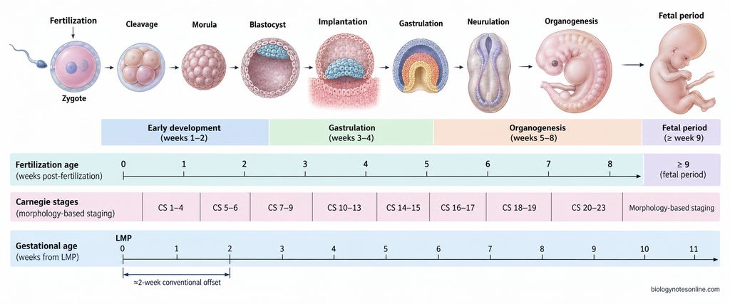
Human embryology is the study of development of human embryo from fertilization and its development into the fetal period. It includes formation of zygote, cleavage, implantation, formation of germ layers and later development of different tissues and organs. The embryonic period is an important period. Most of the major body structures start their formation during this time.
Pre-embryonic and Embryonic Development
The early development of human can be divided into pre-embryonic period and embryonic period. Different developmental changes occur one after another.
- Fertilization- It is the beginning of human development. The male and female gametes fuse together and a diploid zygote is formed.
- The zygote now undergoes repeated mitotic divisions called cleavage. During this process it moves through the uterine tube. The cells formed are increased in number.
- After several divisions, a compact group of cells called morula is formed. It later changes into a blastocyst.
- The blastocyst reaches the uterus and becomes attached with the uterine lining. This is referred to as implantation. The inner cell mass mainly gives rise to embryo proper, while trophoblast takes part in formation of supporting extraembryonic structures.
- During the second week, further organization of the early embryo takes place. Different extraembryonic structures are also formed.
- Gastrulation begins during the third week. In this process, three primary germ layers are formed, ectoderm, mesoderm and endoderm. Different tissues and organs are later developed from these germ layers.
- Neurulation and early organ formation follow after this. From about third to eighth week, most of the major organ systems begin their development. This is the major organ-forming period of the embryo.
Embryonic and Fetal Periods
The embryonic period continues up to the end of 8 weeks after fertilization. During this period, the basic body form is established and the major organs start to develop.
After this period, the developing human is called a fetus. The fetal period starts from week 9 after fertilization and continues up to birth.
The main difference is in the type of development taking place. During embryonic period, formation and early arrangement of major structures occur. In fetal period, growth becomes more prominent. The organs already formed also continue differentiation and functional maturation.
The developmental sequence can be shown as-
Fertilization → Cleavage → Blastocyst → Implantation → Gastrulation → Organ formation → Fetal growth and maturation
Embryonic Age vs Gestational Age
The age of developing human can be expressed in two ways. These are embryonic age and gestational age.
Embryonic age- It is counted from the time of fertilization. It is also called fertilization age or post-fertilization age. Therefore, this age shows the developmental time after the sperm and ovum have fused.
Gestational age- It is commonly used during pregnancy in clinical practice. This age is counted from the first day of the last menstrual period (LMP), and not from the actual day of fertilization.
Ovulation and fertilization normally occur about two weeks after the beginning of the last menstrual period. For this reason, gestational age is generally about 2 weeks more than embryonic age.
For example, an embryo having an age of about 6 weeks after fertilization is usually described clinically as about 8 weeks of gestational age.
Carnegie Stages
Carnegie stages are used to classify the development of human embryos. The system contains 23 stages, beginning with Stage 1 at fertilization and ending with Stage 23 near the end of embryonic period.
These stages are mainly based on the morphological development of embryo. External and internal structures are examined and the embryo is then placed into a particular stage. Age in days or size alone is not taken as the only basis.
Because of this, embryos having nearly the same calendar age may sometimes show a different developmental stage.
The approximate stage range is as follows-
Week 1- Carnegie stages 1-4
Week 2- Stages 5-6
Week 3- Stages 7-9
Week 4- Stages 10-13
Week 5- Stages 14-15
Week 6- Stages 16-17
Week 7- Stages 18-19
Week 8- Stages 20-23
Carnegie staging is mainly used to describe how much morphological development has taken place in the embryo, rather than depending only upon the number of days.
Importance of Embryology
Embryology is important for understanding how a single fertilized cell develops into a complete organism. It also helps to explain the origin of different tissues, organs and many structural abnormalities found at birth. Some of the important uses of embryology are as follows-
- Normal development- Embryology is used to study the normal sequence of development from fertilization onwards. It explains how cells divide, move and become differentiated, and how different tissues and organs are formed from them.
- Understanding adult structures- Many structures of the adult body can be understood properly by knowing how they were formed during embryonic life. Some organs change their position, fuse with other structures or develop from more than one embryonic component. Embryological development therefore gives the developmental basis of anatomy.
- Congenital abnormalities- Abnormality in a normal developmental process can produce congenital defects. Embryology helps to find out which developmental step has been disturbed and how the abnormal structure may have formed. Genetic changes as well as environmental factors can interfere with development.
- Teratology- The effect of harmful environmental agents on the developing embryo or fetus is studied with the help of embryology. The developmental stage is important here. A tissue is usually more sensitive while its major structures are being formed, and different organs have different periods of sensitivity.
- Prenatal study- Knowledge of normal development is also required during prenatal examination. The developmental age, normal formation of structures and abnormal findings can be compared during pregnancy. This information is useful in prenatal screening and diagnosis of some fetal abnormalities.
- Evolutionary relationship- Comparison of embryonic development between different organisms gives information about common developmental patterns and their evolutionary changes. Homologous structures may pass through related developmental processes because they have been inherited from a common ancestral structure. Embryology is therefore also important in evolutionary developmental biology.
- Reproductive medicine- Embryology has an important use in assisted reproductive technology (ART). In procedures such as in vitro fertilization (IVF), oocytes and developing embryos are observed and assessed before further clinical steps are carried out. Knowledge of early embryo development is needed for this work.
- Developmental research- Embryological study provides a basis for finding out how genes, cells and tissues work together during formation of the body. Normal development can first be studied, then changes in that process can be related with developmental abnormalities or differences between organisms.
Historical Development of Embryology
The study of embryonic development has a long history, beginning mainly with observation of developing animals and later changing into experimental and molecular study. Improvement of microscopes and experimental methods played an important role in this development. Some of the major developments in embryology are as follows-
- The earliest systematic studies of embryos are associated with Aristotle during the fourth century BCE. He studied developing chick embryos and also compared the different ways by which animals are produced. He recognized several features of embryonic development including different patterns of cleavage.
- For a long period after Aristotle, very little experimental progress was made. In 1651, William Harvey studied reproduction and development and proposed that animals arise from eggs. His idea was expressed as Ex ovo omnia (“all from the egg”). Harvey also observed the blastoderm of chick embryo and some early blood formation.
- Development of microscope brought out more detailed study of embryo. In 1672, Marcello Malpighi gave one of the first microscopic descriptions of chick development. Structures such as the neural groove, somites and early circulation were observed. This period also brought up a major argument between preformation and epigenesis.
- According to preformation, the organism was already present in a very small form inside the egg or sperm and development was mainly its enlargement. The other view, epigenesis, stated that new structures are gradually formed during development. Kaspar Friedrich Wolff strongly supported epigenesis during the eighteenth century. From chick embryos he showed that structures such as intestine, heart and blood vessels appear during development rather than being present as complete miniature structures from the beginning.
- During the early nineteenth century, embryology developed more rapidly with improved microscopes and methods of observation. Christian Pander described the primary germ layers while studying chick embryos. He also observed that the layers interact with one another during formation of organs. This became an important basis for later studies of tissue interaction and induction.
- Karl Ernst von Baer further developed these studies. He discovered the mammalian egg and described important features of vertebrate embryos. In 1828, he also presented principles showing that general developmental characters appear before more specialized characters. Embryos of different vertebrates may therefore resemble each other in their early organization, but one embryo does not pass through the adult stages of another animal.
- By the later nineteenth century, embryologists began following individual cells and groups of cells to determine what structures they produce. Such studies resulted in the preparation of fate maps. Development was now being studied at cellular level, and experimental embryology started to grow from descriptive embryology.
- Experimental study became especially important during the late nineteenth and early twentieth centuries. Embryonic cells and tissues were removed, separated or transplanted and their further development was then observed. Embryology was no longer based only on describing what structures appeared. Experiments were being used to find out how development is controlled.
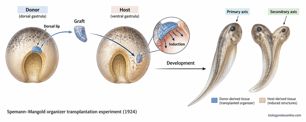
- In 1924, Hans Spemann and Hilde Mangold showed the organizing effect of the dorsal blastopore lip by transplantation experiments in amphibian embryos. The transplanted tissue could induce surrounding host tissues and result in formation of another body axis. This region became known as the Spemann organizer, and the experiment gave an important basis for the concept of embryonic induction.
- During the later twentieth century, embryology became closely connected with genetics, cell biology and molecular biology. Genes involved in cell fate and body pattern formation could now be studied, while molecular markers allowed developing cells and tissues to be followed. Experimental embryology gradually developed into much of what is now called developmental biology.
- Modern embryology uses genetic manipulation, molecular methods, cell-lineage studies and live imaging together with the older methods of observation and embryo manipulation. Development can now be studied from the whole embryo down to genes and individual cells. The basic question, however, remains the same, how a single fertilized cell develops into an organized multicellular organism.
References
- Brandt, C. (2022). Development and heredity in the interwar period: Hans Spemann and Fritz Baltzer on organizers and merogones. Journal of the History of Biology, 55(2), 253–283. https://doi.org/10.1007/s10739-022-09681-w
- Cruz Walma, D. A., & Yamada, K. M. (2020). The extracellular matrix in development. Development, 147(10), dev175596. https://doi.org/10.1242/dev.175596
- Donovan, M. F., & Cascella, M. (2022). Embryology, weeks 6–8. In StatPearls. StatPearls Publishing. https://www.ncbi.nlm.nih.gov/books/NBK563181/
- Elshazzly, M., Lopez, M. J., Reddy, V., & Caban, O. (2023). Embryology, central nervous system. In StatPearls. StatPearls Publishing. https://www.ncbi.nlm.nih.gov/books/NBK526024/
- Ferrer-Vaquer, A., Viotti, M., & Hadjantonakis, A.-K. (2010). Transitions between epithelial and mesenchymal states and the morphogenesis of the early mouse embryo. Cell Adhesion & Migration, 4(3), 447–457. https://doi.org/10.4161/cam.4.3.10771
- Flierman, S., Tijsterman, M., Rousian, M., & de Bakker, B. S. (2023). Discrepancies in embryonic staging: Towards a gold standard. Life, 13(5), 1084. https://doi.org/10.3390/life13051084
- Geng, X., & Oliver, G. (2010). Elucidating the molecular characteristics of organogenesis in human embryos. Genome Biology, 11(8), 130. https://doi.org/10.1186/gb-2010-11-8-130
- Gerrard, D. T., Berry, A. A., Jennings, R. E., Piper Hanley, K., Bobola, N., & Hanley, N. A. (2016). An integrative transcriptomic atlas of organogenesis in human embryos. eLife, 5, e15657. https://doi.org/10.7554/eLife.15657
- Gilbert, S. F. (2000). Developmental biology (6th ed.). Sinauer Associates. https://www.ncbi.nlm.nih.gov/books/NBK9983/
- Grapin-Botton, A. (2008). Endoderm specification. In StemBook. Harvard Stem Cell Institute. https://doi.org/10.3824/stembook.1.30.1
- Gurdon, J. B., & Bourillot, P.-Y. (2001). Morphogen gradient interpretation. Nature, 413(6858), 797–803. https://doi.org/10.1038/35101500
- Kumar, V., Park, S., Lee, U., & Kim, J. (2021). The organizer and its signaling in embryonic development. Journal of Developmental Biology, 9(4), 47. https://doi.org/10.3390/jdb9040047
- Kuratani, S., Uesaka, M., & Irie, N. (2022). How can recapitulation be reconciled with modern concepts of evolution? Journal of Experimental Zoology Part B: Molecular and Developmental Evolution, 338(1–2), 28–35. https://doi.org/10.1002/jez.b.23020
- Lowery, L. A., & Sive, H. (2004). Strategies of vertebrate neurulation and a re-evaluation of teleost neural tube formation. Mechanisms of Development, 121(10), 1189–1197. https://doi.org/10.1016/j.mod.2004.04.022
- Mavrakis, M., Pourquié, O., & Lecuit, T. (2010). Lighting up developmental mechanisms: How fluorescence imaging heralded a new era. Development, 137(3), 373–387. https://doi.org/10.1242/dev.031690
- Miller, C. J., & Davidson, L. A. (2013). The interplay between cell signalling and mechanics in developmental processes. Nature Reviews Genetics, 14(10), 733–744. https://doi.org/10.1038/nrg3513
- Muhr, J., Arbor, T. C., & Ackerman, K. M. (2023). Embryology, gastrulation. In StatPearls. StatPearls Publishing. https://www.ncbi.nlm.nih.gov/books/NBK554394/
- Oliver, R., & Basit, H. (2023). Embryology, fertilization. In StatPearls. StatPearls Publishing. https://www.ncbi.nlm.nih.gov/books/NBK542186/
- Pandit, S., & Sharma, R. (2022). Non invasive assessment of human oocytes and embryos in assisted reproduction: Review on present practices and future trends. Medical Journal Armed Forces India, 78(1), 7–16. https://doi.org/10.1016/j.mjafi.2021.04.002
- Perrimon, N., Pitsouli, C., & Shilo, B.-Z. (2012). Signaling mechanisms controlling cell fate and embryonic patterning. Cold Spring Harbor Perspectives in Biology, 4(8), a005975. https://doi.org/10.1101/cshperspect.a005975
- Rozario, T., & DeSimone, D. W. (2010). The extracellular matrix in development and morphogenesis: A dynamic view. Developmental Biology, 341(1), 126–140. https://doi.org/10.1016/j.ydbio.2009.10.026
- Rugg-Gunn, P. J., Moris, N., & Tam, P. P. L. (2023). Technical challenges of studying early human development. Development, 150(11), dev201797. https://doi.org/10.1242/dev.201797
- Shahbazi, M. N. (2020). Mechanisms of human embryo development: From cell fate to tissue shape and back. Development, 147(14), dev190629. https://doi.org/10.1242/dev.190629
- Tyser, R. C. V., & Srinivas, S. (2022). Recent advances in understanding cell types during human gastrulation. Seminars in Cell & Developmental Biology, 131, 35–43. https://doi.org/10.1016/j.semcdb.2022.05.004
- Wolf, S., Wan, Y., & McDole, K. (2021). Current approaches to fate mapping and lineage tracing using image data. Development, 148(18), dev198994. https://doi.org/10.1242/dev.198994
- Yamada, S., Samtani, R. R., Lee, E. S., Lockett, E., Uwabe, C., Shiota, K., Anderson, S. A., & Lo, C. W. (2010). Developmental atlas of the early first trimester human embryo. Developmental Dynamics, 239(6), 1585–1595. https://doi.org/10.1002/dvdy.22316