

CRISPR-Cas9 is a programmable genome-editing system adapted from a natural microbial defense mechanism. A guide RNA directs the Cas9 nuclease to a matching DNA sequence, where Cas9 cuts the DNA and cellular repair processes determine the resulting genetic change. CRISPR refers to the sequence-based defense system; Cas9 is the protein enzyme that performs the DNA cleavage.
What Is CRISPR-Cas9?
CRISPR-Cas9 is an RNA-guided genome-editing system which is used to target and cut a selected DNA sequence.
It consists of Cas9 nuclease and a guide RNA. The guide RNA provides the sequence information required for recognition of the target DNA. CRISPR-Cas9 is not a single protein. Cas9 is the protein, while CRISPR-Cas9 refers to the functional RNA-guided system containing Cas9, guide RNA, and the DNA being targeted.
CRISPR stands for “Clustered Regularly Interspaced Short Palindromic Repeats.”
The term was originally given to the characteristic DNA regions which are found in bacteria and archaea. These regions contain short repeated DNA sequences arranged in an array. Between these repeats, different non-repetitive sequences called “spacers” are present.
Many of these spacers originate from previously encountered viruses or other foreign genetic elements.
CRISPR originally refers to these repeat-spacer DNA arrays, and not to the Cas9 protein. Together with nearby CRISPR-associated (Cas) genes and proteins, the arrays form CRISPR-Cas adaptive immune systems. These systems help bacteria and archaea to recognize and interfere with invading nucleic acids.
Cas9 means CRISPR-associated protein 9. It is a DNA nuclease found in certain CRISPR-Cas systems, particularly the type II system.
Cas9 is an RNA-guided enzyme. The RNA guides Cas9 towards the complementary DNA sequence. Cas9 can then cleave the DNA with its nuclease domains.
In the natural system, Cas9 is guided by CRISPR RNA (crRNA) along with trans-activating CRISPR RNA (tracrRNA). For genome editing, functions of these two RNAs can also be combined into an engineered single-guide RNA (sgRNA).
CRISPR vs CRISPR-Cas9
CRISPR- It is commonly used as a broad term for CRISPR-based biology and technologies. However, the word CRISPR itself originally describes the repeat-spacer DNA arrays.
Natural CRISPR-Cas systems are much broader. Different CRISPR-Cas types contain different Cas proteins and have different mechanisms for recognizing or destroying foreign nucleic acids.
CRISPR-Cas9- It is one particular CRISPR-Cas system which has been adapted as an RNA-programmable system for genome editing. Its targeting can mainly be changed by changing the guide sequence of RNA, instead of producing a different DNA-binding protein for every target.
Is CRISPR-Cas9 a Protein?
No. CRISPR-Cas9 itself is not a single protein. Cas9 is the protein component.
The working genome-editing complex contains Cas9 together with a guide RNA. This guide RNA directs the nuclease towards a complementary sequence in the target DNA.
The target DNA must also contain the required protospacer adjacent motif (PAM) for Cas9 recognition and cleavage.
Biological Origin of CRISPR-Cas9
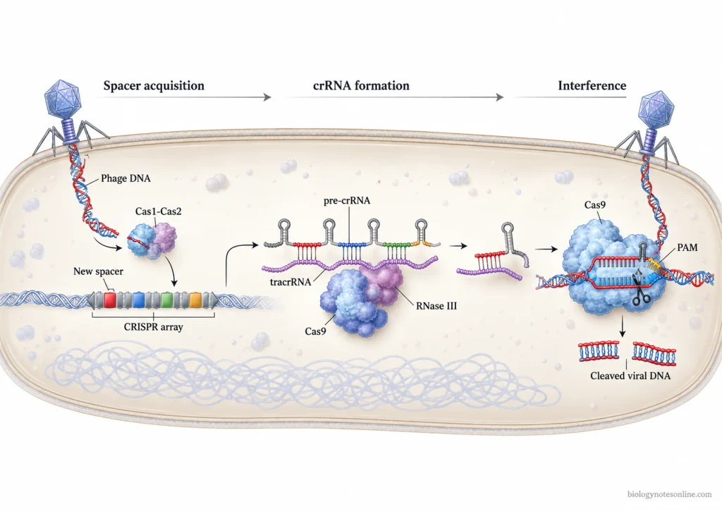
CRISPR-Cas is a natural microbial defense system found in bacteria and archaea which protects the cells against invading genetic materials such as bacteriophages and plasmids.
It was not originally a genome-editing technology. The natural system stores sequences obtained from some previous invaders. Later, RNA molecules and CRISPR-associated (Cas) proteins use these stored sequences for recognizing the related invading nucleic acid. In type II CRISPR-Cas systems, this interference is performed by Cas9.
The natural CRISPR defense mainly occurs through acquisition of foreign sequences, formation of CRISPR RNAs, and interference with the invading genetic material. These biological processes later provided the basis for development of CRISPR-Cas9 as a programmable DNA-editing system.
CRISPR Arrays in Bacterial and Archaeal Genomes
CRISPR arrays are characteristic DNA regions containing short repeated sequences separated by variable sequences called “spacers.”
The repeats occur again and again throughout the array, while the sequence of each spacer can be different. Many of these spacer sequences are derived from viruses, plasmids, and other foreign genetic materials which previously entered the microbial cell.
The CRISPR array is generally associated with cas genes. These genes encode proteins involved in acquisition, RNA processing, or interference. Different CRISPR-Cas systems contain different groups of Cas proteins.
Cas9 is the major interference protein of the type II system. It is not a protein found in every CRISPR-Cas system.
Spacer Acquisition and Molecular Memory
During infection, fragments of the invading genetic element can be taken up and inserted as new spacers into the CRISPR locus. These foreign DNA fragments are obtained from sequences commonly referred to as “protospacers” in the invader.
Cas1 and Cas2 proteins form the conserved adaptation machinery involved in spacer integration in CRISPR-Cas systems.
A newly acquired spacer becomes a part of the microbial genome. During another infection, this stored sequence can provide sequence information for recognition of the same or a closely matching genetic invader.
This stored spacer sequence is referred to as a form of molecular or immunological memory. It can also pass from the microbial cell to its daughter cells during cell division.
CRISPR-Cas is referred to as an adaptive defense system because its targeting information can change following exposure to foreign genetic material. New spacers can be acquired and added to the already existing CRISPR array.
Formation of CRISPR RNAs
The CRISPR array is first transcribed into a long RNA molecule known as precursor CRISPR RNA (pre-crRNA). It contains sequences corresponding to both the repeats and stored spacers.
The pre-crRNA is then processed into shorter mature CRISPR RNAs (crRNAs). Each crRNA carries a spacer-derived sequence which can function as a guide.
In the type II CRISPR-Cas9 system, another small RNA known as trans-activating CRISPR RNA (tracrRNA) also takes part in the process. The tracrRNA pairs with the repeat regions of pre-crRNA.
Processing involving RNase III and Cas9 produces the mature RNA components required for Cas9-mediated immunity.
Recognition and Destruction of Invading Genetic Material
The mature crRNA contains a sequence complementary to the previously encountered foreign DNA.
In type II systems, crRNA and tracrRNA associate with Cas9 and form an RNA-guided complex. When a matching DNA sequence containing an appropriate protospacer adjacent motif (PAM) is encountered, the RNA guides Cas9 towards the target DNA.
Cas9 then cuts both strands of the target DNA. Its HNH nuclease domain cleaves the DNA strand complementary to the guide sequence. The RuvC-like nuclease domain cuts the opposite strand.
During this interference stage, the invading DNA is attacked by the microbial defense system.
Other CRISPR-Cas types use different Cas proteins or protein complexes for recognition of the target and interference. RNA-guided targeting is a central feature, but Cas9 specifically belongs to the type II CRISPR-Cas system.
Adaptation of the Natural System for Genome Editing
Researchers made use of this natural RNA-guided defense mechanism for targeting DNA outside its normal antimicrobial role.
The natural crRNA and tracrRNA of the Cas9 system were experimentally joined together into an engineered single-guide RNA (sgRNA). By changing the guide sequence present in this RNA, Cas9 could be directed towards different selected DNA sequences.
The system was then introduced into mammalian cells. Cas9 together with designed guide RNAs was shown to cut specific endogenous DNA sites in human and mouse genomes, converting the bacterial defense machinery into a programmable genome-editing method.
After formation of the targeted DNA break, the cell’s own DNA repair mechanisms act on the broken site. This can produce sequence alterations which are used for genome editing.
Classification of CRISPR-Cas Systems
CRISPR-Cas systems are classified into two major classes mainly based on the organization of their Cas effector proteins.
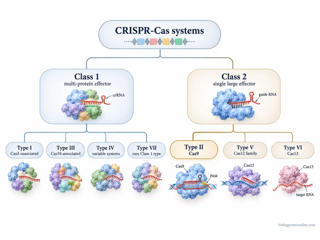
Class 1 systems contain several Cas proteins which work together and form an effector complex. In Class 2 systems, one large Cas protein performs most of the functions involved in target recognition and cleavage.
Types I, III, and IV are included under Class 1, while Types II, V, and VI belong to Class 2. A rare Type VII has also been recognized under Class 1 in the recent classification. CRISPR-Cas9 belongs to Type II.
Class 1 CRISPR-Cas Systems
Class 1 CRISPR-Cas systems- These systems contain a multi-protein effector complex.
Several Cas proteins assemble together with CRISPR RNA (crRNA) for recognition of the target and interference. Types I, III, and IV are the major established types of Class 1. Their effector proteins and mechanisms of interference are different.
Recent classification also includes Type VII under Class 1.
Class 2 CRISPR-Cas Systems
Class 2 CRISPR-Cas systems contain one large multidomain Cas protein as the main effector.
It has a simpler effector arrangement compared to the multi-protein complex of Class 1 systems. Types II, V, and VI are included in this class.
Cas9, Cas12, and Cas13 are the characteristic effector proteins of Type II, Type V, and Type VI, respectively. This single-effector organization has been widely used in developing different CRISPR-based technologies.
Major Types of CRISPR-Cas Systems
Type I
Type I- It is a Class 1 CRISPR-Cas system containing a multi-protein surveillance complex.
The target DNA is recognized with the help of crRNA. Cas3 is generally involved in degradation of the recognized DNA and possesses helicase-nuclease activity.
Type II
Type II- It belongs to Class 2 and contains Cas9 as its major effector protein.
In the natural system, Cas9 works together with crRNA and trans-activating CRISPR RNA (tracrRNA). These RNA components guide Cas9 towards the complementary DNA sequence. The nuclease domains of Cas9 then cut the target DNA strands.
For genome editing, functions of crRNA and tracrRNA can be combined into a single-guide RNA (sgRNA).
Type III
Type III- It is a Class 1 system containing multi-subunit effector machinery. Cas10 is one of its characteristic proteins.
Different Type III systems can act against RNA as well as transcription-associated DNA targets. Some members also contain Cas10-linked signalling mechanisms which take part during the interference process.
Type IV
Type IV- These are Class 1 systems with considerable diversity in their organization.
Many Type IV loci are associated with mobile genetic elements. Some of them lack the complete adaptation machinery found in other CRISPR-Cas systems.
Their mechanism of interference also varies. Some Type IV systems are associated with interactions between mobile genetic elements, while crRNA-guided cleavage of target DNA has been demonstrated in particular Type IV variants.
Type V
Type V- It is a Class 2 CRISPR-Cas system mainly associated with Cas12-family proteins.
Many characterized Cas12 proteins act as RNA-guided DNA nucleases. Cas12 is not simply another name of Cas9. Its guide-RNA requirement, PAM recognition, and DNA-cleavage properties can be different.
Cas12a (Cpf1), for example, uses a crRNA without requiring tracrRNA. It produces staggered cuts in double-stranded DNA.
Type VI
Type VI systems use Cas13 as the major effector protein. It belongs to Class 2.
Unlike Cas9 and commonly used Cas12 enzymes which target DNA, Cas13 mainly targets RNA. A guide RNA directs Cas13 towards a complementary single-stranded RNA sequence.
RNA cleavage is carried out through its HEPN nuclease domains.
Where Does Cas9 Fit in the Classification?
Cas9 is a Class 2, Type II CRISPR-Cas effector protein.
It is an RNA-guided DNA nuclease and forms the major interference machinery of Type II systems. Cas9 itself is not a separate CRISPR class.
Type II became widely used for genome editing because major target-recognition and DNA-cleavage activities are present within one Cas9 protein. The target can mainly be changed by changing the guide sequence of RNA.
The natural crRNA and tracrRNA were also experimentally combined into a single-guide RNA (sgRNA). This made the Cas9 system easier to program for a selected DNA sequence.
Cas9 together with designed guide RNA was later used to produce targeted double-stranded DNA breaks in mammalian genomes. Cellular DNA repair mechanisms can then act on these breaks and produce the required sequence changes.
Cas9 vs Cas12 and Cas13
Cas9- It is the characteristic effector protein of Type II CRISPR-Cas systems. Cas9 mainly functions as an RNA-guided DNA nuclease and cuts a selected DNA target containing the required PAM sequence.
Cas12- Cas12 proteins belong to Type V. Many members of the Cas12 family also target DNA, but their RNA requirement, PAM preference, and mode of DNA cleavage can differ from Cas9.
Cas13- It is a Type VI effector protein. Cas13 primarily targets RNA instead of DNA and functions as an RNA-guided ribonuclease.
Structure of the CRISPR-Cas9 System
CRISPR-Cas9 is structurally made up of Cas9 protein, a guide RNA and the PAM-containing target DNA. The commonly used Streptococcus pyogenes Cas9 (SpCas9) is a large protein of 1,368 amino acids. Its protein structure has two main lobes i.e. recognition (REC) lobe and nuclease (NUC) lobe. The RNA and target DNA are housed in the groove formed between these two lobes.
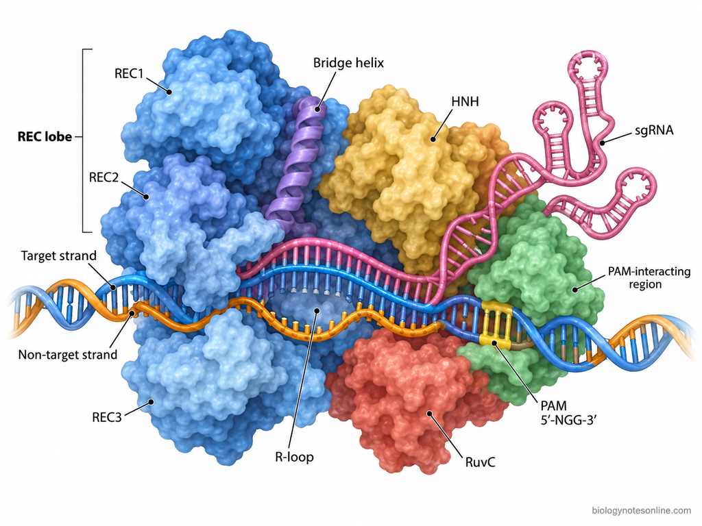
The detailed structural parts of the CRISPR-Cas9 system are as follows-
- Cas9 protein- Cas9 forms the large protein component around which the guide RNA and target DNA are arranged. It has a characteristic bilobed structure. One side forms the recognition (REC) lobe, while the other forms the nuclease (NUC) lobe. A positively charged central channel occurs between these regions, accommodating the negatively charged RNA-DNA structure.
- Recognition (REC) lobe- The REC lobe is mainly an α-helical region of Cas9. It contains the REC1, REC2 and REC3 recognition regions, with the bridge helix present at the nucleic acid interface. These regions make extensive contacts with the guide RNA and the guide RNA-target DNA heteroduplex. REC domains occupy one side of the central channel of Cas9.
- Nuclease (NUC) lobe- It forms the other major part of Cas9 and contains the RuvC domain, HNH domain and PAM-interacting (PI) domain. The RuvC domain is not formed as one continuous region in the primary protein sequence. It contains three separated regions referred to as RuvC I, II and III.The HNH domain lies between portions of the RuvC domain. In the nucleic acid-bound structure, HNH is positioned toward the DNA strand complementary to guide RNA, whereas the RuvC region lies toward the non-target DNA strand. The C-terminal part contains the PAM-interacting domain, which is placed next to the PAM-containing DNA duplex.
- Bridge helix (BH)- This is an arginine-rich α-helix present between the large structural regions of Cas9. It lies close to the bound nucleic acids and forms contacts with guide RNA and the RNA-DNA heteroduplex. The bridge helix is associated with the arrangement of the REC and NUC lobes around the nucleic acid-binding channel.
- Guide RNA (gRNA)- In the natural Type II system, the RNA component is formed from CRISPR RNA (crRNA) and trans-activating CRISPR RNA (tracrRNA). In engineered CRISPR-Cas9 these RNA parts are commonly joined, forming a single-guide RNA (sgRNA).The sgRNA has two major structural regions. A target-specific guide sequence occurs toward its 5′ end, generally about 20 nucleotides in the commonly used SpCas9 system. The remaining portion forms the RNA scaffold which is held by Cas9. This scaffold contains the repeat region, a tetraloop and stem-loop structures. SpCas9 sgRNA contains three stem loops associated with the Cas9-RNA complex.
- Target DNA- The target is a double-stranded DNA containing the selected sequence known as the protospacer. One DNA strand is complementary to the guide region of RNA and is referred to as the target strand. The opposite strand is the non-target strand.When present in the Cas9 complex, the guide region and complementary DNA are arranged as an RNA-DNA heteroduplex. The non-target DNA strand lies displaced from this hybrid, producing an R-loop arrangement inside Cas9. This nucleic acid structure runs through the channel present between the two protein lobes.
- Protospacer Adjacent Motif (PAM)- PAM is a short DNA sequence located immediately beside the protospacer. It is a part of the target DNA, not of the sgRNA. For SpCas9, the common PAM is 5′-NGG-3′, where “N” represents any nucleotide.The PAM remains as a base-paired DNA duplex in the Cas9-bound structure. The GG bases of SpCas9 PAM are present on the non-target strand and are contacted by conserved residues of the PAM-interacting region of Cas9.
- Cas9-sgRNA-target DNA complex- In the complete complex, Cas9 surrounds much of the guide RNA and target DNA. The sgRNA-target DNA heteroduplex occupies the positively charged groove between REC and NUC lobes. The PAM-containing DNA duplex lies near the C-terminal PAM-interacting region.HNH is positioned on the target-strand side. RuvC is arranged toward the displaced non-target strand. The guide RNA scaffold extends through different regions of Cas9, where its repeat duplex and stem-loop structures make contacts with the protein.
Mechanism of CRISPR-Cas9 Genome Editing
CRISPR-Cas9 genome editing is an RNA-guided process in which Cas9 recognizes a selected DNA sequence, cuts the DNA and the broken DNA is then repaired by the cellular DNA repair system.
Cas9 itself does not write a new DNA sequence after cleavage. The genetic changes are produced mainly during repair of the Cas9-induced DNA break through end joining or homology-directed repair (HDR).
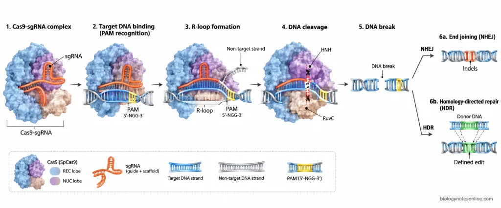
The mechanism of CRISPR-Cas9 genome editing occurs in the following steps-
1. Formation of Cas9-guide RNA Complex
The first step involves association of Cas9 protein with guide RNA (gRNA).
In engineered CRISPR-Cas9 systems, a single-guide RNA (sgRNA) is generally used. It contains the targeting sequence along with the RNA scaffold required for binding with Cas9.
Cas9 binds with the sgRNA and forms a Cas9-sgRNA ribonucleoprotein complex. In the SpCas9 system, the guide region is commonly about 20 nucleotides long and carries the sequence which pairs with the selected target DNA.
2. Recognition of PAM and Target DNA
The Cas9-sgRNA complex searches the DNA for a suitable protospacer adjacent motif (PAM).
PAM recognition takes place before complete pairing between guide RNA and target DNA. For Streptococcus pyogenes Cas9 (SpCas9), the commonly recognized PAM sequence is 5′-NGG-3′.
Cas9 does not open every DNA sequence for checking complete complementarity. It remains associated for a longer period with PAM-containing regions and then checks the nearby DNA sequence against the guide RNA. DNA regions without a suitable PAM are generally released.
3. DNA Unwinding and Guide RNA Pairing
After recognition of PAM, the DNA region immediately adjacent to the PAM begins to open.
In this step, the guide sequence of sgRNA starts pairing with its complementary DNA strand. Pairing begins near the PAM and extends along the target DNA when sufficient sequence complementarity is present.
One DNA strand pairs with the guide RNA, while the opposite non-target DNA strand becomes displaced. This structure is known as an R-loop.
Guide RNA + complementary target DNA → RNA-DNA heteroduplex
Formation of the proper RNA-DNA complex also brings Cas9 into the conformation required for cleavage. A poor sequence match, particularly near the PAM, can prevent proper R-loop formation and DNA cleavage.
4. Cleavage of Target DNA by Cas9
After formation of the correct RNA-DNA complex, two nuclease domains of Cas9 cut the two DNA strands separately.
HNH domain- It cleaves the DNA strand complementary to the guide RNA.
RuvC domain- It cleaves the opposite or non-target DNA strand.
In SpCas9, DNA cleavage generally occurs about 3 base pairs upstream of the PAM. A targeted double-strand DNA break (DSB) is formed.
5. Repair of Cas9-Induced DNA Break
After cleavage, the broken DNA becomes a substrate for the normal DNA repair machinery of the cell.
Most genetic changes produced during conventional CRISPR-Cas9 genome editing arise during this repair process. Two commonly used repair outcomes are non-homologous end joining (NHEJ) and homology-directed repair (HDR).
Non-Homologous End Joining (NHEJ)
NHEJ- It joins the broken DNA ends without requiring a homologous donor template.
Small insertions or deletions (indels) can be generated around the Cas9 cleavage site during repair. When cleavage occurs inside a protein-coding region, these indels can disturb the reading frame or another important sequence and may inactivate the gene.
NHEJ-based editing is commonly used for gene disruption or “knockout”. The repair products formed by end joining are not always identical. Other end-joining processes, such as microhomology-mediated repair, can also contribute to mutations obtained after Cas9 cleavage.
Homology-Directed Repair (HDR)
HDR is used when a defined sequence change is required.
A donor DNA containing the desired sequence along with homologous DNA surrounding the targeted region can be supplied with the CRISPR-Cas9 components.
During repair, sequence information from this donor DNA can be copied into the broken genomic site. Specific nucleotide changes, replacement of a sequence, or insertion of new DNA can be produced by this process.
HDR competes with other cellular DNA repair pathways. Its occurrence strongly depends on the cellular condition and the repair context.
Applications of CRISPR-Cas9
CRISPR-Cas9 is used in different fields including molecular biology, genetics, medicine, agriculture, animal research, and biotechnology. It is used to disrupt, correct, replace, or study a selected gene by targeting its DNA sequence. Modified Cas9 proteins (dCas9) are also used where binding of DNA is required without cutting the target DNA.
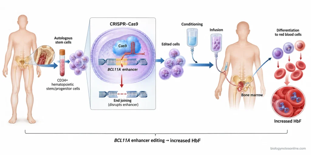
Some of the important applications of CRISPR-Cas9 are as follows-
- Gene Knockout- CRISPR-Cas9 is widely used for knocking out a selected gene and study its function. Cas9 cuts the target DNA and repair by end joining may introduce small insertions or deletions (indels). These changes can disturb the coding sequence, making the gene non-functional. Gene knockout using CRISPR-Cas9 is used in cultured cells, plants, and different experimental animals.
- Gene Insertion and Gene Correction- A selected DNA sequence can be introduced at the Cas9-targeted region when suitable donor DNA is supplied and homology-directed repair (HDR) occurs. It is used for introducing a defined mutation, correcting mutation, and preparation of “knock-in” cells or organisms.
- Functional Genomics and Genetic Screening- CRISPR-Cas9 is used to study functions of genes on a large scale. Libraries containing thousands of guide RNAs can target different genes throughout the genome. Such CRISPR screens are used for finding essential genes, genes of different cellular pathways, and the genes involved in drug response or resistance.
- Preparation of Disease Models- Cells and animals containing specific disease-associated genetic changes can be prepared using CRISPR-Cas9. Mouse models with mutation in single or multiple genes have been produced by introducing Cas9 with required guide RNAs into embryos.Human cells and organoids are also edited for making isogenic disease models and studying the effect of a particular mutation.
- Gene Therapy and Treatment of Genetic Diseases- CRISPR-Cas9 can be used for modification of a patient’s cells outside the body (ex vivo), after which the edited cells are returned to the patient.One of its clinical uses is exagamglogene autotemcel (exa-cel). In this treatment, autologous blood-forming stem and progenitor cells are edited at the erythroid-specific enhancer of the BCL11A gene. The edited cells are transplanted back and increase the production of fetal hemoglobin. It is used for sickle cell disease and transfusion-dependent β-thalassemia.
- Cancer Research and Immunotherapy- Cancer-associated genes are knocked out or altered in cultured cells and animal models for studying tumor development and drug response. CRISPR-Cas9 is also used in engineering immune cells, including T cells, for cancer immunotherapy research. Genes controlling the activity of immune cells can be modified before giving these cells back to the patient.
- Gene Expression Regulation- A nuclease-inactive Cas9, called dead Cas9 (dCas9), can bind the selected DNA but does not cut it. It is used near a gene for repression of transcription, called CRISPR interference (CRISPRi).dCas9 can also be attached with transcriptional activation domains. Selected genes can then be activated by CRISPR activation (CRISPRa).
- Plant Genome Editing and Crop Improvement- CRISPR-Cas9 is widely used in plant genetics and crop breeding. Genes associated with yield, quality, disease resistance, herbicide resistance, and tolerance to environmental stresses can be modified. It is also used to study plant gene functions and for preparation of mutant lines with different agronomically important traits.
- Animal Genetics and Breeding- Targeted mutations can be introduced into animal genomes by Cas9 and specific sgRNAs. More than one gene can also be edited at the same time. It is used in preparation of experimental animal models and in studying genes associated with growth, reproduction, disease resistance, and other animal traits.
- Microbial and Industrial Biotechnology- CRISPR-Cas9 is used to modify bacterial genomes for metabolic engineering. Genes of different cellular pathways can be deleted, inserted, or altered for changing the production of desired metabolites. Engineered microorganisms are studied for increasing production of different value-added biochemicals.
- Genome Imaging and Epigenetic Studies- dCas9 can carry other functional proteins to a selected region of the genome. When attached with fluorescent proteins, it is used for visualization of particular DNA loci in living cells. dCas9 attached with epigenetic-modifying proteins is also used to alter or study chromatin and gene regulation without producing a Cas9 double-strand DNA break.
Advantages of CRISPR-Cas9
CRISPR-Cas9 has several advantages over earlier programmable genome-editing tools such as zinc finger nucleases (ZFNs) and transcription activator-like effector nucleases (TALENs). The major advantage is its RNA-based targeting. For a new DNA target, mainly the guide RNA sequence is changed while Cas9 protein can remain the same. However, the advantage is not same for every target or experiment. PAM requirement, delivery of CRISPR components, and off-target activity can still limit its use.
- Easy Target Design- ZFNs and TALENs recognize DNA using engineered proteins, and new protein components have to be prepared for different target sequences. CRISPR-Cas9 mainly requires designing a guide RNA complementary to the selected DNA. The same Cas9 protein can be used with different guide RNAs.
- Faster and Less Laborious- Guide RNA can be designed and prepared more easily than engineering a new pair of DNA-binding proteins for each target. This reduces the work required for changing from one genomic target to another.
- Lower Cost- CRISPR-Cas9 is comparatively inexpensive for preparing new target-specific reagents. Short guide RNAs are changed instead of repeatedly producing customized DNA-binding proteins, making the technique affordable for many laboratory studies.
- Multiplex Genome Editing- More than one guide RNA can be used with Cas9. Several genomic sites can therefore be targeted within the same experiment. This is referred to as multiplex genome editing, and was demonstrated by using multiple guide sequences for simultaneous editing of different mammalian genomic sites.
- Suitable for Large-Scale Genetic Screening- Thousands of sgRNAs can be prepared as a library for targeting genes throughout the genome. CRISPR-Cas9 is therefore used in high-throughput functional genomic screens for identification of genes associated with a particular cellular phenotype, drug response, or other biological process.
- Wide Range of Applications- CRISPR-Cas9 has been used for genome modification in many different cell types and organisms. It has also allowed targeted modification of organisms and cells which were comparatively difficult to manipulate genetically using earlier methods.
- Good Editing Efficiency in Many Experimental Systems- Targeted DNA modification can be obtained at useful and sometimes high frequencies using CRISPR-Cas9. The efficiency, however, is not fixed. It varies according to guide sequence, genomic target, cell type, delivery method, and other experimental conditions.
- Cas9 Can Be Modified for Other Functions- Cas9 is not limited only to cutting DNA. Nuclease-inactive Cas9 (dCas9) retains RNA-guided DNA binding and can be joined with other functional domains. It is used for gene activation, gene repression, genomic imaging, and other sequence-specific manipulation without making the normal Cas9 double-strand DNA break.
Limitations and Challenges of CRISPR-Cas9
CRISPR-Cas9 is not a completely accurate genome-editing system. It can target a selected DNA sequence with high specificity, but unwanted changes may occur at other DNA sites and also at the correctly targeted site. Editing efficiency is also different with guide RNA, target sequence, cell type, delivery method, and DNA repair.
Some of the important limitations and challenges of CRISPR-Cas9 are-
- Off-target editing can occur. Cas9 may cut DNA sequences which are similar, but not fully identical, to the sequence selected by guide RNA. A number of mismatches can be tolerated depending on their number, position, and the particular guide sequence. This can introduce unwanted mutations at other regions of the genome.
- There is also a chance of unwanted changes at the on-target site. Cas9 produces a DNA double-strand break and its repair is carried out by cellular repair machinery. Besides small insertions or deletions, large deletions and complex genomic rearrangements have been reported after repair of Cas9-induced breaks.
- PAM sequence is required for targeting. Cas9 cannot target every DNA sequence simply because a matching guide RNA can be prepared. A suitable protospacer adjacent motif (PAM) must occur next to the target. For commonly used Streptococcus pyogenes Cas9 (SpCas9), the usual PAM is NGG, which restricts the positions that can be selected for editing.
- Not every guide RNA works with the same efficiency. The sequence of guide RNA and target DNA affects Cas9 activity and specificity. DNA accessibility also has an effect, where closed chromatin can reduce Cas9 binding at some genomic sites.
- Precise editing by homology-directed repair (HDR) is limited. After Cas9 cutting, the cell can repair DNA by different pathways and the required repair pathway cannot always be obtained efficiently. HDR is mainly active during S and G2 phases of the cell cycle, making donor-template based precise changes more difficult in many cells, especially non-dividing cells.
- Delivery of CRISPR-Cas9 components into cells and tissues is another major problem. Cas9 is a large protein (about 160 kDa), while sgRNA is also a large charged molecule. Both must reach the required cells and finally the nucleus. The large size of the SpCas9 gene also creates a packaging problem with vectors such as adeno-associated virus (AAV).
- Mosaicism can occur during embryo editing. If Cas9 continues editing after the first cell division, different cells of the developing embryo may receive different mutations. An edited animal may therefore contain wild-type cells together with cells having one or several different edited genotypes.
- Cas9-induced DNA breaks can activate the cellular DNA damage response. A p53-mediated response and cell-cycle arrest have been observed after CRISPR-Cas9 editing in human cells. This response can reduce recovery of successfully edited cells and can influence which cells survive after editing.
- Immune response against Cas9 is another concern for its therapeutic use. Cas9 proteins commonly used for genome editing are obtained from bacteria such as S. pyogenes and Staphylococcus aureus. Pre-existing antibodies and Cas9-reactive T cells have been detected in human samples, which has to be considered for in vivo delivery and repeated exposure to these proteins.
Ethical and Biosafety Issues in CRISPR-Cas9
The ethical and biosafety issues of CRISPR-Cas9 become particularly important when editing is performed in human cells, embryos, or germline cells. Somatic genome editing is intended to change cells of the treated individual and the changes are generally not inherited by offspring. Editing of sperm, eggs, or embryos can produce heritable (germline) changes, which may pass into future generations.
Some of the important ethical and biosafety issues of CRISPR-Cas9 are-
- Unintended Genetic Changes- CRISPR-Cas9 does not always produce only the desired DNA change. Off-target mutations may occur at other similar DNA sequences. Unwanted changes including larger deletions, chromosomal alterations, or unexpected repair products can also occur at the target region. Such changes become more serious when edited cells are used for human treatment or reproduction.
- Human Germline Editing- Editing of a human embryo, sperm, or egg can introduce a change into the germline. If an edited embryo develops into a person, some genetic changes may be transmitted to its offspring. An unwanted genetic change can also pass on. The effects are therefore not necessarily limited to one individual.
- Consent of Future Generations- A child produced from a germline-edited embryo cannot give prior consent for the genetic modification. The same problem extends to descendants when the edited DNA is inherited. This is one of the major ethical differences between somatic editing and heritable genome editing.
- Mosaicism in Embryo Editing- All cells of an edited embryo do not necessarily receive the same genetic modification. Some cells may remain unedited while others contain one or different edited sequences. This condition is called mosaicism. It can make the biological effect and inheritance of an embryo edit more difficult to predict.
- Therapy and Genetic Enhancement- CRISPR-Cas9 can be considered for correcting or preventing disease-causing genetic changes, but another ethical question comes up when genome editing is proposed for enhancement of healthy individuals. Enhancement may include attempts to change physical or other human traits rather than treating a disease. Many such traits also involve several genes together with environmental factors, making their genetic modification much more complex.
- Equity and Access- Genome-editing treatments can require highly specialized laboratories, clinical facilities, and expensive procedures. Unequal access to such technologies can increase an already existing difference in health care. The concern becomes more prominent if genome editing is used for non-medical enhancement available only to particular groups.
- Long-Term Safety- A genome edit can remain in a cell for its lifetime, and a germline change may continue into later generations. Some unwanted effects may not become clear during the short period of an experiment or clinical study. Long-term observation becomes important for edited human cells and individuals receiving genome-editing treatment.
- CRISPR Gene Drives and Environmental Biosafety- CRISPR-Cas9 can also be used to construct gene drives, where a genetic change is designed to spread through a sexually reproducing population at greater than normal inheritance frequency. Escape or release of such organisms can allow the modification to spread outside an intended population, depending on the drive and the biology of the species. Effects on non-target populations and ecological interactions can be difficult to predict before environmental release.
- Misuse of Genome Editing- The same genome-editing ability which is used in biomedical and biological research can be used for purposes other than the intended scientific or therapeutic application. Concerns over misuse are especially raised for heritable human modification and other applications where the genetic change may have persistent biological effects.
Discovery and Development of CRISPR-Cas9
The discovery of CRISPR-Cas9 occurred through a series of studies carried out by different scientists, first as unusual repeated DNA in microorganisms and later as an RNA-guided genome-editing system. CRISPR itself was not discovered together with Cas9 genome editing at one time. More studies followed for about 25 years before the natural microbial system was converted into a programmable DNA-cutting tool.
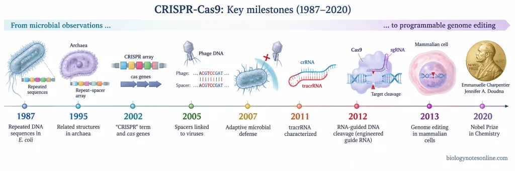
The major events in the discovery and development of CRISPR-Cas9 are as follows-
- 1987- First observation of the repeated DNA sequences– The history of CRISPR dates back to 1987 when Yoshizumi Ishino and his associates were studying the iap gene of Escherichia coli. An unusual arrangement of repeated DNA sequences separated by other sequences was found downstream of the gene. At that time, its biological function was not known and the term “CRISPR” had not been given. These sequences were later recognized as an early example of a CRISPR array.
- 1995- Similar repeat-spacer structures found in Archaea– Francisco Mojica and his associates studied repeated DNA in the archaeal species Haloferax mediterranei and Haloferax volcanii. They described long regions having a repeated 30 bp sequence interspaced with different sequences of about 33-39 bp. The function was still uncertain.
- 2002- The name “CRISPR” and Cas genesIn 2002, Ruud Jansen and associates used the term Clustered Regularly Interspaced Short Palindromic Repeats (CRISPR) for these characteristic repeat-spacer DNA regions.The same study identified four genes, cas1, cas2, cas3, and cas4, which were commonly located next to CRISPR loci. These were called “CRISPR-associated” or cas genes. A functional relationship between the repeats and Cas proteins was suggested, although their actual biological role was not yet clear.
- 2005- Spacer sequences linked with viruses and other foreign DNA– An important development came when different groups studied the sequences present between CRISPR repeats. Francisco Mojica and associates showed that many of these spacers were derived from bacteriophages and other transmissible genetic elements. Similar findings were independently reported by groups including Christine Pourcel and Alexander Bolotin.Mojica and associates also found that some foreign elements matching CRISPR spacers failed to infect the particular spacer-carrying strain. This connected CRISPR with a possible microbial immunity against invading DNA.
- 2007- CRISPR experimentally proved as an acquired defense system– Rodolphe Barrangou, Philippe Horvath, and their associates experimentally studied CRISPR in Streptococcus thermophilus. After bacteriophage exposure, new phage-derived spacers were added into the bacterial CRISPR region.Addition or removal of particular spacers also changed resistance of the bacteria to the corresponding phage. This provided experimental evidence that CRISPR together with Cas proteins acts as an acquired or adaptive microbial defense system.
- 2011- tracrRNA and the Type II CRISPR-Cas system– Elitza Deltcheva, Emmanuelle Charpentier, and associates studied the Type II CRISPR system of Streptococcus pyogenes. They identified trans-activating CRISPR RNA (tracrRNA), which pairs with CRISPR RNA and takes part in its maturation. Cas9 (then also referred to as Csn1 in this work), tracrRNA, crRNA, and host RNase III were associated with this pathway.
- 2012- Cas9 developed as a programmable DNA-cutting system– In 2012, Martin Jinek, Krzysztof Chylinski, Ines Fonfara, Michael Hauer, Jennifer Doudna, Emmanuelle Charpentier, and associates showed that Cas9 could be directed by crRNA and tracrRNA to cut a selected double-stranded DNA sequence. They also joined the two RNA functions into a single engineered RNA chimera, the basis of the later single-guide RNA (sgRNA) design.During the same period, Giedrius Gasiunas, Virginijus Šikšnys, Rodolphe Barrangou, Philippe Horvath, and associates independently demonstrated that a Cas9-crRNA complex functions as an RNA-guided DNA endonuclease and can produce sequence-specific double-strand DNA cleavage.
- 2013- CRISPR-Cas9 editing in mammalian cells– The bacterial system was then brought into mammalian cells. Le Cong, Feng Zhang, and associates showed that Cas9 with short guide RNAs could cut endogenous genomic sites in human and mouse cells. More than one site could also be targeted.At the same time, Prashant Mali, George Church, and associates demonstrated RNA-guided Cas9 genome engineering in human cells. Custom guide RNAs were used for targeting endogenous human DNA sequences.
- 2020- Nobel Prize for CRISPR-Cas9 genome editing– The 2020 Nobel Prize in Chemistry was awarded to Emmanuelle Charpentier and Jennifer A. Doudna “for the development of a method for genome editing.” The award specifically recognized development of the CRISPR-Cas9 genetic-scissors method.The earlier discovery of CRISPR repeats, identification of Cas genes, finding the foreign origin of spacers, demonstration of microbial immunity, characterization of Cas9, and its later use in mammalian cells involved several other scientists and research groups.
CRISPR-Cas9 vs Other Genome-Editing Technologies
CRISPR-Cas9, zinc-finger nucleases (ZFNs), TALENs, base editors, and prime editors are used for targeted changes in DNA, but their targeting and editing systems are different. ZFNs and TALENs depend mainly on engineered DNA-binding proteins. CRISPR-Cas9 uses a guide RNA with Cas9. Base editing and prime editing are newer CRISPR-derived systems where modified Cas proteins are used without making the usual Cas9 double-strand DNA break.
| Genome-editing system | Target recognition | Editing system | Main difference from CRISPR-Cas9 |
|---|---|---|---|
| CRISPR-Cas9 | Guide RNA pairs with complementary DNA, with a suitable PAM | Cas9 nuclease generally produces a targeted double-strand DNA break | Target is mainly changed by changing the guide RNA |
| ZFN | Engineered zinc-finger proteins bind DNA | Two FokI nuclease domains come together and cut DNA | New DNA-binding proteins need to be engineered for different targets |
| TALEN | Engineered TALE repeat proteins recognize DNA bases | FokI nuclease is used for DNA cleavage | Target recognition is protein-based, not guide RNA-based |
| Base Editing | Guide RNA directs a modified CRISPR-Cas protein | A deaminase directly converts selected DNA bases without the usual double-strand break | Used mainly for defined base conversions |
| Prime Editing | Prime-editing guide RNA (pegRNA) specifies target and contains editing information | Cas9 nickase is joined with reverse transcriptase | Can introduce substitutions, small insertions, and deletions without making a double-strand break or using a donor DNA template |
CRISPR-Cas9 vs Zinc-Finger Nucleases
Zinc-finger nucleases (ZFNs) contain engineered zinc-finger DNA-binding domains joined with the FokI nuclease. Individual zinc fingers recognize short DNA sequences, and two ZFNs are generally required around the target so that FokI can dimerize and cut DNA.
CRISPR-Cas9 recognizes the selected sequence mainly through RNA-DNA base pairing. For another target, the guide RNA can be changed while the Cas9 protein remains the same. In ZFNs, the protein DNA-binding region has to be engineered according to the new target, which makes their reprogramming more complex.
CRISPR-Cas9 vs TALENs
TALENs also use protein-based DNA recognition. The TALE DNA-binding region contains repeated modules, where individual repeat-variable diresidues (RVDs) determine recognition of DNA bases. This region is attached with a FokI nuclease domain. Two TALEN molecules are used for cleavage of the target DNA.
In CRISPR-Cas9, a new target generally needs a new guide RNA sequence. TALEN targeting needs construction of a different arrangement of TALE protein repeats. Both systems can make targeted double-strand DNA breaks, but one depends on RNA-guided recognition and the other on engineered protein-DNA recognition.
CRISPR-Cas9 vs Base Editing
Conventional Cas9 genome editing commonly starts by producing a double-strand DNA break, after which cellular DNA repair gives the genetic change. Base editing works differently. It uses a modified CRISPR-Cas protein together with a base-modifying enzyme (deaminase) for direct conversion of particular DNA bases without producing the usual double-strand break.
The first cytosine base editor was developed for conversion of a target C to T (or G to A on the opposite strand). Other base-editor systems have expanded the types of base changes possible, but base editing remains dependent on the particular chemistry and editing window of the editor being used.
CRISPR-Cas9 vs Prime Editing
Prime editing is also derived from CRISPR technology but has a different editing arrangement. It uses a Cas9 nickase attached to an engineered reverse transcriptase (RT) and a prime-editing guide RNA (pegRNA). The pegRNA carries both the target-recognition sequence and information for the required edit.
Unlike conventional Cas9 nuclease editing, prime editing does not normally produce a double-strand DNA break. It can introduce all types of base substitutions and also small insertions or deletions without requiring a separate donor DNA template. Its editing architecture is therefore more complex than the standard Cas9 + sgRNA system.
CRISPR-Cas9 At a Glance
The important points of CRISPR-Cas9 can be summarized as follows-
| Feature | CRISPR-Cas9 |
|---|---|
| Full form of CRISPR | Clustered Regularly Interspaced Short Palindromic Repeats |
| Natural origin | Derived from the CRISPR-Cas adaptive defense systems of bacteria and archaea |
| Natural function | Protection against invading genetic materials such as bacteriophages and plasmids |
| Classification | Class 2, Type II CRISPR-Cas system |
| Major effector protein | Cas9 (CRISPR-associated protein 9) |
| Nature of Cas9 | RNA-guided DNA nuclease |
| Main components for genome editing | Cas9 protein, guide RNA (gRNA), target DNA, and PAM |
| Natural guide RNA | crRNA + tracrRNA |
| Engineered guide RNA | Single-guide RNA (sgRNA), combining crRNA and tracrRNA functions |
| Guide sequence | Generally about 20 nucleotides for commonly used Streptococcus pyogenes Cas9 (SpCas9) |
| Main DNA target | A DNA sequence (protospacer) complementary to the guide RNA |
| PAM | Protospacer Adjacent Motif, a short DNA sequence present next to the target |
| PAM of SpCas9 | Usually 5′-NGG-3′, where N can be any nucleotide |
| Cas9 nuclease domains | HNH and RuvC |
| DNA cleavage | HNH cuts the guide-complementary strand and RuvC cuts the opposite strand |
| Common cleavage position | Around 3 base pairs upstream of the PAM in SpCas9 |
| Result of Cas9 activity | Targeted DNA double-strand break (DSB) |
| Major DNA repair routes | End joining, commonly NHEJ, and homology-directed repair (HDR) |
| NHEJ outcome | Frequently produces small insertions or deletions (indels), useful for gene knockout |
| HDR outcome | Can introduce a defined DNA change when a suitable donor template is available |
| Major applications | Gene knockout, gene correction, disease models, functional genomics, crop editing, animal research, and gene therapy |
| Major advantage | Target can generally be changed by changing the guide RNA rather than engineering a new DNA-binding protein |
| Major limitations | Off-target editing, unwanted on-target changes, PAM requirement, variable editing efficiency, delivery problems, and unpredictable DNA repair |
| Cas9 vs Cas12 | Cas9 is a Type II effector. Cas12 belongs to Type V and many Cas12 proteins also target DNA |
| Cas9 vs Cas13 | Cas9 primarily targets DNA, whereas Type VI Cas13 primarily targets RNA |
| First CRISPR repeats reported | 1987, during studies by Yoshizumi Ishino and associates in E. coli |
| CRISPR term introduced | 2002 by Ruud Jansen and associates |
| Adaptive defense experimentally demonstrated | 2007 by Rodolphe Barrangou, Philippe Horvath, and associates |
| Programmable Cas9 DNA cleavage demonstrated | 2012, including work by Jennifer Doudna, Emmanuelle Charpentier and associates, with independent related Cas9 work by Virginijus Šikšnys and associates |
| Nobel Prize | 2020 Nobel Prize in Chemistry to Emmanuelle Charpentier and Jennifer A. Doudna for development of a method for genome editing |
Common Misconceptions about CRISPR-Cas9
Some common misconceptions about CRISPR-Cas9 and their actual facts are summarized below-
| Common misconception | Actual fact |
|---|---|
| CRISPR-Cas9 is a single protein. | No. Cas9 is the protein. The genome-editing system contains Cas9 together with a guide RNA, while target DNA and its PAM are required for targeting. |
| CRISPR and Cas9 mean the same thing. | CRISPR refers to the broader microbial CRISPR-Cas system and its repeat-spacer arrays. Cas9 is an effector protein of the Class 2, Type II CRISPR-Cas system. Other CRISPR systems contain different Cas proteins. |
| Guide RNA cuts the DNA. | The guide RNA does not cut DNA. It provides the sequence for recognition of the target. DNA cleavage is carried out by the HNH and RuvC nuclease domains of Cas9. |
| Cas9 can cut any DNA sequence selected by the researcher. | A matching guide sequence alone is not enough. A suitable protospacer adjacent motif (PAM) must occur beside the target sequence. PAM requirements depend on the Cas protein being used. |
| PAM is a part of the guide RNA. | PAM is present in the target DNA, adjacent to the protospacer. For commonly used Streptococcus pyogenes Cas9 (SpCas9), the usual PAM is 5′-NGG-3′. |
| CRISPR-Cas9 is 100% accurate. | It is not completely accurate. Cas9 can sometimes act at similar non-target sequences, producing off-target changes. Unwanted changes may also occur at the intended target during DNA repair. |
| Cas9 itself produces the final genetic change. | Cas9 mainly makes the targeted DNA break. The cell then repairs this break. NHEJ or other end-joining repair and HDR are responsible for many of the final sequence changes obtained after conventional Cas9 editing. |
| CRISPR-Cas9 can only knock out genes. | Gene knockout is one common use, generally through indels produced during end joining. With a suitable donor template and HDR, selected insertions, substitutions, or other defined changes can also be introduced. |
| Every CRISPR system uses Cas9. | Cas9 belongs specifically to Type II. Type V systems commonly contain Cas12 proteins, whereas Type VI contains Cas13 effectors. Cas13 mainly targets RNA rather than DNA. |
| All edited cells receive exactly the same DNA change. | The editing outcome is not always identical. Repair of a Cas9-induced break can produce different insertions, deletions, or other repair products among cells. |
References
- Abudayyeh, O. O., Gootenberg, J. S., Konermann, S., Joung, J., Slaymaker, I. M., Cox, D. B. T., Shmakov, S., Makarova, K. S., Semenova, E., Minakhin, L., Severinov, K., Regev, A., Lander, E. S., Koonin, E. V., & Zhang, F. (2016). C2c2 is a single-component programmable RNA-guided RNA-targeting CRISPR effector. Science, 353(6299), aaf5573. https://doi.org/10.1126/science.aaf5573
- Anders, C., Niewoehner, O., Duerst, A., & Jinek, M. (2014). Structural basis of PAM-dependent target DNA recognition by the Cas9 endonuclease. Nature, 513(7519), 569–573. https://doi.org/10.1038/nature13579
- Anzalone, A. V., Randolph, P. B., Davis, J. R., Sousa, A. A., Koblan, L. W., Levy, J. M., Chen, P. J., Wilson, C., Newby, G. A., Raguram, A., & Liu, D. R. (2019). Search-and-replace genome editing without double-strand breaks or donor DNA. Nature, 576(7785), 149–157. https://doi.org/10.1038/s41586-019-1711-4
- Babu, K., Kathiresan, V., Kumari, P., Newsom, S., Parameshwaran, H. P., Chen, X., Liu, J., Qin, P. Z., & Rajan, R. (2021). Coordinated actions of Cas9 HNH and RuvC nuclease domains are regulated by the bridge helix and the target DNA sequence. Biochemistry, 60(49), 3783–3800. https://doi.org/10.1021/acs.biochem.1c00354
- Barrangou, R., Fremaux, C., Deveau, H., Richards, M., Boyaval, P., Moineau, S., Romero, D. A., & Horvath, P. (2007). CRISPR provides acquired resistance against viruses in prokaryotes. Science, 315(5819), 1709–1712. https://doi.org/10.1126/science.1138140
- Brokowski, C., & Adli, M. (2019). CRISPR ethics: Moral considerations for applications of a powerful tool. Journal of Molecular Biology, 431(1), 88–101. https://doi.org/10.1016/j.jmb.2018.05.044
- Brouns, S. J. J., Jore, M. M., Lundgren, M., Westra, E. R., Slijkhuis, R. J. H., Snijders, A. P. L., Dickman, M. J., Makarova, K. S., Koonin, E. V., & van der Oost, J. (2008). Small CRISPR RNAs guide antiviral defense in prokaryotes. Science, 321(5891), 960–964. https://doi.org/10.1126/science.1159689
- Chandrasegaran, S., & Carroll, D. (2016). Origins of programmable nucleases for genome engineering. Journal of Molecular Biology, 428(5, Part B), 963–989. https://doi.org/10.1016/j.jmb.2015.10.014
- Charlesworth, C. T., Deshpande, P. S., Dever, D. P., Camarena, J., Lemgart, V. T., Cromer, M. K., Vakulskas, C. A., Collingwood, M. A., Zhang, L., Bode, N. M., Behlke, M. A., Dejene, B., Cieniewicz, B., Romano, R., Lesch, B. J., Gomez-Ospina, N., Mantri, S., Pavel-Dinu, M., Weinberg, K. I., & Porteus, M. H. (2019). Identification of preexisting adaptive immunity to Cas9 proteins in humans. Nature Medicine, 25, 249–254. https://doi.org/10.1038/s41591-018-0326-x
- Chen, K., Wang, Y., Zhang, R., Zhang, H., & Gao, C. (2019). CRISPR/Cas genome editing and precision plant breeding in agriculture. Annual Review of Plant Biology, 70, 667–697. https://doi.org/10.1146/annurev-arplant-050718-100049
- Cho, S., Shin, J., & Cho, B.-K. (2018). Applications of CRISPR/Cas system to bacterial metabolic engineering. International Journal of Molecular Sciences, 19(4), 1089. https://doi.org/10.3390/ijms19041089
- Coller, B. S. (2019). Ethics of human genome editing. Annual Review of Medicine, 70, 289–305. https://doi.org/10.1146/annurev-med-112717-094629
- Cong, L., Ran, F. A., Cox, D., Lin, S., Barretto, R., Habib, N., Hsu, P. D., Wu, X., Jiang, W., Marraffini, L. A., & Zhang, F. (2013). Multiplex genome engineering using CRISPR/Cas systems. Science, 339(6121), 819–823. https://doi.org/10.1126/science.1231143
- Deltcheva, E., Chylinski, K., Sharma, C. M., Gonzales, K., Chao, Y., Pirzada, Z. A., Eckert, M. R., Vogel, J., & Charpentier, E. (2011). CRISPR RNA maturation by trans-encoded small RNA and host factor RNase III. Nature, 471(7340), 602–607. https://doi.org/10.1038/nature09886
- Effah, S. N., Barrera, S. C., Urturi Ortiz, N., Dampier, W., Nonnemacher, M. R., & Wigdahl, B. (2026). CRISPR/Cas9-based genome editing: Understanding differences in DNA repair pathways, profiles, and outcomes. International Journal of Molecular Sciences, 27(13), 5905. https://doi.org/10.3390/ijms27135905
- Esvelt, K. M., Smidler, A. L., Catteruccia, F., & Church, G. M. (2014). Concerning RNA-guided gene drives for the alteration of wild populations. eLife, 3, e03401. https://doi.org/10.7554/eLife.03401
- Fu, Y., Foden, J. A., Khayter, C., Maeder, M. L., Reyon, D., Joung, J. K., & Sander, J. D. (2013). High-frequency off-target mutagenesis induced by CRISPR-Cas nucleases in human cells. Nature Biotechnology, 31(9), 822–826. https://doi.org/10.1038/nbt.2623
- Gaj, T., Gersbach, C. A., & Barbas, C. F., III. (2013). ZFN, TALEN, and CRISPR/Cas-based methods for genome engineering. Trends in Biotechnology, 31(7), 397–405. https://doi.org/10.1016/j.tibtech.2013.04.004
- Gaj, T., Sirk, S. J., Shui, S.-L., & Liu, J. (2016). Genome-editing technologies: Principles and applications. Cold Spring Harbor Perspectives in Biology, 8(12), a023754. https://doi.org/10.1101/cshperspect.a023754
- Gasiunas, G., Barrangou, R., Horvath, P., & Siksnys, V. (2012). Cas9-crRNA ribonucleoprotein complex mediates specific DNA cleavage for adaptive immunity in bacteria. Proceedings of the National Academy of Sciences of the United States of America, 109(39), E2579–E2586. https://doi.org/10.1073/pnas.1208507109
- Guo, C., Ma, X., Gao, F., & Guo, Y. (2023). Off-target effects in CRISPR/Cas9 gene editing. Frontiers in Bioengineering and Biotechnology, 11, 1143157. https://doi.org/10.3389/fbioe.2023.1143157
- Haapaniemi, E., Botla, S., Persson, J., Schmierer, B., & Taipale, J. (2018). CRISPR–Cas9 genome editing induces a p53-mediated DNA damage response. Nature Medicine, 24, 927–930. https://doi.org/10.1038/s41591-018-0049-z
- Hryhorowicz, M., Lipiński, D., & Zeyland, J. (2023). Evolution of CRISPR/Cas systems for precise genome editing. International Journal of Molecular Sciences, 24(18), 14233. https://doi.org/10.3390/ijms241814233
- Hsu, P. D., Lander, E. S., & Zhang, F. (2014). Development and applications of CRISPR-Cas9 for genome engineering. Cell, 157(6), 1262–1278. https://doi.org/10.1016/j.cell.2014.05.010
- Ishino, Y., Shinagawa, H., Makino, K., Amemura, M., & Nakata, A. (1987). Nucleotide sequence of the iap gene, responsible for alkaline phosphatase isozyme conversion in Escherichia coli, and identification of the gene product. Journal of Bacteriology, 169(12), 5429–5433. https://doi.org/10.1128/jb.169.12.5429-5433.1987
- Jansen, R., van Embden, J. D. A., Gaastra, W., & Schouls, L. M. (2002). Identification of genes that are associated with DNA repeats in prokaryotes. Molecular Microbiology, 43(6), 1565–1575. https://doi.org/10.1046/j.1365-2958.2002.02839.x
- Jiang, F., & Doudna, J. A. (2017). CRISPR-Cas9 structures and mechanisms. Annual Review of Biophysics, 46, 505–529. https://doi.org/10.1146/annurev-biophys-062215-010822
- Jiang, F., Taylor, D. W., Chen, J. S., Kornfeld, J. E., Zhou, K., Thompson, A. J., Nogales, E., & Doudna, J. A. (2016). Structures of a CRISPR-Cas9 R-loop complex primed for DNA cleavage. Science, 351(6275), 867–871. https://doi.org/10.1126/science.aad8282
- Jinek, M., Chylinski, K., Fonfara, I., Hauer, M., Doudna, J. A., & Charpentier, E. (2012). A programmable dual-RNA-guided DNA endonuclease in adaptive bacterial immunity. Science, 337(6096), 816–821. https://doi.org/10.1126/science.1225829
- Jinek, M., East, A., Cheng, A., Lin, S., Ma, E., & Doudna, J. (2013). RNA-programmed genome editing in human cells. eLife, 2, e00471. https://doi.org/10.7554/eLife.00471
- Komor, A. C., Kim, Y. B., Packer, M. S., Zuris, J. A., & Liu, D. R. (2016). Programmable editing of a target base in genomic DNA without double-stranded DNA cleavage. Nature, 533(7603), 420–424. https://doi.org/10.1038/nature17946
- Kosicki, M., Tomberg, K., & Bradley, A. (2018). Repair of double-strand breaks induced by CRISPR–Cas9 leads to large deletions and complex rearrangements. Nature Biotechnology, 36, 765–771. https://doi.org/10.1038/nbt.4192
- Larson, M. H., Gilbert, L. A., Wang, X., Lim, W. A., Weissman, J. S., & Qi, L. S. (2013). CRISPR interference (CRISPRi) for sequence-specific control of gene expression. Nature Protocols, 8(11), 2180–2196. https://doi.org/10.1038/nprot.2013.132
- Liao, H., Wu, J., VanDusen, N. J., Li, Y., & Zheng, Y. (2024). CRISPR-Cas9-mediated homology-directed repair for precise gene editing. Molecular Therapy – Nucleic Acids, 35(4), 102344. https://doi.org/10.1016/j.omtn.2024.102344
- Liu, M., Rehman, S., Tang, X., Gu, K., Fan, Q., Chen, D., & Ma, W. (2019). Methodologies for improving HDR efficiency. Frontiers in Genetics, 9, 691. https://doi.org/10.3389/fgene.2018.00691
- Locatelli, F., Lang, P., Wall, D., Meisel, R., Corbacioglu, S., Li, A. M., de la Fuente, J., Shah, A. J., Carpenter, B., Kwiatkowski, J. L., Mapara, M., Liem, R. I., Cappellini, M. D., Algeri, M., Kattamis, A., Sheth, S., Grupp, S., Handgretinger, R., Kohli, P., . . . Frangoul, H. (2024). Exagamglogene autotemcel for transfusion-dependent β-thalassemia. The New England Journal of Medicine, 390(18), 1663–1676. https://doi.org/10.1056/NEJMoa2309673
- Makarova, K. S., Wolf, Y. I., Alkhnbashi, O. S., Costa, F., Shah, S. A., Saunders, S. J., Barrangou, R., Brouns, S. J. J., Charpentier, E., Haft, D. H., Horvath, P., Moineau, S., Mojica, F. J. M., Terns, R. M., Terns, M. P., White, M. F., Yakunin, A. F., Garrett, R. A., van der Oost, J., . . . Koonin, E. V. (2015). An updated evolutionary classification of CRISPR–Cas systems. Nature Reviews Microbiology, 13(11), 722–736. https://doi.org/10.1038/nrmicro3569
- Makarova, K. S., Shmakov, S. A., Wolf, Y. I., Mutz, P., Altae-Tran, H., Beisel, C. L., Brouns, S. J. J., Charpentier, E., Cheng, D., Doudna, J., Haft, D. H., Horvath, P., Moineau, S., Mojica, F. J. M., Pausch, P., Pinilla-Redondo, R., Shah, S. A., Siksnys, V., Terns, M. P., . . . Koonin, E. V. (2025). An updated evolutionary classification of CRISPR–Cas systems including rare variants. Nature Microbiology, 10(12), 3346–3361. https://doi.org/10.1038/s41564-025-02180-8
- Mali, P., Yang, L., Esvelt, K. M., Aach, J., Guell, M., DiCarlo, J. E., Norville, J. E., & Church, G. M. (2013). RNA-guided human genome engineering via Cas9. Science, 339(6121), 823–826. https://doi.org/10.1126/science.1232033
- Marraffini, L. A. (2015). CRISPR-Cas immunity in prokaryotes. Nature, 526, 55–61. https://doi.org/10.1038/nature15386
- Mariano Junior, C. G., de Oliveira, V. C., & Ambrósio, C. E. (2024). Gene editing in small and large animals for translational medicine: A review. Animal Reproduction, 21(1), e20230089. https://doi.org/10.1590/1984-3143-AR2023-0089
- Martínez-Gálvez, G., Ata, H., Campbell, J. M., & Ekker, S. C. (2016). ssDNA and the Argonautes: The quest for the next golden editor. Human Gene Therapy, 27(6), 419–422. https://doi.org/10.1089/hum.2016.071
- Mojica, F. J. M., Díez-Villaseñor, C., García-Martínez, J., & Soria, E. (2005). Intervening sequences of regularly spaced prokaryotic repeats derive from foreign genetic elements. Journal of Molecular Evolution, 60(2), 174–182. https://doi.org/10.1007/s00239-004-0046-3
- Mojica, F. J. M., Ferrer, C., Juez, G., & Rodríguez-Valera, F. (1995). Long stretches of short tandem repeats are present in the largest replicons of the Archaea Haloferax mediterranei and Haloferax volcanii and could be involved in replicon partitioning. Molecular Microbiology, 17(1), 85–93. https://doi.org/10.1111/j.1365-2958.1995.mmi_17010085.x
- Mout, R., Ray, M., Lee, Y.-W., Scaletti, F., & Rotello, V. M. (2017). In vivo delivery of CRISPR/Cas9 for therapeutic gene editing: Progress and challenges. Bioconjugate Chemistry, 28(4), 880–884. https://doi.org/10.1021/acs.bioconjchem.7b00057
- Nidhi, S., Anand, U., Oleksak, P., Tripathi, P., Lal, J. A., Thomas, G., Kuca, K., & Tripathi, V. (2021). Novel CRISPR-Cas systems: An updated review of the current achievements, applications, and future research perspectives. International Journal of Molecular Sciences, 22(7), 3327. https://doi.org/10.3390/ijms22073327
- Nishimasu, H., Cong, L., Yan, W. X., Ran, F. A., Zetsche, B., Li, Y., Kurabayashi, A., Ishitani, R., Zhang, F., & Nureki, O. (2015). Crystal structure of Staphylococcus aureus Cas9. Cell, 162(5), 1113–1126. https://doi.org/10.1016/j.cell.2015.08.007
- Nishimasu, H., Ran, F. A., Hsu, P. D., Konermann, S., Shehata, S. I., Dohmae, N., Ishitani, R., Zhang, F., & Nureki, O. (2014). Crystal structure of Cas9 in complex with guide RNA and target DNA. Cell, 156(5), 935–949. https://doi.org/10.1016/j.cell.2014.02.001
- Ran, F. A., Hsu, P. D., Wright, J., Agarwala, V., Scott, D. A., & Zhang, F. (2013). Genome engineering using the CRISPR-Cas9 system. Nature Protocols, 8(11), 2281–2308. https://doi.org/10.1038/nprot.2013.143
- Rui, Y., Wilson, D. R., & Green, J. J. (2019). Non-viral delivery to enable genome editing. Trends in Biotechnology, 37(3), 281–293. https://doi.org/10.1016/j.tibtech.2018.08.010
- Saifaldeen, M., Al-Ansari, D. E., Ramotar, D., & Aouida, M. (2020). CRISPR FokI dead Cas9 system: Principles and applications in genome engineering. Cells, 9(11), 2518. https://doi.org/10.3390/cells9112518
- Sander, J. D., & Joung, J. K. (2014). CRISPR-Cas systems for editing, regulating and targeting genomes. Nature Biotechnology, 32, 347–355. https://doi.org/10.1038/nbt.2842
- Shalem, O., Sanjana, N. E., & Zhang, F. (2015). High-throughput functional genomics using CRISPR–Cas9. Nature Reviews Genetics, 16(5), 299–311. https://doi.org/10.1038/nrg3899
- Sorek, R., Kunin, V., & Hugenholtz, P. (2008). CRISPR—A widespread system that provides acquired resistance against phages in bacteria and archaea. Nature Reviews Microbiology, 6, 181–186. https://doi.org/10.1038/nrmicro1793
- Sternberg, S. H., Redding, S., Jinek, M., Greene, E. C., & Doudna, J. A. (2014). DNA interrogation by the CRISPR RNA-guided endonuclease Cas9. Nature, 507(7490), 62–67. https://doi.org/10.1038/nature13011
- Sun, L., Lutz, B. M., & Tao, Y.-X. (2016). The CRISPR/Cas9 system for gene editing and its potential application in pain research. Translational Perioperative and Pain Medicine, 1(3), 22–33. https://doi.org/10.31480/2330-4871/040
- Tsai, S. Q., & Joung, J. K. (2016). Defining and improving the genome-wide specificities of CRISPR-Cas9 nucleases. Nature Reviews Genetics, 17(5), 300–312. https://doi.org/10.1038/nrg.2016.28
- van der Oost, J., Westra, E. R., Jackson, R. N., & Wiedenheft, B. (2014). Unravelling the structural and mechanistic basis of CRISPR–Cas systems. Nature Reviews Microbiology, 12, 479–492. https://doi.org/10.1038/nrmicro3279
- Wang, H., La Russa, M., & Qi, L. S. (2016). CRISPR/Cas9 in genome editing and beyond. Annual Review of Biochemistry, 85, 227–264. https://doi.org/10.1146/annurev-biochem-060815-014607
- Wang, H., Yang, H., Shivalila, C. S., Dawlaty, M. M., Cheng, A. W., Zhang, F., & Jaenisch, R. (2013). One-step generation of mice carrying mutations in multiple genes by CRISPR/Cas-mediated genome engineering. Cell, 153(4), 910–918. https://doi.org/10.1016/j.cell.2013.04.025
- Wiley, L., Cheek, M., LaFar, E., Ma, X., Sekowski, J., Tanguturi, N., & Iltis, A. (2025). The ethics of human embryo editing via CRISPR-Cas9 technology: A systematic review of ethical arguments, reasons, and concerns. HEC Forum, 37(2), 267–303. https://doi.org/10.1007/s10730-024-09538-1
- Wu, X., Kriz, A. J., & Sharp, P. A. (2014). Target specificity of the CRISPR-Cas9 system. Quantitative Biology, 2(2), 59–70. https://doi.org/10.1007/s40484-014-0030-x
- Xiong, X., Chen, M., Lim, W. A., Zhao, D., & Qi, L. S. (2016). CRISPR/Cas9 for human genome engineering and disease research. Annual Review of Genomics and Human Genetics, 17, 131–154. https://doi.org/10.1146/annurev-genom-083115-022258
- Xue, C., & Greene, E. C. (2021). DNA repair pathway choices in CRISPR-Cas9-mediated genome editing. Trends in Genetics, 37(7), 639–656. https://doi.org/10.1016/j.tig.2021.02.008
- Zetsche, B., Gootenberg, J. S., Abudayyeh, O. O., Slaymaker, I. M., Makarova, K. S., Essletzbichler, P., Volz, S. E., Joung, J., van der Oost, J., Regev, A., Koonin, E. V., & Zhang, F. (2015). Cpf1 is a single RNA-guided endonuclease of a class 2 CRISPR-Cas system. Cell, 163(3), 759–771. https://doi.org/10.1016/j.cell.2015.09.038
- Zhou, Y., Zhu, S., Cai, C., Yuan, P., Li, C., Huang, Y., & Wei, W. (2014). High-throughput screening of a CRISPR/Cas9 library for functional genomics in human cells. Nature, 509, 487–491. https://doi.org/10.1038/nature13166
- Zhu, Y., & Huang, Z. (2019). Recent advances in structural studies of the CRISPR-Cas-mediated genome editing tools. National Science Review, 6(3), 438–451. https://doi.org/10.1093/nsr/nwy150