

A cancer cell is an abnormal body cell in which normal control of cell growth, survival, and other cellular behavior has been disturbed. Cancer cells develop from normal cells after accumulation of mutations or other genetic alterations that change normal cellular regulation. The cell may continue proliferation when it should stop, survive when it should undergo programmed cell death (apoptosis), and no longer respond properly to signals coming from surrounding cells and tissue.
The changes in a cancer cell can affect the cell cycle, differentiation, apoptosis, maintenance of genome integrity, and interaction of the cell with its surrounding tissue. These changes are not same in every cancer cell. Different cancers can acquire different combinations of genetic alterations during their development.
A cancer cell is also commonly referred to as a malignant cell. The term “neoplastic cell” is broader and does not always mean cancer, because a neoplasm may be benign or malignant.
A cancer cell and a tumor are not the same thing. A tumor is a mass of tissue. In a cancerous tumor, cancer cells occur along with other components such as stromal cells, immune cells, blood vessels, and extracellular material. Some cancers do not produce a solid tumor. Leukemias, for example, develop from blood-forming cells and generally do not form solid tumors.
Morphology of Cancer Cells
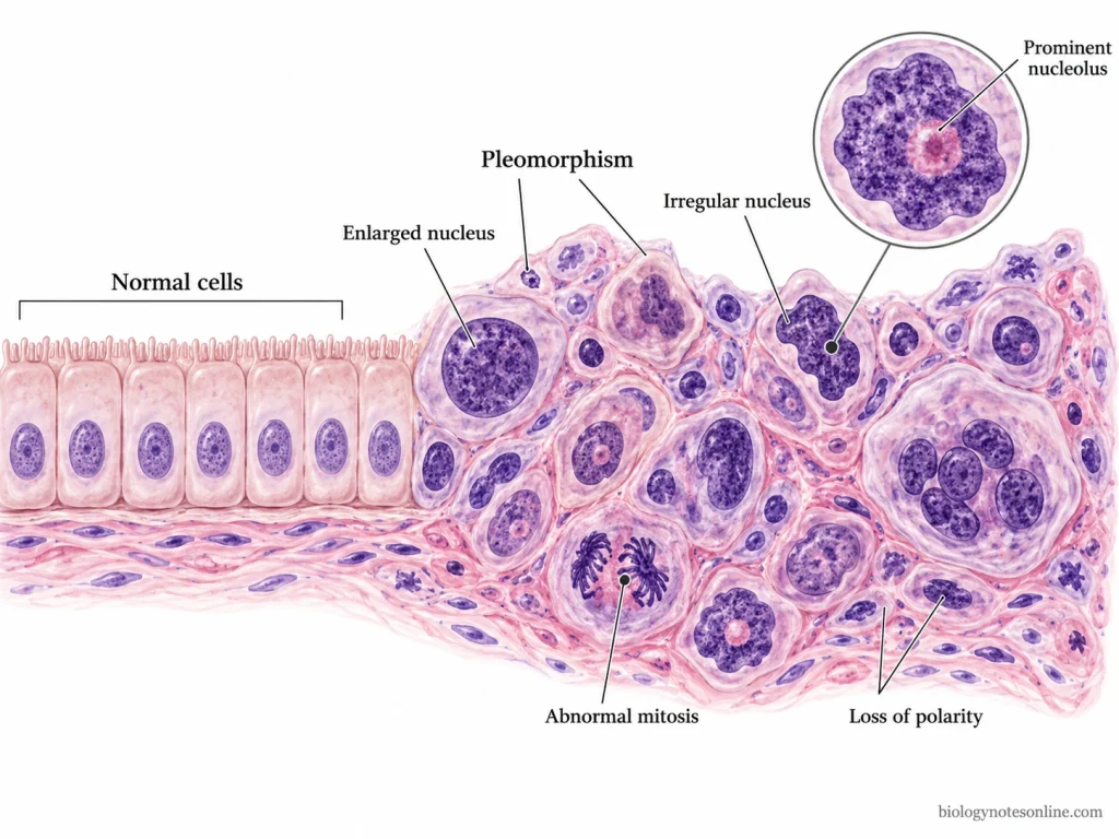
- Cancer cells do not have one fixed morphology. Their appearance differs with type of tumor and degree of differentiation. Some well-differentiated malignant cells can look very similar to the normal cells from which they developed.
- Variation in size and shape is frequently seen among cancer cells. This is referred to as pleomorphism. The cells may vary widely from one another, and variation can also occur in their nuclei. It is generally more prominent in poorly differentiated or anaplastic tumors.
- The nucleus is often enlarged in malignant cells. Nuclear shape may become irregular and the nucleus can occupy a much larger part of the cell. The nuclear-to-cytoplasmic ratio is increased in many cancers. This feature is not present to same degree in every tumor.
- Nuclear chromatin may become coarse, clumped or distributed abnormally. The nuclei can also stain deeply, a condition known as hyperchromasia. Irregular nuclear membrane is another common morphological abnormality.
- The nucleoli may become enlarged and prominent. More than one prominent nucleolus can also be present in a cancer cell.
- Cytoplasm of cancer cells is variable. Some malignant cells contain very little cytoplasm around a large nucleus, while others contain more cytoplasm and retain features of their tissue of origin. The cytoplasm may also differ in staining and appearance between different tumor cells.
- Mitotic figures are often increased in actively proliferating malignant tumors. Abnormal or bizarre mitotic figures can also occur because cell division becomes abnormal. These include unusual spindle arrangements and irregular chromosome separation.
- Some malignant tumors contain very large tumor giant cells. They may have one very large abnormal nucleus or several nuclei. Multinucleated cancer cells are also found in some tumors.
- The normal polarity and arrangement of cells may be lost. Cancer cells can become crowded and show abnormal relation with neighboring cells. These changes are more related to tissue organization, while nuclear shape, cell size, cytoplasm and nucleoli are features of individual cancer cell morphology.
- No single morphological feature is found in every cancer. Some of these abnormalities can also occur in non-neoplastic or reactive cells. Cancer morphology is examined from a combination of cellular and tissue features rather than one abnormal feature alone.
Types of Cancer Cells
Cancer cells are classified according to the normal cell or tissue from which they develop. The major types are given below.
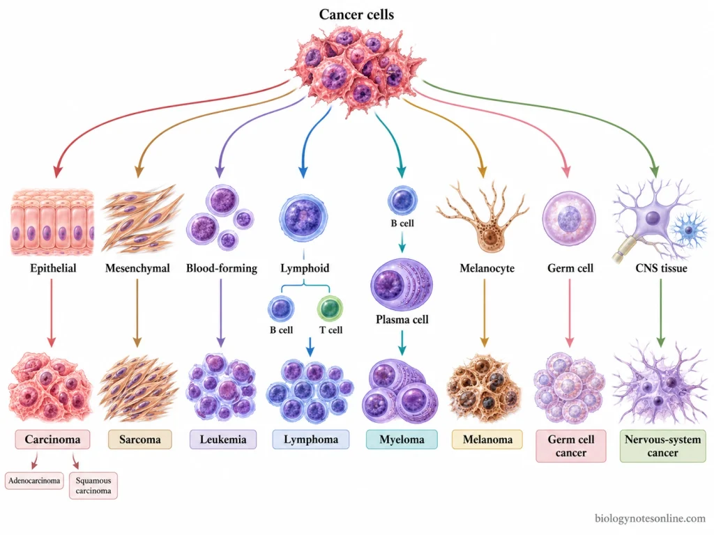
- Carcinoma cells- These cancer cells develop from epithelial cells. Epithelial cells cover body surfaces and also form lining and glandular parts of many organs. Many cancers of the skin, lung, breast and gastrointestinal tract are carcinomas. Carcinoma cells are further divided according to their epithelial cell type. Adenocarcinoma and squamous cell carcinoma are two types.
- Sarcoma cells- Sarcoma cells arise from connective or mesenchymal tissues. They may develop from bone, cartilage, muscle, fat, fibrous tissue or blood vessels. Sarcomas are much less common than carcinomas.
- Leukemia cells- These are malignant cells formed from hematopoietic or lymphoid cell lineages. The abnormal cells mainly proliferate in bone marrow. They may also be present in peripheral blood and other tissues. Different subtypes of leukemia are present depending on cell of origin and other biological features.
- Lymphoma cells- Lymphoma cells develop from cells of the lymphoid system. They can originate from different lymphocyte lineages. B-cell and T-cell neoplasms are major groups, each containing many separate types of lymphoma.
- Myeloma cells- Myeloma is formed from malignant plasma cells of the B-cell lineage. These cells commonly proliferate within bone marrow.
- Melanoma cells- Melanoma cells arise from melanocytes, the pigment-producing cells. Their cellular origin is different from epithelial cells of carcinomas and connective-tissue cells of sarcomas.
- Germ cell cancer cells- They develop from germ-cell lineage. Germ cell tumors may arise in the gonads and also at some extragonadal sites. The malignant germ cell tumors contain cells with different patterns and degrees of differentiation.
- Nervous-system cancer cells- Cancer cells arising from nervous system form another group. They do not come under the broad carcinoma or sarcoma classification. These cancers are named and further classified according to the particular nervous-system cell type from which they develop.
How Normal Cells Become Cancer Cells
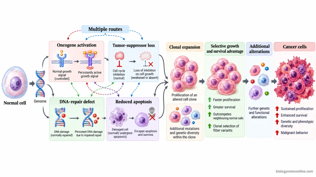
- A normal cell becomes a cancer cell through accumulation of genetic and regulatory changes. These changes occur in deoxyribonucleic acid (DNA) and affect the systems controlling cell growth, survival and other cell activities. Usually several changes are involved. Cancer development is a multistep process.
- Some genetic changes affect genes that normally stimulate cell proliferation. A normal proto-oncogene can become abnormally active and is then called an oncogene. The cell can receive growth-promoting signals even when normal stimulation is absent.
- Other changes inactivate tumor suppressor genes. These genes normally restrict cell proliferation or stop damaged cells from continuing through the cell cycle. When their function is lost, this control becomes reduced or absent.
- DNA repair genes can also become defective. Normally these genes recognize or repair DNA damage. Their loss allows mutations to remain and more genetic changes may accumulate in the cell. Genetic instability can increase during this process.
- Changes affecting apoptosis allow abnormal cells to survive when they would normally undergo cell death. The damaged cell is not removed. It can continue dividing and may acquire further genetic alterations.
- The changes do not have to occur in one fixed order. Different cancers can develop through different combinations of altered genes. Even cancers of the same type may contain different combinations of these changes.
- A cell having a growth or survival advantage can produce more daughter cells. The altered cell population then expands. Further changes can arise within these cells, and some of these changes may give another advantage during tumor development.
- During transformation, the normal controls over proliferation, differentiation and survival are progressively disturbed. DNA repair may also become defective. Accumulated abnormalities in several genes finally produce the altered cellular behavior associated with cancer cells.
Loss of Normal Growth and Survival Controls
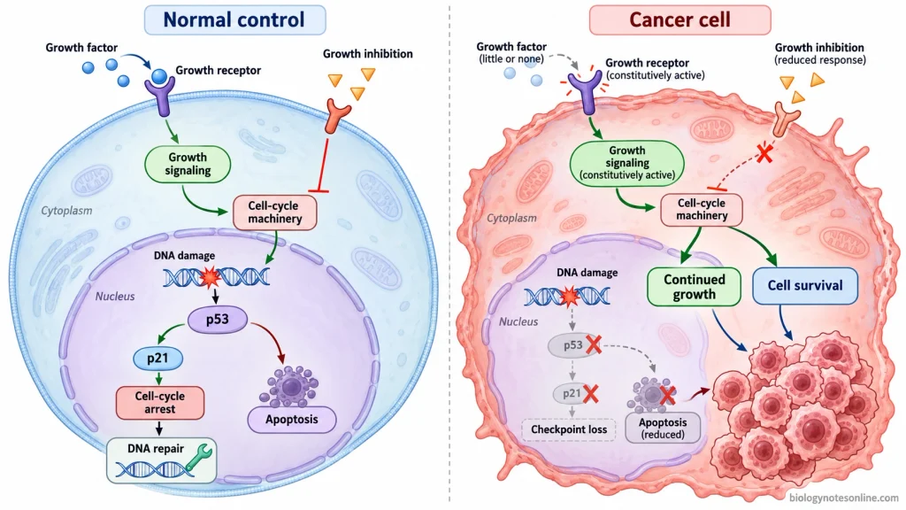
- Normal cells require external growth signals for entering and continuing the cell cycle. Cancer cells may gradually become less dependent on these normal signals. Some cancer cells produce their own growth factors. Nearby stromal cells can also be stimulated to supply growth factors. The growth factor receptors may increase in number or become active without normal stimulation. Proteins present in the signaling pathway below these receptors can also remain active, so the proliferative signal continues inside the cell.
- Growth-inhibitory signals normally restrict unnecessary cell proliferation. When tumor suppressor activity is lost, cancer cells may no longer respond properly to these signals and the cell cycle continues when a normal cell would stop.
- The cell-cycle checkpoints stop cells containing serious damage. The p53 protein can produce cell-cycle arrest after deoxyribonucleic acid (DNA) damage. DNA repair can take place during this arrest. If this control is lost, the damaged cell continues through the cell cycle. More genetic changes may remain in the dividing cells.
- Cell number is controlled by both cell division and cell loss. The cell population can increase when proliferation becomes higher, or when fewer abnormal cells are removed. Cancer cells commonly acquire both of these properties during tumor development.
- Apoptosis removes many cells having severe damage or other abnormal conditions. It can also take place when a cell loses the survival signals required for its survival. In cancer cells, this cell-death response may become defective. The abnormal cell remains alive instead of being removed.
- Changes in p53 and other regulators of apoptosis can increase cancer cell survival. Anti-apoptotic signaling may also become stronger. These cells can then survive DNA damage, growth-factor deprivation and some other unfavorable conditions that normally produce cell death.
- Loss of growth and survival controls is not same in every cancer. One tumor may show strong abnormal growth-factor signaling, while another may have major defects in growth suppression or cell-death pathways. Several altered controls may also occur together during development of cancer.
Cancer Cell Metabolism and Survival in Low Oxygen
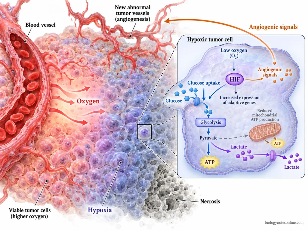
- Many cancer cells can survive when oxygen becomes very low. This low oxygen condition is known as hypoxia. It is common in many solid tumors. Tumor cells located farther from functional blood vessels receive less oxygen, because oxygen can move only a limited distance through the tissue. Some regions of the tumor become severely hypoxic.
- Cancer cells cannot be considered as cells which can always live indefinitely without oxygen. Very severe and prolonged oxygen deprivation can produce cell death and necrotic areas inside the tumor. Some cancer cells, however, may remain alive at extremely low or locally undetectable oxygen if sufficient nutrients are still available.
- Hypoxia-inducible factors (HIFs) become activated when oxygen level falls. HIFs control genes involved in cell growth and survival. The cancer cells can then adjust to the hypoxic condition. Some cancer cells also produce higher levels of these proteins.
- The metabolism of hypoxic cancer cells is changed by HIF activity. Glucose uptake can increase. More glucose passes through glycolysis and the expression of several glycolytic enzymes also becomes increased. Adenosine triphosphate (ATP) can still be produced when oxygen-dependent energy production becomes limited. Lactate production also rises.
- Cancer cells do not use nutrients in exactly the same way as most normal cells. They can use different nutrients and energy may be produced through altered metabolic pathways. These metabolic changes provide energy and cellular materials required by the growing cancer cells.
- Under hypoxia, mitochondrial metabolism and other metabolic pathways can be rearranged. Cancer cells may change the use of glucose, amino acids and lipids according to available oxygen and nutrients. This metabolic response is not same in every cancer cell or in every region of a tumor.
- HIFs also can activate genes involved in formation of new blood vessels. The new blood vessels can increase oxygen and nutrient supply toward the tumor. Hypoxic regions may still remain. Tumor blood vessels are often abnormal and oxygen is not distributed equally throughout the tumor.
- Hypoxia creates a strong selection condition inside the tumor. Cells unable to tolerate low oxygen may die, while cells having better hypoxic and metabolic adaptation can remain alive. A hypoxic tumor can contain viable cancer cells as well as necrotic regions at different distances from the blood supply.
Cancer Cells vs. Normal Cells
| Feature | Normal Cells | Cancer Cells |
|---|---|---|
| Cell growth and division | Normal cells divide according to the requirement of the tissue. Cell proliferation is controlled by several regulatory mechanisms. | Cancer cells continue to proliferate when normal growth controls are lost or altered. Uncontrolled proliferation is one of the main properties of cancer cells. |
| Response to growth signals | The cells usually require proper extracellular growth signals for proliferation. They also respond to signals that restrict cell division. | Cancer cells can become less dependent on normal growth signals. The response to growth-inhibitory signals may also be reduced or lost. |
| Cell death | Abnormal or severely damaged cells may undergo apoptosis. It removes cells that should not remain in the tissue. | Cancer cells can develop resistance to apoptosis. The abnormal cells then survive under conditions in which normal cells may undergo cell death. |
| Differentiation | Normal cells undergo differentiation and acquire the specialized structure and function of their tissue. Many fully differentiated cells divide rarely or stop dividing. | Cancer cells often show defective differentiation. Poorly differentiated cancer cells may lose many of the specialized features of the normal cell from which they developed. |
| Cell morphology | Normal cells of a particular tissue generally have a more regular size, shape and nuclear appearance. | Cancer cells may show variation in cell and nuclear size and shape. Enlarged nuclei, increased nuclear-to-cytoplasmic ratio, irregular nuclei and prominent nucleoli can occur. These features are not same in every cancer. |
| Cell arrangement | Normal cells maintain the organization and cellular relationships of their tissue. | The normal arrangement and polarity may become altered in cancer. Cancer cells can also lose normal inhibition produced by cell-cell contact. |
| Replicative lifespan | Most normal human somatic cells have a limited proliferative capacity. Replicative senescence can stop further cell division. | Many cancer cells overcome this limitation. Most human cancer cells maintain telomere function, commonly through telomerase activity, and can continue dividing for very long periods. |
| Genome | Normal cells contain systems for maintaining and repairing their genetic material. | Cancer cells commonly accumulate genetic alterations. Genetic instability is also present in many cancers and allows additional changes to arise during tumor progression. |
| Metabolism | Cellular metabolism is regulated according to the normal function and nutrient requirement of the tissue. | Cancer cells can rearrange their metabolism for continued growth and proliferation. The metabolic changes differ according to cell of origin, genetic alterations and the tissue environment. |
| Invasion and spread | Normal cells remain within their proper tissue boundaries and do not normally invade surrounding tissues. | Malignant cancer cells can invade nearby tissue. Some cancer cells can also spread and grow at distant sites, which is called metastasis. |
Cancer Cell at a Glance
| Feature | Quick Summary |
|---|---|
| Definition | Cancer cells are abnormal cells that grow and divide without normal cellular control. |
| Origin | They develop from normal cells after accumulation of genetic and epigenetic alterations. |
| Cell division | Cell proliferation becomes increased or uncontrolled. |
| Growth signals | Cancer cells may become less dependent on normal external growth signals. |
| Growth inhibition | Normal growth-inhibitory signals may be ignored or become defective. |
| Apoptosis | Cancer cells can develop resistance to programmed cell death (apoptosis). |
| Differentiation | Many cancer cells show reduced or abnormal differentiation. |
| Shape and size | Cells often show variation in size and shape, known as pleomorphism. |
| Nucleus | The nucleus may be enlarged and irregular with prominent nucleoli and increased nuclear-to-cytoplasmic ratio. |
| Cell arrangement | Normal cell polarity and tissue arrangement may be lost. |
| Genetic changes | Mutations and other genetic alterations accumulate in cancer cells. |
| Replicative capacity | Many cancer cells can continue dividing for very long periods. |
| Metabolism | Nutrient and energy metabolism is altered to support cancer cell growth and survival. |
| Hypoxia | Many cancer cells can adapt to low oxygen conditions, but they cannot universally survive indefinitely without oxygen. |
| Tumor formation | Continued survival and proliferation of altered cells can produce a tumor. |
| Invasion | Malignant cancer cells can enter and grow into surrounding tissues. |
| Metastasis | Some cancer cells spread through blood or lymph and form tumors at distant sites. |
| Major types | Carcinoma, sarcoma, leukemia, lymphoma, myeloma, melanoma, germ cell cancers and nervous-system cancers. |
References
- Al-Abbadi, M. A. (2011). Basics of cytology. Avicenna Journal of Medicine, 1(1), 18–28. https://doi.org/10.4103/2231-0770.83719
- Alberts, B., Johnson, A., Lewis, J., Raff, M., Roberts, K., & Walter, P. (2002a). Cancer as a microevolutionary process. In Molecular biology of the cell (4th ed.). Garland Science. https://www.ncbi.nlm.nih.gov/books/NBK26891/
- Alberts, B., Johnson, A., Lewis, J., Raff, M., Roberts, K., & Walter, P. (2002b). The molecular basis of cancer-cell behavior. In Molecular biology of the cell (4th ed.). Garland Science. https://www.ncbi.nlm.nih.gov/books/NBK26902/
- Andreeff, M., Goodrich, D. W., & Pardee, A. B. (2003). Cell proliferation and differentiation. In D. W. Kufe, R. E. Pollock, R. R. Weichselbaum, R. C. Bast Jr., T. S. Gansler, J. F. Holland, & E. Frei III (Eds.), Holland-Frei cancer medicine (6th ed.). BC Decker. https://www.ncbi.nlm.nih.gov/books/NBK13866/
- Baba, A. I., & Câtoi, C. (2007a). Tumor cell morphology. In Comparative oncology. The Publishing House of the Romanian Academy. https://www.ncbi.nlm.nih.gov/books/NBK9553/
- Baba, A. I., & Câtoi, C. (2007b). Tumors of hematopoietic and lymphoid tissues. In Comparative oncology. The Publishing House of the Romanian Academy. https://www.ncbi.nlm.nih.gov/books/NBK9562/
- Bellizzi, A. M. (2020). An algorithmic immunohistochemical approach to define tumor type and assign site of origin. Advances in Anatomic Pathology, 27(3), 114–163. https://doi.org/10.1097/PAP.0000000000000256
- Bertout, J. A., Patel, S. A., & Simon, M. C. (2008). The impact of O₂ availability on human cancer. Nature Reviews Cancer, 8(12), 967–975. https://doi.org/10.1038/nrc2540
- Chitty, J. L., Filipe, E. C., Lucas, M. C., Herrmann, D., Cox, T. R., & Timpson, P. (2018). Recent advances in understanding the complexities of metastasis (Version 2). F1000Research, 7, 1169. https://doi.org/10.12688/f1000research.15064.2
- Connolly, J. L., Schnitt, S. J., Wang, H. H., Longtine, J. A., Dvorak, A., & Dvorak, H. F. (2003). Role of the surgical pathologist in the diagnosis and management of the cancer patient. In D. W. Kufe, R. E. Pollock, R. R. Weichselbaum, R. C. Bast Jr., T. S. Gansler, J. F. Holland, & E. Frei III (Eds.), Holland-Frei cancer medicine (6th ed.). BC Decker. https://www.ncbi.nlm.nih.gov/books/NBK13237/
- Cooper, G. M. (2000a). The development and causes of cancer. In The cell: A molecular approach (2nd ed.). Sinauer Associates. https://www.ncbi.nlm.nih.gov/books/NBK9963/
- Cooper, G. M. (2000b). Tumor suppressor genes. In The cell: A molecular approach (2nd ed.). Sinauer Associates. https://www.ncbi.nlm.nih.gov/books/NBK9894/
- Dentro, S. C., Wedge, D. C., & Van Loo, P. (2017). Principles of reconstructing the subclonal architecture of cancers. Cold Spring Harbor Perspectives in Medicine, 7(8), a026625. https://doi.org/10.1101/cshperspect.a026625
- Eales, K. L., Hollinshead, K. E. R., & Tennant, D. A. (2016). Hypoxia and metabolic adaptation of cancer cells. Oncogenesis, 5(1), e190. https://doi.org/10.1038/oncsis.2015.50
- Fischer, E. G. (2020). Nuclear morphology and the biology of cancer cells. Acta Cytologica, 64(6), 511–519. https://doi.org/10.1159/000508780
- Fouad, Y. A., & Aanei, C. (2017). Revisiting the hallmarks of cancer. American Journal of Cancer Research, 7(5), 1016–1036. https://pmc.ncbi.nlm.nih.gov/articles/PMC5446472/
- Gay, L., Baker, A.-M., & Graham, T. A. (2016). Tumour cell heterogeneity. F1000Research, 5, 238. https://doi.org/10.12688/f1000research.7210.1
- He, X., Lee, B., & Jiang, Y. (2022). Extracellular matrix in cancer progression and therapy. Medical Review, 2(2), 125–139. https://doi.org/10.1515/mr-2021-0028
- Hinck, L., & Näthke, I. (2014). Changes in cell and tissue organization in cancer of the breast and colon. Current Opinion in Cell Biology, 26, 87–95. https://doi.org/10.1016/j.ceb.2013.11.003
- Infantino, V., Santarsiero, A., Convertini, P., Todisco, S., & Iacobazzi, V. (2021). Cancer cell metabolism in hypoxia: Role of HIF-1 as key regulator and therapeutic target. International Journal of Molecular Sciences, 22(11), 5703. https://doi.org/10.3390/ijms22115703
- Li, W. (Ed.). (2022). Leukemia. Exon Publications. https://doi.org/10.36255/exon-publications-leukemia
- Liao, C., Liu, X., Zhang, C., & Zhang, Q. (2023). Tumor hypoxia: From basic knowledge to therapeutic implications. Seminars in Cancer Biology, 88, 172–186. https://doi.org/10.1016/j.semcancer.2022.12.011
- Lu, P., Weaver, V. M., & Werb, Z. (2012). The extracellular matrix: A dynamic niche in cancer progression. Journal of Cell Biology, 196(4), 395–406. https://doi.org/10.1083/jcb.201102147
- Maeda, H., & Kakiuchi, N. (2024). Clonal expansion in normal tissues. Cancer Science, 115(7), 2117–2124. https://doi.org/10.1111/cas.16183
- Nakahara, R., Maeda, K., Aki, S., & Osawa, T. (2023). Metabolic adaptations of cancer in extreme tumor microenvironments. Cancer Science, 114(4), 1200–1207. https://doi.org/10.1111/cas.15722
- National Research Council Committee on Diet, Nutrition, and Cancer. (1982). Cancer: Its nature and relationship to diet. In Diet, nutrition, and cancer. National Academies Press. https://www.ncbi.nlm.nih.gov/books/NBK216637/
- Paolillo, M., & Schinelli, S. (2019). Extracellular matrix alterations in metastatic processes. International Journal of Molecular Sciences, 20(19), 4947. https://doi.org/10.3390/ijms20194947
- Paredes, F., Williams, H. C., & San Martin, A. (2021). Metabolic adaptation in hypoxia and cancer. Cancer Letters, 502, 133–142. https://doi.org/10.1016/j.canlet.2020.12.020
- Pavlova, N. N., Zhu, J., & Thompson, C. B. (2022). The hallmarks of cancer metabolism: Still emerging. Cell Metabolism, 34(3), 355–377. https://doi.org/10.1016/j.cmet.2022.01.007
- Raskin, R. E. (2010). General categories of cytologic interpretation. In R. E. Raskin & D. J. Meyer (Eds.), Canine and feline cytology: A color atlas and interpretation guide (2nd ed., pp. 15–25). Saunders Elsevier. https://doi.org/10.1016/B978-141604985-2.50007-4
- Royer, C., & Lu, X. (2011). Epithelial cell polarity: A major gatekeeper against cancer? Cell Death & Differentiation, 18(9), 1470–1477. https://doi.org/10.1038/cdd.2011.60
- Ruddon, R. W. (2003). What makes a cancer cell a cancer cell? In D. W. Kufe, R. E. Pollock, R. R. Weichselbaum, R. C. Bast Jr., T. S. Gansler, J. F. Holland, & E. Frei III (Eds.), Holland-Frei cancer medicine (6th ed.). BC Decker. https://www.ncbi.nlm.nih.gov/books/NBK12516/
- Weston, A., & Harris, C. C. (2003). Multistage carcinogenesis. In D. W. Kufe, R. E. Pollock, R. R. Weichselbaum, R. C. Bast Jr., T. S. Gansler, J. F. Holland, & E. Frei III (Eds.), Holland-Frei cancer medicine (6th ed.). BC Decker. https://www.ncbi.nlm.nih.gov/books/NBK13982/
- Zhang, S., Xiao, X., Yi, Y., Wang, X., Zhu, L., Shen, Y., Lin, D., & Wu, C. (2024). Tumor initiation and early tumorigenesis: Molecular mechanisms and interventional targets. Signal Transduction and Targeted Therapy, 9(1), 149. https://doi.org/10.1038/s41392-024-01848-7