

Necrosis is irreversible death of cells or tissues following severe injury such as ischemia, infection, toxins, or physical damage. Necrotic cells typically swell, lose plasma-membrane integrity, and release their contents into surrounding tissue, often triggering inflammation.
What Is Necrosis?
Necrosis is the morphological change that occurs in cells or tissues following irreversible cell injury and cell death. It is commonly associated with loss of cell membrane integrity, leakage of intracellular contents and inflammation in the surrounding tissue.
Necrosis as Irreversible Cell Injury
Cell injury can remain reversible for some time. But when the damaging stimulus is severe or prolonged, the cell reaches a point where recovery is not possible. This is referred to as irreversible cell injury.
During this process, severe mitochondrial damage and loss of membrane integrity develop. The plasma membrane finally breaks and cellular contents are released outside the cell. These changes produce the morphological appearance called necrosis.
Necrosis therefore describes the structural changes seen after cell death rather than only one single molecular pathway. Different regulated forms of necrotic cell death are also known.
Key Characteristics of a Necrotic Cell
Some of the important characteristics of a necrotic cell are-
- Cell swelling occurs with swelling of intracellular organelles.
- The plasma membrane loses its integrity and finally ruptures.
- Cellular contents leak into the surrounding tissue, commonly producing an inflammatory response.
- The cytoplasm becomes more eosinophilic because of protein denaturation and loss of cytoplasmic RNA.
- The nucleus may undergo pyknosis, in which the nucleus becomes small and condensed.
- Karyorrhexis causes fragmentation of the nucleus.
- Karyolysis is the gradual dissolution and disappearance of the nucleus.
- Cellular organelles become damaged and the normal structural details of the cell are finally lost.
What Causes Necrosis?
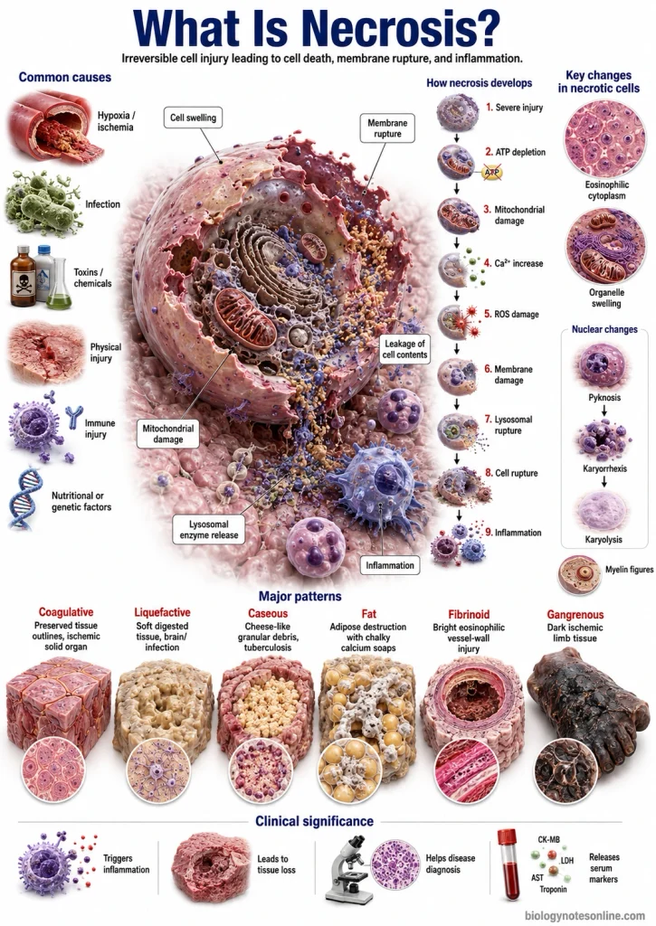
Necrosis is caused by severe injury to the cells, when the injury becomes irreversible. Different agents can produce this type of injury. Hypoxia, chemicals, physical agents and infections are some of the common causes.
The following are the major causes of necrosis-
- Hypoxia– It is the deficiency of oxygen in the cells. Reduced oxygen affects aerobic respiration and formation of ATP. If hypoxia is severe or continues for longer period, the cells become injured and may finally die.
- Ischemia– It occurs due to reduction or complete loss of blood supply to a tissue. In this condition, both oxygen and nutrients are reduced. Removal of metabolic wastes is also affected. Ischemia is an important cause of necrosis, such as during myocardial infarction and infarction of different tissues.
- Physical Agents– Mechanical trauma, excessive heat, severe cold, radiation and electric shock can cause cell injury. These agents can damage cellular structures directly. Severe injury results in death of the affected cells.
- Chemical Agents– Different poisons and toxic chemicals can produce necrosis. Some chemicals alter membrane permeability or damage important enzymes and cellular organelles. Environmental and occupational chemicals are also included in this group.
- Drugs– Certain drugs can cause cell and tissue injury when used in excess or in susceptible individuals. Some drugs act directly on cellular components while others form toxic metabolites. The severity depends on the drug, dose and affected tissue.
- Infectious Agents– Viruses, bacteria, fungi and parasites can cause necrosis. They may directly damage the cells or produce different toxins. Tissue injury can also occur due to inflammatory reaction against the infecting organism.
- Immune Reactions– The immune system can also cause injury to normal cells. It occurs in autoimmune reactions and different hypersensitivity reactions. Immune-mediated vascular injury can produce severe tissue damage and necrosis.
- Genetic Defects– Some genetic abnormalities affect the production or function of important cellular proteins. Defective enzymes, damaged proteins or other inherited abnormalities can cause cell injury. If the damage is beyond repair, cell death can occur.
- Nutritional Imbalance– Deficiency of protein, calories or particular vitamins can injure the cells. Excess nutrients can also produce different metabolic abnormalities. Severe or persistent nutritional disturbances therefore can contribute to cell injury and death.
How Does Necrosis Develop?
Necrosis develops when cell injury becomes severe and reaches the stage of irreversible cell injury. The exact changes may differ with the type of injury and tissue. In general, mitochondrial damage, loss of ATP, calcium disturbance and membrane damage are important events in this process.
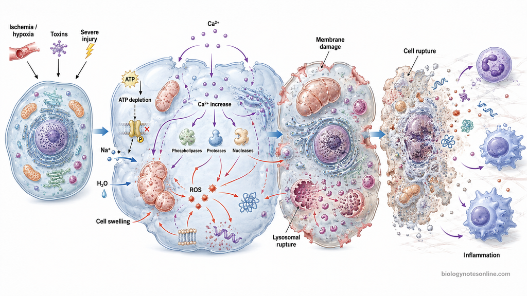
The development of necrosis can be described as follows-
- ATP Depletion– Severe injury decreases the formation of ATP, particularly during hypoxia and ischemia. Energy-dependent ion pumps cannot work properly. Sodium and water enter inside the cell and cellular swelling now starts.
- Mitochondrial Damage– Mitochondria become damaged when the injury continues. Oxidative phosphorylation is affected and formation of ATP decreases further. Severe mitochondrial permeability changes can finally make the cellular injury irreversible.
- Calcium Increase– Failure of membrane ion pumps and membrane injury causes an increase of cytoplasmic Ca²⁺. The increased calcium activates phospholipases, proteases, endonucleases and other enzymes. These enzymes damage membrane phospholipids, proteins and nucleic acids.
- Oxidative Damage– Reactive oxygen species (ROS) are produced during different types of cellular injury. Excess ROS damages lipids, proteins and nucleic acids. Mitochondrial and cellular membrane injury is also increased during this process.
- Membrane Damage– The plasma membrane gradually loses its normal integrity. Ions and water enter the cell, while different cellular components begin to leak outside. Damage to mitochondrial and lysosomal membranes also takes place.
- Lysosomal Rupture– Injury to lysosomal membrane releases different hydrolytic enzymes into the cytoplasm. These enzymes digest proteins, nucleic acids and other cellular components. The structural organization of the cell is progressively lost.
- Cell Rupture– The cell and its organelles become swollen. Finally, the plasma membrane ruptures and intracellular materials are released into the surrounding tissue. At this stage, the cell cannot recover.
- Inflammation– Materials released from the necrotic cell act as danger signals in the surrounding tissue. These stimulate inflammatory cells and inflammatory mediators. Therefore, necrosis is commonly associated with an inflammatory response.
Morphological Changes in Necrotic Cells
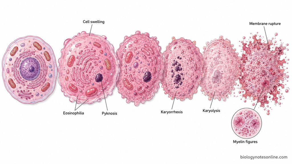
The necrotic cells show different structural changes in the cytoplasm and nucleus. These changes develop due to denaturation of cellular proteins and enzymatic digestion of the dead cell. Cell swelling, increased cytoplasmic eosinophilia and nuclear changes are commonly observed.
Cytoplasmic Changes
The necrotic cell generally becomes swollen and the normal cellular details are gradually lost. The cytoplasm appears more eosinophilic or pink during staining with hematoxylin and eosin (H&E). This occurs due to loss of cytoplasmic RNA and denaturation of cytoplasmic proteins.
The cytoplasm may become glassy and more homogeneous than that of normal cells. Vacuoles can also develop after digestion of cytoplasmic organelles. Damaged cell membranes may form whorled phospholipid masses called myelin figures. These are more prominent in irreversibly injured cells.
With further damage, the plasma membrane loses its integrity. Cell organelles become swollen and the membrane finally ruptures. Cellular components are then released into the surrounding tissue.
Nuclear Changes
The nucleus of a necrotic cell shows three major morphological changes. These are pyknosis, karyorrhexis and karyolysis. They develop due to breakdown and alteration of the nuclear chromatin.
Pyknosis is the shrinkage of the nucleus with marked condensation of chromatin. The nucleus becomes smaller and deeply basophilic. This change can be followed by fragmentation of the pyknotic nucleus.
Karyorrhexis refers to fragmentation of the nucleus. The condensed nuclear material breaks into several small fragments. These fragments are gradually lost as degradation continues.
Karyolysis is the fading and dissolution of the nucleus. The basophilia of chromatin decreases due to degradation of DNA by nucleases. Finally, the nucleus may completely disappear from the necrotic cell.
Major Patterns of Necrosis
Necrosis can show different morphological patterns in tissues. The pattern depends on the cause of injury, tissue involved and the way dead cells are digested. Some patterns can be seen grossly, while fibrinoid necrosis is mainly identified by microscopic examination.
| Pattern | Characteristic appearance | Typical cause/site | Representative example |
|---|---|---|---|
| Coagulative necrosis | Tissue architecture remains preserved for some time. Cells become eosinophilic and nuclei are lost. | Ischemia in most solid organs, except brain. | Myocardial infarction |
| Liquefactive necrosis | Dead tissue becomes soft and liquid due to enzymatic digestion. Normal tissue structure is lost. | Bacterial or fungal infections and ischemic injury of CNS. | Brain infarction |
| Caseous necrosis | Soft, friable and cheese-like material is formed. Microscopically, granular amorphous debris is present. | Commonly seen in tuberculosis and some fungal infections. | Tuberculous granuloma |
| Fat necrosis | Chalky white deposits may be formed due to fat breakdown and calcium soap formation. | Acute pancreatitis or traumatic injury to fatty tissue. | Pancreatic fat necrosis |
| Fibrinoid necrosis | Vessel wall shows bright pink, amorphous fibrin-like material on H&E staining. | Immune-mediated vascular injury. | Vasculitis |
| Gangrenous necrosis | Tissue becomes dry and dark in dry gangrene. Liquefaction develops when secondary infection occurs. | Severe ischemia of limb or other extremity. | Gangrene of lower limb |
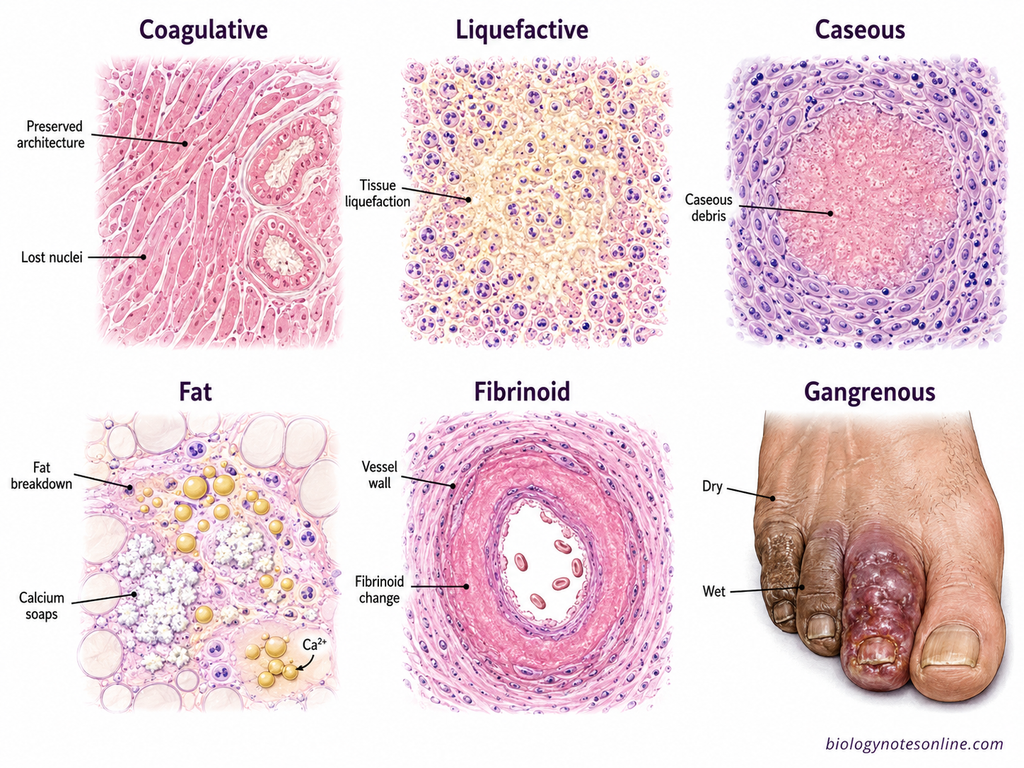
1. Coagulative Necrosis
Coagulative necrosis is commonly seen after ischemic injury in most solid organs, except the brain. The basic tissue architecture remains preserved for some days even though the cells are dead. The affected area is generally firm.
Microscopically, the cells become more eosinophilic and lose their nuclei. The cell outlines are still visible. This pattern is commonly present in infarction of the heart, kidney and other solid organs.
2. Liquefactive Necrosis
In liquefactive necrosis, the dead tissue is digested by hydrolytic enzymes. The normal tissue structure is lost and the area changes into a soft or liquid viscous mass. It is commonly seen in bacterial infections and sometimes fungal infections.
This type of necrosis also occurs after ischemic injury of the central nervous system (CNS). During bacterial infection, accumulation of dead cells and inflammatory material can form pus.
3. Caseous Necrosis
Caseous necrosis is most commonly associated with tuberculosis. The necrotic tissue has a soft, friable and cheese-like appearance. The term caseous is based on this gross appearance.
Under the microscope, the normal tissue architecture is completely lost. The area contains granular and amorphous cellular debris, generally surrounded by a granulomatous inflammatory reaction. Certain fungal infections may also show this pattern.
4. Fat Necrosis
Fat necrosis mainly involves adipose tissue. It can occur in acute pancreatitis, where pancreatic lipases are released and digest fat cells. The released fatty acids combine with calcium and form calcium soaps. This process is referred to as saponification.
The affected areas may appear as chalky white deposits. Fat necrosis can also develop in fatty tissues such as the breast following trauma.
5. Fibrinoid Necrosis
Fibrinoid necrosis is generally seen in the walls of blood vessels. It is associated particularly with immune-mediated vascular injury. Immune complexes and plasma proteins, including fibrin, accumulate in the damaged vessel wall.
On H&E staining, the affected vessel wall appears as bright pink and amorphous material. Unlike most other patterns, it is mainly a microscopic finding.
6. Gangrenous Necrosis
Gangrenous necrosis is not a separate microscopic pattern of necrosis. It is a clinical term generally used for ischemic necrosis involving a limb or other extremity. Loss of blood supply initially produces mainly coagulative necrosis.
In dry gangrene, coagulative necrosis is more prominent. When bacterial infection occurs in the dead tissue, liquefactive changes are added and this is referred to as wet gangrene.
Necrosis vs Apoptosis
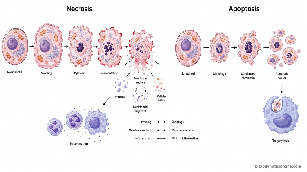
Necrosis and apoptosis are two different forms of cell death with different morphological changes. Necrosis generally shows cell swelling, membrane rupture and release of cellular contents. In apoptosis, the cell becomes smaller and cellular components are removed without early rupture of plasma membrane.
The differences between necrosis and apoptosis are-
| Characteristics | Necrosis | Apoptosis |
|---|---|---|
| Cell size | Cell becomes swollen. The organelles also show swelling. | Cell shrinkage occurs and the cell becomes smaller. |
| Nuclear changes | Pyknosis, karyorrhexis and karyolysis can be seen. The nucleus finally disappears in many necrotic cells. | Chromatin becomes condensed. Nuclear fragmentation occurs and fragments can be included within apoptotic bodies. |
| Plasma membrane | Plasma membrane integrity is lost. Finally the membrane ruptures. | Plasma membrane generally remains intact during the main process, although membrane blebbing occurs. |
| Cellular leakage | Cellular contents leak into the surrounding tissue after membrane damage. | Cellular contents are generally retained within membrane-bound fragments until their removal by phagocytes. |
| Inflammation | An inflammatory response is commonly produced due to release of intracellular materials. | Usually does not produce significant inflammation when apoptotic cells are rapidly cleared. |
| Cell involvement | Commonly involves groups or contiguous areas of cells, such as during tissue ischemia. Single-cell necrosis can also occur. | Usually affects individual or scattered cells rather than a large continuous area. |
| Occurrence | Mostly associated with pathological injury such as ischemia, toxins, infections and severe physical damage. | Occurs during normal physiological processes and also in pathological conditions, such as irreparable DNA damage. |
| Molecular regulation | Necrotic cell death is not always unregulated. Accidental necrosis occurs after overwhelming injury, while regulated necrotic forms such as necroptosis, pyroptosis and ferroptosis involve specific molecular mechanisms. | It is a highly regulated form of cell death. Caspases and intrinsic or extrinsic signalling pathways have important roles in classical apoptosis. |
The classical morphological differences include swelling and membrane rupture in necrotic cells, whereas shrinkage and maintenance of membrane integrity are typical of apoptosis. The older idea that necrosis is always accidental or unregulated is not completely correct. Different regulated forms of necrotic cell death are now well established.
Necrosis, Infarction, and Gangrene: What Is the Difference?
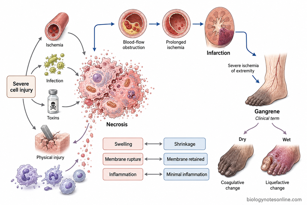
Necrosis is the morphological changes that occur after death of cells in a living tissue. Infarction is an area of ischemic necrosis which is produced due to obstruction of blood supply. Gangrene is a clinical term used for necrosis of a large tissue area, mostly an extremity.
Necrosis and Infarction
Necrosis can be produced by ischemia, infections, chemicals, physical agents and other severe cellular injuries. Infarction is mainly related to loss of blood supply.
When blood flow is obstructed, the tissue becomes ischemic. If ischemia continues, the cells die and an area of infarction is formed. In most solid tissues this produces coagulative necrosis, but infarction of brain shows liquefactive necrosis.
Thus, infarction is ischemic necrosis. All necrosis however does not develop due to infarction. Myocardial infarction is an example where prolonged ischemia results in necrosis of heart muscle.
Necrosis and Gangrene
Gangrene is not a specific microscopic pattern of necrosis. The term is generally applied when a considerable tissue mass undergoes necrosis, particularly a limb.
It commonly occurs due to severe loss of blood supply. The affected tissue dies and later may also be infected with microorganisms. Based on these changes, gangrene is commonly described as dry and wet gangrene.
Dry and Wet Gangrene in Relation to Necrotic Patterns
Dry gangrene occurs due to loss of arterial blood supply, commonly in distal parts of limb. The affected tissue shows mainly coagulative necrosis. It becomes dry, dark and shrunken.
Wet gangrene develops when bacterial infection is added to the necrotic tissue. The dead tissue is digested and liquefactive changes occur. The affected part becomes swollen and moist, and infection may spread rapidly.
Dry gangrene therefore mainly contains coagulative type of necrosis. In wet gangrene, the ischemic necrosis is complicated by bacterial infection and liquefaction.
| Term | Meaning | Main cause/feature | Typical example |
|---|---|---|---|
| Necrosis | Death of cells or tissue following irreversible injury. | Can occur due to ischemia, infection, chemicals, toxins or physical injury. | Coagulative necrosis in heart tissue |
| Infarction | Area of ischemic necrosis produced due to obstruction of blood supply. | Loss of arterial supply is most common, but venous obstruction can also produce infarction. | Myocardial infarction |
| Gangrene | Clinical term used for extensive tissue necrosis, commonly in an extremity. | Severe ischemia, with or without secondary infection. | Gangrene of lower limb |
| Dry gangrene | Ischemic tissue becomes dry, dark and shrunken. | Mainly associated with coagulative necrosis due to reduced arterial supply. | Dry gangrene of toes |
| Wet gangrene | Necrotic tissue becomes swollen, moist and infected. | Ischemic necrosis with bacterial infection and added liquefactive changes. | Wet gangrene of foot |
Biological and Clinical Significance of Necrosis
Necrosis is important because it indicates irreversible injury and death of cells in a tissue. It is seen in many pathological conditions such as infarction, infections, toxic injury and trauma. The released cellular materials also produce changes in the surrounding tissue.
Some of the important biological and clinical significance of necrosis are-
- Inflammation– Necrotic cells lose membrane integrity and their intracellular contents come outside the cells. These materials produce an inflammatory reaction in the surrounding tissue. Leukocytes are then recruited to the damaged area.
- Dead Cell Removal– The inflammatory cells help in removal of necrotic cells and cellular debris. The dead material is digested and removed mainly by phagocytic cells. This is an important process before repair of the damaged tissue can occur.
- Tissue Repair– After removal of necrotic tissue, repair of the injured area can begin. Some tissues may regenerate if surviving cells are capable of division. In severe tissue destruction, healing may occur by formation of fibrous scar tissue.
- Loss of Function– Extensive necrosis can decrease or completely destroy the function of an affected tissue. Its effect depends on the organ and amount of tissue involved. Necrosis of cardiac muscle during myocardial infarction, for example, reduces functional myocardium.
- Disease Diagnosis– The type and distribution of necrosis can provide information about the underlying disease. Caseous necrosis is commonly related with tuberculosis, while liquefactive necrosis is seen in brain infarction and many bacterial infections. Therefore, its morphological pattern is useful during pathological examination.
- Serum Markers– Necrotic cells release intracellular proteins and enzymes into the blood due to loss of plasma membrane integrity. Measurement of such substances can help to detect tissue injury. Cardiac troponins are used during myocardial injury, while increased transaminases can indicate hepatocellular injury.
- Extent of Injury– Presence of necrosis indicates that cellular injury has crossed the reversible stage. The cells cannot return to their normal condition. Amount and distribution of necrosis are therefore useful for assessing severity of tissue damage.
- Complications– Large areas of necrosis can produce important complications. Dead tissue may become infected, undergo liquefaction or later develop dystrophic calcification. Extensive tissue destruction can also result in permanent loss of tissue structure and function.
References
- Adigun, R., Basit, H., Zubair, M., & Murray, J. (2025). Cell liquefactive necrosis. In StatPearls. StatPearls Publishing. https://www.ncbi.nlm.nih.gov/books/NBK430935/
- Kalogeris, T., Baines, C. P., Krenz, M., & Korthuis, R. J. (2012). Cell biology of ischemia/reperfusion injury. International Review of Cell and Molecular Biology, 298, 229–317. https://doi.org/10.1016/B978-0-12-394309-5.00006-7
- Khalid, N., & Azimpouran, M. (2023). Necrosis pathology. In StatPearls. StatPearls Publishing. https://www.ncbi.nlm.nih.gov/books/NBK557627/
- Kim, E. H., Wong, S.-W., & Martinez, J. (2019). Programmed necrosis and disease: We interrupt your regular programming to bring you necroinflammation. Cell Death & Differentiation, 26(1), 25–40. https://doi.org/10.1038/s41418-018-0179-3
- Kumar, V., Abbas, A. K., Fausto, N., & Aster, J. C. (Eds.). (2010). Robbins and Cotran pathologic basis of disease (8th ed.). Saunders/Elsevier. https://shop.elsevier.com/books/robbins-and-cotran-pathologic-basis-of-disease-professional-edition/kumar/978-1-4377-0792-2
- Kumar, V., Abbas, A. K., & Aster, J. C. (2013). Robbins basic pathology (9th ed.). Elsevier/Saunders. https://www.ncbi.nlm.nih.gov/nlmcatalog/101573511
- Land, W. G. (2018). Regulated cell death. In Damage-associated molecular patterns in human diseases: Volume 1: Injury-induced innate immune responses (pp. 427–466). Springer International Publishing. https://doi.org/10.1007/978-3-319-78655-1_19
- Marzetti, E., Calvani, R., Landi, F., Coelho-Júnior, H. J., & Picca, A. (2024). Mitochondrial quality control processes at the crossroads of cell death and survival: Mechanisms and signaling pathways. International Journal of Molecular Sciences, 25(13), 7305. https://doi.org/10.3390/ijms25137305
- McCall, K. (2010). Genetic control of necrosis—Another type of programmed cell death. Current Opinion in Cell Biology, 22(6), 882–888. https://doi.org/10.1016/j.ceb.2010.09.002
- Mechanic, O. J., Gavin, M., Shams, P., & Grossman, S. A. (2023). Acute myocardial infarction. In StatPearls. StatPearls Publishing. https://www.ncbi.nlm.nih.gov/books/NBK459269/
- Miller, M. A., & Zachary, J. F. (2017). Mechanisms and morphology of cellular injury, adaptation, and death. In J. F. Zachary (Ed.), Pathologic basis of veterinary disease (6th ed., pp. 2–43.e19). Elsevier. https://doi.org/10.1016/B978-0-323-35775-3.00001-1
- Noh, M. R., & Padanilam, B. J. (2024). Cell death induced by acute renal injury: A perspective on the contributions of accidental and programmed cell death. American Journal of Physiology-Renal Physiology, 327(1), F4–F20. https://doi.org/10.1152/ajprenal.00275.2023
- Zhang, Y., Chen, X., Gueydan, C., & Han, J. (2018). Plasma membrane changes during programmed cell deaths. Cell Research, 28(1), 9–21. https://doi.org/10.1038/cr.2017.133