

Inclusion bodies are the different materials or structures present inside a cell. These are mainly found in the cytoplasm, although some inclusion bodies can also occur inside nucleus or in the periplasmic region of bacteria. They may be formed normally in the cell or appear during some particular conditions.
The composition of inclusion bodies is not always same. Cytoplasmic inclusions in eukaryotic cells may contain glycogen, lipid droplets, pigments and some other stored materials. Protein aggregates can also form inclusion bodies, particularly when proteins are produced in large amounts or become improperly folded.
Inclusion bodies are generally different from normal cell organelles. Organelles are organized cellular structures having particular functions, whereas many inclusions are stored materials or deposits present within the cell. However, this difference is not absolute in prokaryotes. Some bacterial inclusions such as carboxysomes and magnetosomes are highly organized structures and can perform specialized cellular functions.
Inclusion bodies occur in both prokaryotic and eukaryotic cells. Bacterial cells may contain glycogen granules, polyphosphate granules, sulfur granules, gas vesicles, magnetosomes and carboxysomes. In eukaryotic cells, inclusions commonly occur as lipid droplets, glycogen particles and different pigments. Protein inclusion bodies are also found in different organisms.
Key definition: Inclusion bodies are intracellular materials or structures found mainly in the cytoplasm, which may contain stored substances, pigments, specialized cellular components or aggregated proteins.
What Does the Term “Inclusion Body” Mean?
The term inclusion body is used for different types of structures present inside the cells. It does not always indicate the same material. Its meaning changes according to the organism and the condition in which the inclusion is present.
In prokaryotic cells, inclusion bodies may be normal storage or specialized structures. These include granules used for storing carbon, phosphate or sulfur and other inclusions with particular functions. Some bacterial inclusions are highly organized within the cell.
During viral infection, the term is used for viral inclusion bodies formed inside the infected host cell. These structures contain viral and host components and are associated with different stages of viral replication. They may occur in cytoplasm or nucleus depending on the virus.
In recombinant protein production, inclusion bodies usually refer to dense protein aggregates formed inside the expression host. They are commonly seen during high-level expression of foreign proteins in Escherichia coli. The proteins present in these bodies are often partially or improperly folded, although inclusion bodies are not always made only of completely inactive protein.
The term is also used in pathology for abnormal intracellular inclusions. These may contain accumulated or aggregated proteins and other cellular substances. Different characteristic inclusions are found in several neurodegenerative diseases, where disease-associated proteins accumulate within affected cells.
Therefore, Inclusion body is a broad term. It may indicate a normal bacterial inclusion, a virus-associated structure, an aggregate formed during recombinant protein expression or an abnormal cellular deposit found during disease.
Type of Inclusion Body
| Type of Inclusion Body | Description | Examples / Main Role |
|---|---|---|
| Storage inclusions | These mainly store reserve materials inside bacterial cells. The stored material can be used during nutrient limitation. | Glycogen granules, PHA granules, polyphosphate granules, sulfur granules and cyanophycin. |
| Gas vesicles | These are hollow gas-containing structures. Their wall is made up of proteins and lipid membrane is absent. | Found in some aquatic bacteria and archaea. Mainly used for buoyancy. |
| Carboxysomes | These are polyhedral bacterial microcompartments with a protein shell. RuBisCO and carbonic anhydrase are present inside. | Involved in carbon dioxide concentration and carbon fixation. |
| Magnetosomes | These are specialized bacterial inclusions containing magnetic mineral crystals. A membrane is present around each crystal. | Common minerals are magnetite (Fe₃O₄) and greigite (Fe₃S₄). Help in magnetic orientation. |
| Viral inclusion bodies | These are formed inside cells infected with certain viruses. They may occur in nucleus or cytoplasm. | Some act as viral factories for replication or assembly, while others contain accumulated viral materials. |
| Recombinant protein inclusion bodies | These are protein-rich aggregates formed during high-level expression of foreign proteins, especially in Escherichia coli. | May contain misfolded, partially folded and sometimes biologically active recombinant proteins. |
| Pathological inclusion bodies | These are abnormal intracellular accumulations formed during some disease conditions. | Lewy bodies, neurofibrillary tangles and other abnormal protein inclusions. |
| Nuclear inclusions | These inclusion bodies are present inside the nucleus. | Seen in some viral infections and disorders involving abnormal protein accumulation. |
| Cytoplasmic inclusions | These are present within the cytoplasm. Their composition is different according to the type. | Bacterial storage inclusions, many viral inclusions and recombinant protein aggregates. |
General Characteristics of Inclusion Bodies
- Inclusion bodies are intracellular structures or materials found mainly within the cytoplasm. Some types can also occur inside the nucleus or other cellular regions.
- They are not made up of one particular substance. Different inclusions may contain proteins, lipids, glycogen, polyphosphate, sulfur, pigments, viral components or other stored materials.
- Inclusion bodies are generally different from regular cell organelles. Many are deposits or storage materials and do not have the common organization of organelles. However, some bacterial inclusions such as magnetosomes and carboxysomes are specialized and highly organized cellular structures.
- They may be membrane-bound or non-membrane-bound, depending on the type of inclusion. Thus, presence of a surrounding membrane is not a general feature for all inclusion bodies.
- Inclusion bodies are found in both prokaryotic and eukaryotic cells. Bacteria contain several storage and specialized inclusions, whereas eukaryotic cells may contain glycogen, lipid, pigments and different protein inclusions.
- The size, shape and number of inclusion bodies are not fixed. These vary according to the type of inclusion, cell and physiological condition.
- Some inclusion bodies are normal components of the cell. They can act mainly in storage of nutrients or other substances. Bacterial storage inclusions are examples of these structures.
- Other inclusion bodies are formed during particular conditions. Recombinant protein inclusion bodies, for example, are commonly formed by aggregation of highly expressed proteins in Escherichia coli. These aggregates may also contain some biologically active protein.
- Viral inclusion bodies are formed in cells infected with certain viruses. They may contain viral proteins, nucleic acids and host factors and can serve as sites associated with viral replication and assembly.
- Inclusion bodies are not always inactive structures. Some bacterial inclusions perform specialized functions, and recombinant protein inclusion bodies can contain proteins having native-like structure or biological activity.
- Some abnormal protein inclusions are also associated with cellular stress and disease conditions. In this case, proteins or other cellular materials accumulate within the affected cells.
- Inclusion bodies can often be identified by light microscopy, special staining or electron microscopy, depending on their composition and size. Their appearance is also different for different types of inclusions.
Structure of Inclusion Bodies in Prokaryotic Cells
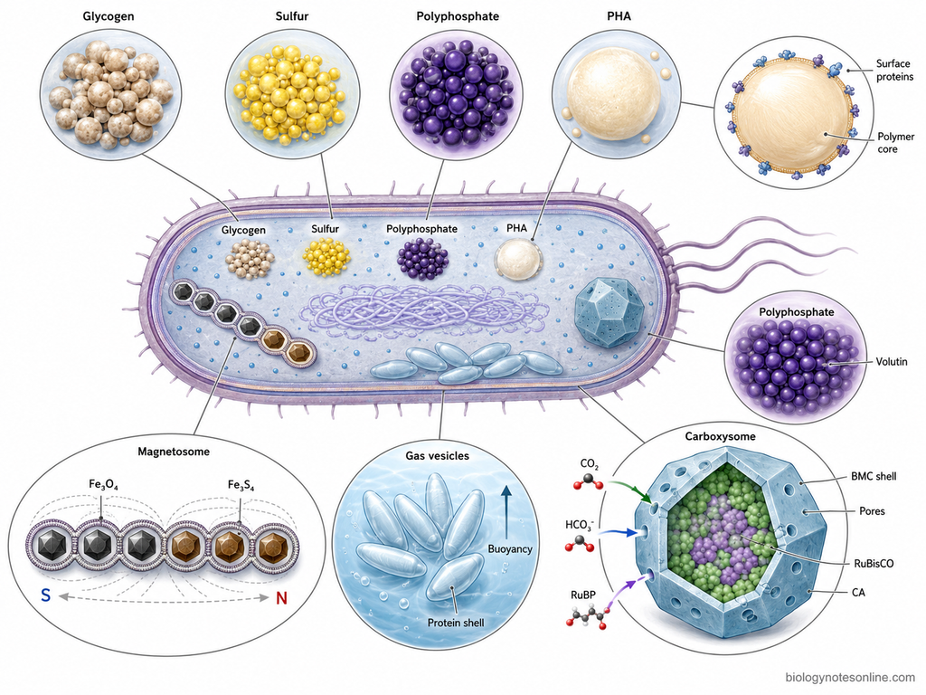
- Inclusion bodies are structures found inside the cytoplasm of prokaryotic cells. These are not same in all bacteria. Their shape, composition and internal arrangement varies with the type of inclusion.
- Many bacterial inclusion bodies occur as granules. Glycogen, polyphosphate, sulfur and polyhydroxyalkanoates (PHA) are some of the common materials stored in these granules.
- Some inclusions are without any typical membrane surrounding them. The material is accumulated directly within the cytoplasm and remains in the form of dense particles or granules.
- Polyhydroxyalkanoate granules are mainly used for carbon storage. The center contains polymer material. Different proteins are found associated with the surface of these granules and help in their formation and utilization.
- Polyphosphate granules contain long chains of inorganic phosphate. These are also called volutin granules and, after staining, may show metachromatic appearance.
- In some sulfur bacteria, sulfur is deposited inside the cell as sulfur granules. These granules mainly contain elemental sulfur which can later be used during the oxidation process.
- Gas vesicles are quite different from the storage granules. These are hollow gas-containing structures with a thin protein covering. Lipid membrane is absent. They are commonly present in some aquatic bacteria and archaea and help the cells in buoyancy.
- Magnetosomes are membrane-covered structures containing magnetic mineral crystals. Magnetite (Fe₃O₄) is common, while greigite (Fe₃S₄) is present in some species. The magnetosomes are often arranged in chains inside the bacterial cell.
- Carboxysomes are polyhedral bacterial microcompartments. A protein shell is present around the internal enzymes, mainly RuBisCO and carbonic anhydrase. They are especially associated with carbon dioxide fixation in many autotrophic bacteria.
- The carboxysome shell is made up of different bacterial microcompartment (BMC) proteins. These proteins form the outer covering with small pores. Through these pores, selected small molecules can pass.
- The structural organization is therefore not uniform among bacterial inclusions. Some remain as simple storage deposits, while gas vesicles, magnetosomes and carboxysomes show a more specialized structure.
Types and Classification of Inclusion Bodies in Bacteria
Different inclusion bodies are present in bacterial cells. These differ in their composition, structure and function. Some mainly store reserve materials, while some perform specialized functions.
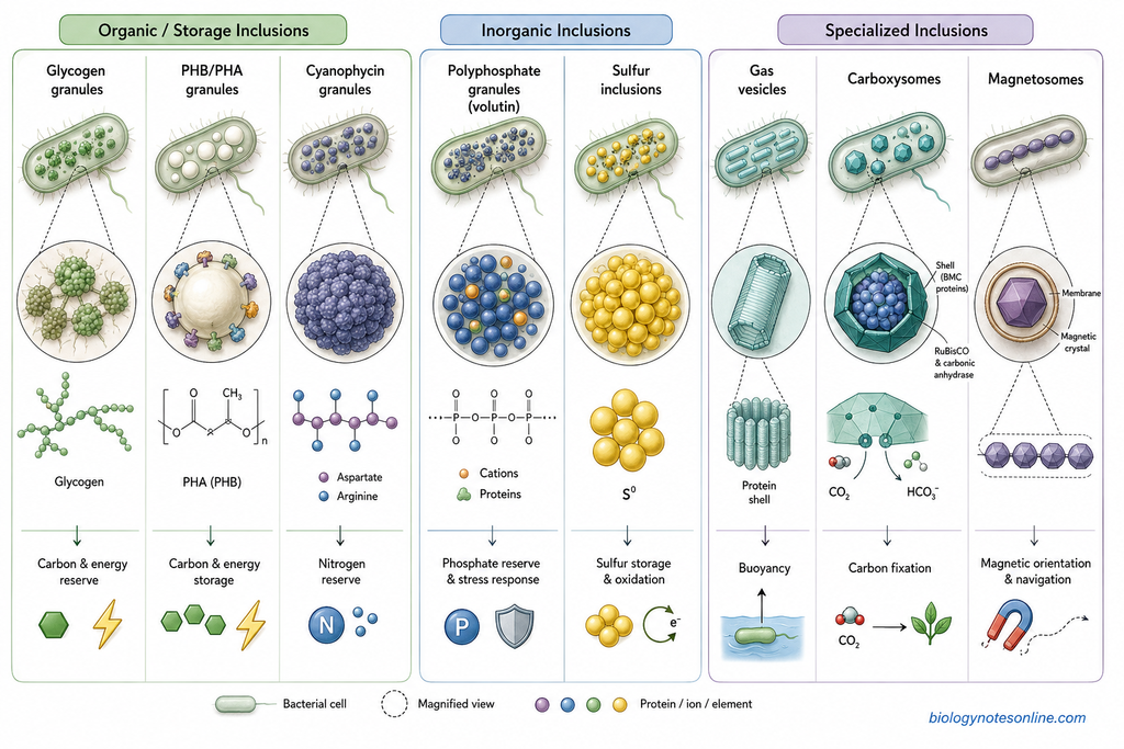
Organic/Storage Inclusions
- Glycogen granules
- Example- Glycogen granules found in many bacteria and cyanobacteria.
- Composition- These are made up of glycogen, a branched polymer of glucose.
- Structure- Glycogen occurs as small granules inside the cytoplasm. Typical lipid membrane is absent.
- Function- It mainly acts as carbon and energy reserve. Stored glycogen is used when sufficient carbon source is not available.
- PHB/PHA granules
- Example- PHB granules of Cupriavidus necator.
- Composition- These contain polyhydroxyalkanoates (PHA). Polyhydroxybutyrate (PHB) is one of its common forms.
- Structure- Usually present as rounded granules. PHA polymer remains in the central part and different proteins are associated with its surface.
- Function- These are mainly used for storage of carbon and energy. Large amount can accumulate when carbon is available in excess.
- Cyanophycin granules
- Example- Cyanophycin granules of cyanobacteria.
- Composition- Cyanophycin is mainly made up of an aspartate backbone with arginine residues attached to it.
- Structure- It occurs as dense granules within the cytoplasm. Lipid membrane is generally absent around them.
- Function- It mainly acts as nitrogen reserve. The stored material is utilized when nitrogen becomes limited.
Inorganic Inclusions
- Polyphosphate granules
- Example- Polyphosphate or volutin granules of different bacteria.
- Composition- These contain long chains of inorganic phosphate (polyP).
- Structure- Polyphosphate remains concentrated as dense intracellular granules. Different cations and proteins may also remain associated.
- Function- These mainly serve as phosphate reserve. Polyphosphate also takes part in stress response and some other cellular processes.
- Sulfur inclusions
- Example- Sulfur globules of Beggiatoa and other sulfur-oxidizing bacteria.
- Composition- These contain mainly elemental sulfur (S⁰).
- Structure- Sulfur is accumulated as rounded globules. Their location is not same in all bacteria and may be cytoplasmic, periplasmic or sometimes extracellular.
- Function- Stored sulfur is used during oxidation of reduced sulfur compounds and energy metabolism.
Specialized Inclusions
- Gas vesicles
- Example- Gas vesicles found in cyanobacteria and some aquatic bacteria.
- Composition- Their wall is made up of gas-vesicle proteins. Lipid membrane is absent.
- Structure- These are hollow and gas-filled structures with a thin rigid protein shell.
- Function- Gas vesicles provide buoyancy. They help the cells to maintain a suitable position in water.
- Carboxysomes
- Example- Carboxysomes of cyanobacteria and some chemoautotrophic bacteria.
- Composition- These contain RuBisCO, carbonic anhydrase and bacterial microcompartment proteins.
- Structure- Carboxysomes are polyhedral structures surrounded by a protein shell. Enzymes remain enclosed inside.
- Function- These are involved in carbon fixation. Carbon dioxide is concentrated around RuBisCO.
- Magnetosomes
- Example- Magnetosomes of magnetotactic bacteria.
- Composition- These contain magnetic crystals of magnetite (Fe₃O₄) or greigite (Fe₃S₄).
- Structure- Each mineral crystal is enclosed by a membrane. Several magnetosomes are commonly arranged in a chain.
- Function- Magnetosomes help the bacterial cells in magnetic orientation and movement along magnetic field lines.
Functions of Inclusion Bodies in Bacteria
The functions of bacterial inclusion bodies are different according to the type of inclusion. The following are some of the important functions-
- Storage of carbon – Glycogen granules and polyhydroxyalkanoate (PHA) granules store carbon inside bacterial cells. PHA also acts as an energy reserve and can be utilized when nutrients become limited.
- Storage of phosphate – Polyphosphate granules are used for storing inorganic phosphate. The stored polyphosphate can be used during phosphate deficiency and also takes part in different cellular processes.
- Storage of nitrogen – Cyanophycin granules mainly act as nitrogen reserve. They also contain carbon and are especially common in cyanobacteria and some other bacteria.
- Storage of sulfur – Some sulfur-oxidizing bacteria accumulate elemental sulfur as sulfur globules. The stored sulfur can later be oxidized and used in energy metabolism.
- Energy reserve – Some inclusions store compounds that can later supply energy to the bacterial cell. PHA is an important example. Polyphosphate can also contribute phosphate and energy for different cellular reactions.
- Buoyancy – Gas vesicles help some aquatic bacteria and cyanobacteria to regulate their position in water. By changing the amount of gas-filled vesicles, the cells can remain at regions having suitable light and nutrients.
- Carbon dioxide fixation – Carboxysomes help in carbon fixation. They contain RuBisCO and carbonic anhydrase within a protein shell, which helps in increasing carbon dioxide around RuBisCO.
- Orientation in magnetic field – Magnetosomes help magnetotactic bacteria to orient along magnetic field lines. This can assist the cells in reaching suitable regions of aquatic sediments or water columns.
- Protection during unfavorable conditions – Stored materials present in inclusion bodies can support bacterial survival when nutrients become limited. Polyphosphate has also been linked with resistance to starvation, oxidative stress, heavy metals and some other stress conditions.
- Maintaining cellular reserves – Inclusion bodies allow excess nutrients to be accumulated when these are available in high amount. The stored material can then be utilized later rather than being lost from the cell.
- Specialized metabolic functions – Not all bacterial inclusions are only storage structures. Carboxysomes perform a role in carbon-concentrating mechanism, gas vesicles in buoyancy and magnetosomes are associated with magnetic orientation.
Viral Inclusion Bodies
Viral inclusion bodies are the distinct areas having altered staining appearance in a virus-infected cell. These may develop inside the nucleus or cytoplasm. Some inclusion bodies contain viral nucleic acids, proteins and different host components and act as sites of viral replication or assembly. These are commonly referred to as virus factories. But every viral inclusion is not a virus factory. Some may represent accumulated viral materials or changes produced during fixation and staining.
Their size and appearance are not same for all viruses. They may be single or multiple, round or irregular and their staining may be acidophilic or basophilic. Some viruses produce inclusions mainly at one cellular location, while a few can produce both nuclear and cytoplasmic inclusions.
Classification According to Location
Intranuclear inclusion bodies
- Intranuclear inclusion bodies are formed within the nucleus of infected cells. These are particularly seen in infections caused by several DNA viruses.
- Herpesviruses commonly produce intranuclear inclusions. Cowdry type A inclusions are one characteristic form and generally appear as eosinophilic nuclear inclusions with chromatin pushed toward the nuclear margin.
- Adenoviruses also produce intranuclear inclusion bodies. These may have a dense or smudgy appearance and are commonly amphophilic to basophilic on staining.
- Cytomegalovirus (CMV) forms prominent intranuclear inclusions. The infected cells become enlarged and the characteristic inclusion can give an owl’s-eye appearance.
Intracytoplasmic inclusion bodies
- These inclusion bodies occur within the cytoplasm of virus-infected cells. Poxviruses, reoviruses and rabies virus are some important examples producing cytoplasmic inclusions.
- Negri bodies are intracytoplasmic inclusions associated with rabies virus infection. They are found particularly in infected neurons and are sites associated with rabies virus RNA synthesis and replication.
- Poxvirus-infected cells can also contain cytoplasmic inclusion bodies. Some basophilic inclusions represent sites of viral synthesis, while other acidophilic inclusions may contain accumulated viral proteins and mature virions.
- In many RNA viruses, cytoplasmic inclusions can develop as membrane-less compartments. Viral proteins and genome become concentrated within these structures during replication.
Nuclear and cytoplasmic inclusions
- Some viruses can produce inclusion bodies in both the nucleus and cytoplasm of the same infected cell.
- Measles virus and other morbilliviruses can show both intranuclear and intracytoplasmic inclusions. These inclusions are commonly acidophilic.
- Cytomegalovirus infection may also show nuclear together with cytoplasmic inclusions. The intranuclear inclusion is usually the more characteristic microscopic feature.
Classification According to Staining
Eosinophilic/acidophilic inclusions
- Eosinophilic or acidophilic inclusion bodies take up acidic dyes such as eosin and therefore appear pink to red in routine hematoxylin and eosin (H&E) staining.
- Negri bodies of rabies are a common example of acidophilic cytoplasmic inclusion bodies. Herpesvirus Cowdry type A inclusions are another example, but these occur within the nucleus.
- Acidophilic inclusions may contain viral proteins, nucleocapsids or other infection-associated material. Their exact composition varies between different viruses.
Basophilic inclusions
- Basophilic inclusion bodies stain more strongly with basic dyes and appear blue to purple in routine stained preparations.
- Adenovirus commonly forms basophilic or amphophilic intranuclear inclusions. These may become large and occupy much of the infected nucleus.
- Basophilic cytoplasmic inclusions can occur in poxvirus infection. Some of these represent active sites where viral components are being synthesized.
- The staining property is not fixed for every viral inclusion. It can vary with the virus, stage of infection and the staining or fixation method used.
Examples of Viral Inclusion Bodies
Different viral inclusion bodies are produced during viral infection. Some are present inside nucleus, while others occur in the cytoplasm. Their staining and appearance also vary according to the virus.
- Negri bodies – These are intracytoplasmic inclusion bodies found in neurons infected with rabies virus. They are mainly seen in neurons of the hippocampus and cerebellar Purkinje cells. Negri bodies contain viral ribonucleoprotein components and are associated with viral replication. They usually appear eosinophilic on staining.
- Guarnieri bodies – These are cytoplasmic inclusion bodies produced during infection with poxviruses. They represent sites associated with viral replication in the cytoplasm. Guarnieri bodies can be demonstrated in infected epithelial cells.
- Henderson-Paterson bodies – These are large intracytoplasmic inclusions found in molluscum contagiosum. They are also called molluscum bodies. The inclusions enlarge within infected epidermal cells and can push the nucleus toward the periphery. Their staining may change from eosinophilic to basophilic as the infected keratinocyte moves toward the granular layer.
- Cowdry type A inclusion bodies – These are intranuclear inclusions commonly associated with herpesvirus infections. They are especially described in herpes simplex virus infection and can also occur with varicella-zoster virus. The inclusions are usually eosinophilic and may be surrounded by a clear halo with chromatin present near the nuclear margin.
- Owl’s-eye inclusion bodies – These are characteristic inclusions seen in cytomegalovirus (CMV) infection. The infected cell becomes enlarged and contains a prominent intranuclear inclusion, producing the typical owl’s-eye appearance. Cytoplasmic inclusions may also be present in CMV-infected cells.
- Adenoviral inclusions – Adenoviruses produce intranuclear inclusion bodies in infected cells. These may become large and occupy much of the nucleus, giving a dense basophilic or smudged nuclear appearance. Unlike Cowdry type A bodies, they are not generally referred to by one commonly used eponym.
How Do Viral Inclusion Bodies Form?
The formation of viral inclusion bodies is different among different groups of viruses. In many viruses, these structures develop after viral components become concentrated at a particular region of the infected cell. The general process can be given as follows-
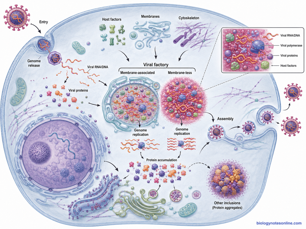
- Entry of virus into the cell – The virus first enters into a suitable host cell. After entry, the viral genome is released and the early stages of viral infection are started.
- Accumulation of viral components – Viral nucleic acid and newly formed viral proteins begin to accumulate inside the infected cell. These components may remain in the cytoplasm or nucleus, depending on the virus.
- Reorganization of cellular components – In this step, viral proteins interact with different host-cell components. Cellular membranes, cytoskeleton or nuclear structures may be rearranged and host proteins can also be recruited to the developing site. This process is especially prominent in many viruses forming replication compartments.
- Formation of a replication compartment – Viral and cellular materials become concentrated within a particular intracellular region. This organized region may be referred to as a viral factory, viroplasm or viral replication compartment. Some of these structures are membrane-associated, while others can form as membrane-less compartments.
- Viral genome replication – In many viral inclusion bodies, the viral genome is replicated within the formed compartment. Required viral proteins and some host factors are concentrated at this site, which helps in carrying out the replication process.
- Protein synthesis and assembly – Some viral factories are also associated with synthesis, processing or accumulation of viral proteins. Assembly of new virus particles may occur at or near these compartments in certain viruses. It is not the same for every virus.
- Formation of other viral inclusions – All viral inclusion bodies are not active sites of virus replication. Some inclusions can develop later by accumulation of viral proteins or other infection-associated materials which are not incorporated into newly formed virus particles.
Significance of Viral Inclusion Bodies
The significance of viral inclusion bodies depends on the virus and the type of inclusion formed. Some are active sites of viral multiplication, whereas others are mainly accumulated viral materials. The following are some of the important significance-
- Sites of viral replication – In many viruses, inclusion bodies act as viral factories. Viral genome, proteins and required host factors become concentrated in these regions where genome replication can take place.
- Viral assembly – Some viral inclusion bodies are also involved in assembly of newly formed virus particles. Replication and assembly may occur within the same compartment or at nearby regions of the infected cell.
- Concentration of viral components – Inclusion bodies bring viral nucleic acids and proteins together at one particular site. This can increase the efficiency of different steps of viral multiplication.
- Reorganization of host cell – Formation of viral factories is often associated with changes in cellular membranes, cytoskeleton and different organelles. Mitochondria and cellular membranes are recruited around some viral factories.
- Protection of viral replication machinery – Some replication compartments separate viral components from the remaining cytoplasm. This can help the virus to organize replication and may also reduce exposure of viral material to some host antiviral mechanisms. This function is not present in every inclusion body.
- Diagnostic significance – Characteristic inclusion bodies can give an important clue during microscopic examination of infected tissues. Negri bodies in rabies, cytomegalovirus inclusions and adenoviral nuclear inclusions are some examples. However, absence of an inclusion body does not exclude viral infection.
- Identification of infected cells – Their characteristic location, shape and staining help in recognizing cells affected by some viral infections. Nuclear or cytoplasmic inclusions are especially useful during histopathological examination.
- Study of virus-host interaction – Viral inclusion bodies are also useful for studying how viruses use host proteins, membranes and cytoskeletal components. Their formation gives information about different stages of the viral replication cycle.
- Targets for antiviral studies – Proteins and interactions required for formation of viral replication compartments are studied as possible antiviral targets. Membrane-less viral inclusions and their formation by phase separation are also being investigated for this purpose.
- Not all inclusions are functional factories – Some viral inclusions are formed by accumulation of excess structural proteins or nucleocapsids during later stages of infection. These may represent inactive or dead-end material rather than active sites of replication.
Recombinant Protein Inclusion Bodies
Recombinant protein inclusion bodies are different from the normal storage inclusions of bacteria. These are protein-rich aggregates formed during expression of foreign or recombinant proteins, most commonly in Escherichia coli. They are mainly produced when the newly synthesized protein fails to remain properly soluble inside the bacterial cell.
What Are Recombinant Protein Inclusion Bodies?
- Recombinant protein inclusion bodies are dense intracellular aggregates containing a high amount of the expressed recombinant protein.
- These are commonly formed during high-level heterologous protein expression in E. coli. The recombinant polypeptide becomes accumulated mainly in insoluble form within the cytoplasm.
- Inclusion bodies were earlier considered simply as deposits of completely misfolded and inactive proteins. This view is not fully correct now. Some proteins present within inclusion bodies can retain native-like structure and biological activity.
- These inclusion bodies should not be confused with glycogen, PHA, polyphosphate or other bacterial storage inclusions. Recombinant protein inclusion bodies are mainly formed by aggregation of expressed polypeptides.
How Are Protein Inclusion Bodies Formed?
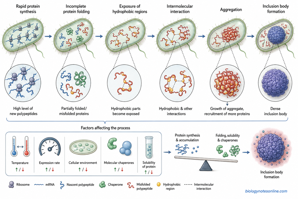
- Rapid protein synthesis – During strong recombinant expression, a large amount of newly synthesized polypeptide is produced within a short time. The cellular folding machinery may not handle all these proteins properly.
- Incomplete protein folding – Newly formed polypeptides normally pass through different folding intermediates. Some recombinant proteins fail to reach the correct three-dimensional structure and remain partially folded or misfolded.
- Exposure of hydrophobic regions – Hydrophobic parts of a protein are normally buried after proper folding. During improper folding, these regions can remain exposed to the aqueous cytoplasm.
- Intermolecular interaction – Exposed hydrophobic regions of one protein can interact with similar regions of another protein molecule. Other intermolecular contacts are also involved.
- Aggregation – More and more polypeptides become associated with the developing protein aggregate. Small aggregates can also act as sites where additional molecules become deposited.
- The accumulated proteins finally develop into dense inclusion bodies. Their formation is therefore related to the balance between protein synthesis, folding, solubility and aggregation.
- Expression conditions also affect this process. Temperature, expression rate, cellular environment and availability of molecular chaperones can change the amount and properties of inclusion bodies.
Factors That Promote Inclusion Body Formation
- High expression rate – Excessive synthesis of recombinant protein increases its intracellular concentration. This increases the possibility of aggregation.
- Strong promoter – Strong expression systems can produce large amounts of protein very rapidly. The folding capacity of the bacterial cell may become insufficient under such conditions.
- High cultivation temperature – Higher temperature generally increases protein aggregation in many recombinant expression systems. Lower expression temperatures are therefore commonly used to improve soluble protein production.
- Intrinsic properties of the protein – Large multidomain proteins, proteins having long hydrophobic regions and some intrinsically disordered proteins are more prone to inclusion body formation.
- Insufficient chaperone capacity – Molecular chaperones help newly formed proteins to fold correctly. During excessive protein production, the available chaperones may become insufficient and aggregation increases.
- Inappropriate cellular environment – The bacterial cytoplasm may not provide the same pH, redox condition, cofactors, post-translational modifications or folding machinery present in the original organism from which the protein was obtained.
- Codon and expression-related factors – Improper codon usage, problems during translation and very rapid transcription or translation may disturb normal protein folding. These conditions can promote formation of insoluble protein.
Structure and Composition
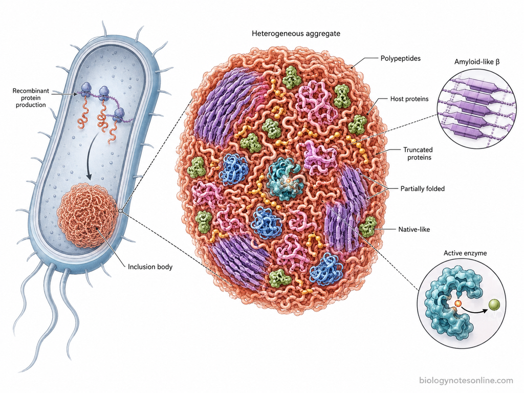
- Recombinant inclusion bodies contain mainly the expressed polypeptide chains. Host-cell proteins, truncated protein products and some other cellular components may also become associated with the aggregates.
- The proteins are not always present only in a completely unfolded state. Partially folded molecules and protein regions having native-like conformation can also occur within the inclusion body.
- Some bacterial inclusion bodies show an ordered amyloid-like structure. Their internal organization can contain cross-β type protein arrangements rather than being only an amorphous mass of denatured protein.
- Native-like protein molecules can remain trapped together with the more aggregated protein fraction. The amount is different according to the protein and expression conditions.
- Some inclusion bodies therefore show measurable biological activity without complete solubilization and refolding. Active enzyme molecules have been demonstrated within several bacterial recombinant inclusion bodies.
- The older concept that every inclusion body contains only inactive and completely denatured protein is now considered incomplete. Their structure can range from poorly folded aggregates to more ordered protein assemblies containing functional protein species.
| Advantages of Recombinant Inclusion Bodies | Disadvantages of Recombinant Inclusion Bodies |
|---|---|
| High amount of recombinant protein can accumulate inside the bacterial cell, particularly during strong expression. | The protein present in inclusion bodies is often insoluble and cannot be used directly in many applications. |
| Inclusion bodies can contain a relatively high proportion of the target recombinant protein, which can make the initial separation from soluble host proteins easier. | Solubilization and refolding are commonly required to recover the protein in soluble and active form. |
| Aggregated protein is sometimes less exposed to cellular proteases, and this may reduce degradation of the recombinant product. | During solubilization and refolding, some protein may be lost due to precipitation or formation of incorrect structures. |
| Inclusion body formation can allow production of proteins that may otherwise be toxic to the host cell when present in soluble active form. | Refolding is not always successful. Some proteins do not return completely to their native conformation after solubilization. |
| The dense aggregates can often be collected by centrifugation after cell disruption. | Additional downstream steps increase the time, cost and complexity of recombinant protein purification. |
| Inclusion bodies may protect some proteins from unwanted interactions within the bacterial cytoplasm. | Strong denaturing agents such as urea or guanidinium salts may be required for solubilization of tightly aggregated proteins. |
| Some modern studies have shown that inclusion bodies may contain partially folded or native-like protein molecules. | Denaturants and refolding conditions have to be removed or carefully controlled before further use of the protein. |
| Certain inclusion bodies can retain measurable biological activity, so they are not always completely inactive protein deposits. | Inclusion body composition is not always pure. Host proteins, membrane material, nucleic acids and other cellular components may remain associated with them. |
| Their formation can be useful when very high recombinant expression is required and soluble production is difficult. | Formation of inclusion bodies usually indicates that the cellular folding capacity was not sufficient for the amount or type of protein being produced. |
| Inclusion bodies can also be studied or used as functional protein materials in some biotechnological applications. | The amount, structure and activity of the aggregated protein can vary greatly with host strain, temperature, promoter strength and expression conditions. |
Importance of Inclusion Bodies
The importance of inclusion bodies is different according to their type. In bacteria, many inclusions are normal cellular structures, whereas viral and recombinant protein inclusion bodies have different significance. The following are some of the important functions and uses-
- Storage of reserve materials – Bacterial inclusion bodies store different materials inside the cell. Carbon is stored as glycogen or polyhydroxyalkanoates (PHA), phosphate as polyphosphate and nitrogen in the form of cyanophycin. These reserves can be used when external nutrients become limited.
- Survival during nutrient limitation – Stored substances present in bacterial inclusions provide materials for cellular metabolism during unfavorable conditions. Polyphosphate also takes part in different stress responses of bacteria.
- Carbon fixation – Carboxysomes are important bacterial microcompartments involved in carbon dioxide fixation. They contain RuBisCO and carbonic anhydrase and increase the concentration of CO₂ around RuBisCO.
- Buoyancy in aquatic microorganisms – Gas vesicles help some bacteria and archaea to maintain their position in water. This allows the cells to reach regions having more suitable light, oxygen or nutrients.
- Magnetic orientation – Magnetosomes allow magnetotactic bacteria to orient and move along magnetic field lines. These structures contain magnetic mineral crystals and commonly occur in chains inside the bacterial cell.
- Organization of metabolic reactions – Some bacterial microcompartments keep particular enzymes and metabolic reactions together. The protein shell can also separate toxic or volatile intermediates from the remaining cytoplasm.
- Sites of viral multiplication – Some viral inclusion bodies act as virus factories. Viral genome replication and assembly of virus particles may take place in these intracellular regions. Viral and host components become concentrated at the same site.
- Diagnosis of viral infections – Characteristic viral inclusions can help during microscopic identification of some infections. Their location, staining and appearance may provide an important diagnostic clue. However, all viral inclusion bodies are not specific enough to be used alone for diagnosis.
- Recombinant protein production – During high-level expression in Escherichia coli, recombinant proteins can accumulate as protein inclusion bodies. These aggregates may contain a high proportion of the target protein and can often be separated easily from soluble cellular proteins.
- Source of biologically active proteins – Recombinant inclusion bodies are not always completely inactive protein deposits. Some contain native-like or partially folded proteins with measurable biological activity. This has increased their importance in biotechnology.
- Use as protein and biomaterial systems – Bacterial recombinant inclusion bodies are being studied as immobilized enzymes, protein delivery materials and other biological materials. Their dense and mechanically stable structure is useful for such applications.
- Study of protein aggregation – Recombinant inclusion bodies are also useful for understanding protein folding, aggregation and cellular protein-quality control. Their formation provides a model for studying how partially folded proteins become associated inside cells.
Inclusion Bodies vs Cell Organelles
| Inclusion Bodies | Cell Organelles |
|---|---|
| Inclusion bodies are intracellular materials or structures present mainly in the cytoplasm. | Cell organelles are organized cellular structures that perform particular functions inside the cell. |
| They may be used for storage, aggregation or some specialized cellular functions. | Organelles take part in essential processes such as energy production, protein synthesis, transport and digestion. |
| Their composition is not same in all types. Glycogen, polyphosphate, sulfur, proteins or other materials may be present. | Each organelle has a more definite structural composition related to its function. |
| Many inclusion bodies are not surrounded by a lipid membrane. | Some organelles are membrane-bound, while others such as ribosomes are non-membranous. |
| They are often simple granules, deposits or aggregates. Some bacterial inclusions are more highly organized. | Organelles generally have a more organized and stable structure within the cell. |
| Inclusion bodies may be temporary and their amount can change according to nutrient availability or cellular condition. | Cell organelles are normally maintained as regular components of the cell, although their number may also change. |
| Some inclusion bodies mainly store reserve materials. Glycogen granules, PHA granules and polyphosphate granules are examples. | Organelles usually perform active metabolic or structural functions rather than only storing reserve materials. |
| Some specialized bacterial inclusions such as carboxysomes, gas vesicles and magnetosomes perform particular functions. | Examples include mitochondria, chloroplasts, endoplasmic reticulum, Golgi apparatus, lysosomes and ribosomes. |
| Inclusion bodies occur in both prokaryotic and eukaryotic cells, but their types are different. | Membrane-bound organelles are mainly characteristic of eukaryotic cells. Prokaryotes lack the classical membrane-bound organelles. |
| The boundary between inclusion body and organelle is not always completely strict in prokaryotes. Some specialized bacterial compartments show organelle-like organization. | Organelles are generally considered more defined functional cellular compartments with specific organization. |
Inclusion Bodies vs Storage Granules
| Inclusion Bodies | Storage Granules |
|---|---|
| Inclusion bodies is a broad term used for different intracellular materials or structures present inside cells. | Storage granules are inclusions mainly formed for storage of reserve materials inside the cell. |
| They may be associated with storage, protein aggregation, viral infection or some specialized cellular functions. | Their main function is storage of carbon, phosphate, sulfur, nitrogen or other reserve substances. |
| Inclusion bodies may contain proteins, glycogen, lipids, polyphosphate, sulfur, pigments, viral components or other materials. | Storage granules usually contain one main reserve material or a particular class of stored compound. |
| They are found in both prokaryotic and eukaryotic cells. | Storage granules are especially common in bacteria and other microorganisms, but storage inclusions are also present in eukaryotic cells. |
| Some inclusion bodies are normal cellular structures, while others are formed during infection, stress or recombinant protein expression. | Storage granules are generally normal cellular inclusions formed when nutrients are available in excess. |
| Not all inclusion bodies are used for storage. Gas vesicles, carboxysomes, magnetosomes, viral inclusions and recombinant protein inclusion bodies have other roles. | Storage granules are mainly concerned with accumulation and later utilization of stored material. |
| Their structure is highly variable. Some are simple deposits and some have a protein shell or membrane. | Many storage granules occur as dense or rounded particles within the cytoplasm. Their covering depends on the type. |
| Inclusion bodies may be temporary or persistent according to their origin and function. | Storage granules increase during nutrient-rich conditions and can decrease when the stored material is utilized. |
| Examples include glycogen granules, PHA granules, polyphosphate granules, gas vesicles, carboxysomes, magnetosomes, viral inclusion bodies and recombinant protein aggregates. | Examples include glycogen granules, polyhydroxyalkanoate (PHA) granules, polyphosphate granules, sulfur granules and cyanophycin granules. |
| Therefore, storage granules form only one group under the wider term inclusion bodies. | Every storage granule can be considered a type of cellular inclusion, but every inclusion body is not a storage granule. |
References
- Achbergerová, L., & Nahálka, J. (2011). Polyphosphate—An ancient energy source and active metabolic regulator. Microbial Cell Factories, 10, 63. https://doi.org/10.1186/1475-2859-10-63
- Bhatwa, A., Wang, W., Hassan, Y. I., Abraham, N., Li, X.-Z., & Zhou, T. (2021). Challenges associated with the formation of recombinant protein inclusion bodies in Escherichia coli and strategies to address them for industrial applications. Frontiers in Bioengineering and Biotechnology, 9, 630551. https://doi.org/10.3389/fbioe.2021.630551
- Dahl, C., & Prange, A. (2006). Bacterial sulfur globules: Occurrence, structure and metabolism. In J. M. Shively (Ed.), Inclusions in prokaryotes (pp. 21–51). Springer. https://doi.org/10.1007/3-540-33774-1_2
- Docampo, R. (2006). Acidocalcisomes and polyphosphate granules. In J. M. Shively (Ed.), Inclusions in prokaryotes (pp. 53–70). Springer. https://doi.org/10.1007/3-540-33774-1_3
- Federici, B. A., Park, H.-W., & Sakano, Y. (2006). Insecticidal protein crystals of Bacillus thuringiensis. In J. M. Shively (Ed.), Inclusions in prokaryotes (pp. 195–236). Springer. https://doi.org/10.1007/3-540-33774-1_8
- Fernández de Castro, I., Tenorio, R., & Risco, C. (2021). Virus factories. In D. H. Bamford & M. Zuckerman (Eds.), Encyclopedia of virology (4th ed., pp. 495–500). Academic Press. https://doi.org/10.1016/B978-0-12-814515-9.00001-1
- Fernández de Castro, I., Volonté, L., & Risco, C. (2013). Virus factories: Biogenesis and structural design. Cellular Microbiology, 15(1), 24–34. https://doi.org/10.1111/cmi.12029
- Fuller, R. C. (1999). Microbial inclusions with special reference to PHA inclusions and intracellular boundary envelopes. International Journal of Biological Macromolecules, 25(1–3), 21–29. https://doi.org/10.1016/S0141-8130(99)00011-2
- Giessen, T. W. (2022). Encapsulins. Annual Review of Biochemistry, 91, 353–380. https://doi.org/10.1146/annurev-biochem-040320-102858
- Grage, K., Jahns, A. C., Parlane, N., Palanisamy, R., Rasiah, I. A., Atwood, J. A., & Rehm, B. H. A. (2009). Bacterial polyhydroxyalkanoate granules: Biogenesis, structure, and potential use as nano-/micro-beads in biotechnological and biomedical applications. Biomacromolecules, 10(4), 660–669. https://doi.org/10.1021/bm801394s
- Jendrossek, D. (Ed.). (2021). Bacterial organelles and organelle-like inclusions (2nd ed.). Springer. https://doi.org/10.1007/978-3-030-60173-7
- Kerfeld, C. A., Aussignargues, C., Zarzycki, J., Cai, F., & Sutter, M. (2018). Bacterial microcompartments. Nature Reviews Microbiology, 16(5), 277–290. https://doi.org/10.1038/nrmicro.2018.10
- Kerfeld, C. A., Heinhorst, S., & Cannon, G. C. (2010). Bacterial microcompartments. Annual Review of Microbiology, 64, 391–408. https://doi.org/10.1146/annurev.micro.112408.134211
- Knipe, D. M., Prichard, A., Sharma, S., & Pogliano, J. (2022). Replication compartments of eukaryotic and bacterial DNA viruses: Common themes between different domains of host cells. Annual Review of Virology, 9(1), 307–327. https://doi.org/10.1146/annurev-virology-012822-125828
- Li, Z., Zheng, M., He, Z., Qin, Y., & Chen, M. (2023). Morphogenesis and functional organization of viral inclusion bodies. Cell Insight, 2(3), 100103. https://doi.org/10.1016/j.cellin.2023.100103
- Mattes, F. M., McLaughlin, J. E., Emery, V. C., Clark, D. A., & Griffiths, P. D. (2000). Histopathological detection of owl’s eye inclusions is still specific for cytomegalovirus in the era of human herpesviruses 6 and 7. Journal of Clinical Pathology, 53(8), 612–614. https://doi.org/10.1136/jcp.53.8.612
- McDowell, H. B., & Hoiczyk, E. (2022). Bacterial nanocompartments: Structures, functions and applications. Journal of Bacteriology, 204(3), e00346-21. https://doi.org/10.1128/JB.00346-21
- Nikolic, J., Lagaudrière-Gesbert, C., Scrima, N., Blondel, D., & Gaudin, Y. (2019). Structure and function of Negri bodies. In Current laboratory techniques in rabies diagnosis, research and prevention (Vol. 1215, pp. 111–127). Springer. https://doi.org/10.1007/978-3-030-14741-9_6
- Novoa, R. R., Calderita, G., Arranz, R., Fontana, J., Granzow, H., & Risco, C. (2005). Virus factories: Associations of cell organelles for viral replication and morphogenesis. Biology of the Cell, 97(2), 147–172. https://doi.org/10.1042/BC20040058
- Ramón, A., Señorale-Pose, M., & Marín, M. (2014). Inclusion bodies: Not that bad…. Frontiers in Microbiology, 5, 56. https://doi.org/10.3389/fmicb.2014.00056
- Rosano, G. L., & Ceccarelli, E. A. (2014). Recombinant protein expression in Escherichia coli: Advances and challenges. Frontiers in Microbiology, 5, 172. https://doi.org/10.3389/fmicb.2014.00172
- Sanz-Luque, E., Bhaya, D., & Grossman, A. R. (2020). Polyphosphate: A multifunctional metabolite in cyanobacteria and algae. Frontiers in Plant Science, 11, 938. https://doi.org/10.3389/fpls.2020.00938
- Sathyanarayanan, V., Razak, A., Prabhu, M. M., Saravu, K., Ganesh, P. C., & Rao, A. K. (2011). A case report of herpetic and candidal esophagitis in an immunocompetent adult. Asian Pacific Journal of Tropical Biomedicine, 1(3), 251–252. https://doi.org/10.1016/S2221-1691(11)60037-3
- Schüler, D., & Müller, F.-D. (2021). Biosynthesis and intracellular organization of magnetosomes in magnetotactic bacteria. In D. Jendrossek (Ed.), Bacterial organelles and organelle-like inclusions (2nd ed., pp. 53–70). Springer. https://doi.org/10.1007/978-3-030-60173-7_3
- Shively, J. M. (Ed.). (2006). Inclusions in prokaryotes. Springer. https://doi.org/10.1007/3-540-33774-1
- Shively, J. M. (2006). Prokaryote inclusions: Descriptions and discoveries. In J. M. Shively (Ed.), Inclusions in prokaryotes (pp. 3–20). Springer. https://doi.org/10.1007/3-540-33774-1_1
- Singh, A., Upadhyay, V., Singh, A., & Panda, A. K. (2020). Structure-function relationship of inclusion bodies of a multimeric protein. Frontiers in Microbiology, 11, 876. https://doi.org/10.3389/fmicb.2020.00876
- Su, J. M., Wilson, M. Z., Samuel, C. E., & Ma, D. (2021). Formation and function of liquid-like viral factories in negative-sense single-stranded RNA virus infections. Viruses, 13(1), 126. https://doi.org/10.3390/v13010126
- Takalo, M., Salminen, A., Soininen, H., Hiltunen, M., & Haapasalo, A. (2013). Protein aggregation and degradation mechanisms in neurodegenerative diseases. American Journal of Neurodegenerative Disease, 2(1), 1–14. PubMed record
- Tumlirsch, T., & Jendrossek, D. (2017). Proteins with CHADs (conserved histidine α-helical domains) are attached to polyphosphate granules in vivo and constitute a novel family of polyphosphate-associated proteins (phosins). Applied and Environmental Microbiology, 83(7), e03399-16. https://doi.org/10.1128/AEM.03399-16
- Wang, L., Maji, S. K., Sawaya, M. R., Eisenberg, D., & Riek, R. (2008). Bacterial inclusion bodies contain amyloid-like structure. PLoS Biology, 6(8), e195. https://doi.org/10.1371/journal.pbio.0060195
- Yeates, T. O., Crowley, C. S., & Tanaka, S. (2010). Bacterial microcompartment organelles: Protein shell structure and evolution. Annual Review of Biophysics, 39, 185–205. https://doi.org/10.1146/annurev.biophys.093008.131418