

Microbodies are small, single-membrane organelles found in eukaryotic cells that compartmentalize specialized metabolic reactions. The term is most closely associated with peroxisomes and specialized peroxisomal forms such as glyoxysomes, glycosomes, and Woronin bodies. Depending on the organism and cell type, these organelles participate in fatty-acid metabolism, hydrogen peroxide handling, photorespiration, the glyoxylate pathway, compartmentalized glycolysis, or fungal septal-pore sealing.
| Quick fact | Answer |
|---|---|
| Cell type | Eukaryotic cells |
| Boundary | Usually one membrane in the peroxisomal microbody family |
| Own DNA | No |
| Own ribosomes | No |
| Core example | Peroxisome |
| Specialized examples | Glyoxysome, glycosome, Woronin body |
| Important molecules | Catalase, oxidases, PEX proteins |
| Major processes | Fatty-acid metabolism, H₂O₂ metabolism, photorespiration and specialized lineage-specific pathways |
What are Microbodies?
Microbodies are small cytoplasmic organelles having a single membrane, found in almost all eukaryotic cells. They are generally spherical in shape. The organelles contain different oxidative enzymes which take part in several metabolic reactions of the cell. Peroxisomes are the major microbodies present in eukaryotes.
During some oxidation reactions, hydrogen peroxide (H₂O₂) is produced inside the microbody. This is broken down by the enzyme catalase into water and oxygen. Thus, production as well as degradation of hydrogen peroxide takes place in these organelles.
In plants, specialized microbodies known as glyoxysomes are present mainly in germinating oil-rich seeds. They contain enzymes of fatty acid metabolism and glyoxylate cycle, helping in the utilization of stored lipids during early seedling growth.
Characteristics of Microbodies
The following are the important characteristics of microbodies-
- They occur as separate structures within the cytoplasm. Generally spherical or oval, but their shape is not always fixed.
- A single membrane forms the outer boundary. It separates the enzymes of microbody from rest of the cytoplasmic contents and forms a separate region for different metabolic reactions.
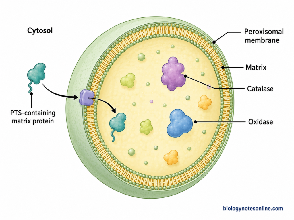
- The internal part is referred to as matrix. It is finely granular and contains different enzymes. Many of these are concerned with oxidation reactions.
- DNA is absent. They also have no ribosomes of their own. Thus, proteins required for formation and functioning of microbodies are encoded by nuclear genes, formed on cytosolic ribosomes and then transported into the organelle.
- A definite size and number is not found in all cells. Both can change considerably with cell type and different metabolic or environmental conditions.
- The membrane provides compartmentalization of reactions. Due to this, several oxidative reactions can remain confined within the microbody rather than taking place throughout the cytoplasm.
Structure of Microbodies
- Microbodies are small membrane-bound cell organelles. They are generally spherical or oval in shape. Their size is variable, commonly about 0.5-1.5 µm in diameter.
- A microbody is surrounded by a single membrane. This membrane encloses the dense internal matrix of the organelle.
- The matrix is granular in appearance and contains different proteins and enzymes. Catalase and several oxidases are present in peroxisomal matrix. The enzyme content can differ with the type of microbody and tissue.
- In some microbodies, a dense crystalline or crystalloid core may also be present inside the matrix. In liver peroxisomes of some mammals, this core contains urate oxidase. Such a core is not found in all peroxisomes.
- Microbodies do not contain their own DNA. They also do not have their own protein synthesizing system. The peroxisomal proteins are encoded by nuclear DNA and are synthesized outside the organelle, which are then transported into it.
- The surrounding membrane also contains specific membrane proteins. Some of these proteins called peroxins (PEX proteins) are involved in formation of the organelle and transport of proteins into its matrix.
- Glyoxysomes have the same general microbody structure, having a single surrounding membrane and an enzyme-containing matrix. Their enzyme composition is different from other peroxisomes, especially the presence of enzymes associated with the glyoxylate cycle in young seedlings.
Enzymes and Genetic Material of Microbodies
Enzymes of Microbodies
Microbodies contain a number of enzymes. These enzymes are not same in all forms, and differ according to the metabolic activities of the organelle. Some of the important enzymes are-
- Catalase
It is one of the important enzymes of peroxisomes. Catalase breaks hydrogen peroxide (H₂O₂) into water and oxygen. It is mainly present in the peroxisomal matrix. - Oxidases
Different hydrogen peroxide-producing oxidases are found in peroxisomes. Urate oxidase, D-amino acid oxidase, α-hydroxy acid oxidase and some other oxidases have been detected, but their occurrence differs with organism and tissue. - Enzymes of β-oxidation
Peroxisomes also contain a fatty acid β-oxidation system. Acyl-CoA oxidase, multifunctional protein and 3-ketoacyl-CoA thiolase are some of its important enzymes. This pathway is involved in oxidation of different fatty acids. - Isocitrate lyase and Malate synthase
These are characteristic enzymes of glyoxysomes. Both take part in the glyoxylate cycle and are especially prominent in glyoxysomes of germinating oil-rich seeds. - Glycolytic enzymes
The glycosomes of trypanosomes contain several enzymes of glycolysis. In Trypanosoma brucei, the first part of glycolysis takes place within these microbody-like organelles.
Genetic Material of Microbodies
- Microbodies do not contain their own DNA or RNA. Thus, an independent genetic system is absent in them.
- They do not have their own protein synthesizing system. Peroxisomal matrix proteins are encoded by genes present in the nucleus.
- Most matrix enzymes are synthesized on free ribosomes in the cytosol and after their synthesis, these are transported into the microbody. Experiments with catalase and urate oxidase also showed their synthesis on free polysomes rather than inside peroxisomes.
- The transport of these proteins is carried out with the help of specific peroxisomal targeting signals and PEX proteins. So, unlike mitochondria and chloroplasts, microbodies are not provided with a separate genetic machinery for making their own proteins.
Types of Microbodies
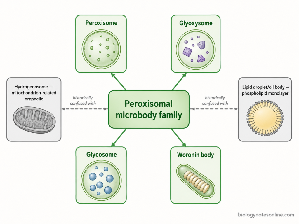
The major types of microbodies include peroxisomes, glyoxysomes, glycosomes and Woronin bodies. They differ in occurrence and the metabolic reactions taking place inside them.
1. Peroxisomes
Peroxisomes are the most widely occurring microbodies of eukaryotic cells. The organelles contain different oxidases together with catalase. Catalase is concerned with the removal of hydrogen peroxide formed during oxidation reactions.
Fatty acid β-oxidation also occurs in them. Very-long-chain fatty acids are particularly degraded in animal peroxisomes. In plants the function is somewhat different, and leaf peroxisomes participate in photorespiration. Liver cells contain a large number of peroxisomes because of their active oxidative metabolism.
2. Glyoxysomes
Glyoxysomes– These are specialized peroxisomes occurring mainly in germinating oil seeds. Castor bean is a common example. They contain enzymes of β-oxidation as well as enzymes required for the glyoxylate cycle.
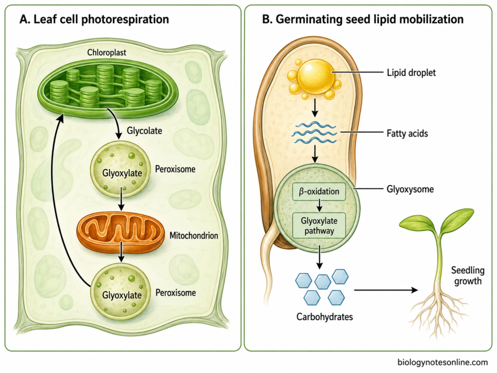
The characteristic enzymes are isocitrate lyase and malate synthase. Stored fats are first degraded to acetyl-CoA. Carbon present in acetyl-CoA can then be conserved through the glyoxylate cycle and ultimately used in carbohydrate formation, which is important during the early growth of seedling.
3. Glycosomes
These are specialized microbodies found mainly in kinetoplastid protozoans such as Trypanosoma brucei and Leishmania spp. Glycosomes contain most of the enzymes concerned with glycolysis. Thus, a major part of glycolytic pathway takes place inside these organelles rather than freely in cytoplasm.
They also contain enzymes associated with some other metabolic processes. Purine salvage and parts of lipid metabolism are included. Proteins required by glycosomes are imported with the help of peroxisomal targeting signals (PTS) and different peroxins.
4. Woronin Bodies
Woronin bodies occur in filamentous ascomycete fungi, especially members of Pezizomycotina. They are derived from peroxisomes. Usually these bodies remain close to the septal pore present between adjoining hyphal compartments.
A dense core made mainly of Hex1 protein is present inside. During injury, the body moves towards the septal pore and blocks it. Loss of cytoplasm from the damaged part is therefore restricted. Neurospora crassa is one of the well-known examples having Woronin bodies.
Major Types of Peroxisomal Microbodies
Microbodies occur in different specialized forms in eukaryotic cells. They differ mainly in their enzymes, metabolic pathways and cellular functions. The major types of peroxisomal microbodies are given below-
| Type | Main organisms/cells | Membrane | Characteristic proteins/enzymes | Major pathway or activity | Main function |
|---|---|---|---|---|---|
| Peroxisome | Widespread in eukaryotic cells | Single bilayer | Catalase, oxidases | Fatty-acid oxidation and H₂O₂ metabolism | Oxidative and lipid metabolism |
| Glyoxysome | Plants and some fungi | Single bilayer | Isocitrate lyase, malate synthase | Glyoxylate pathway | Seed and fungal carbon metabolism |
| Glycosome | Kinetoplastids and diplonemids | Single bilayer | Glycolytic enzymes, peroxins | Compartmentalized glycolysis | Carbohydrate metabolism |
| Woronin body | Filamentous ascomycetes | Peroxisome-derived | Hex1 | Septal-pore response | Prevents cytoplasmic leakage |
Occurrence of Microbodies
Microbodies are found in eukaryotic cells. Their occurrence is wide, although the same specialized form is not present in all organisms.
- Animal cells– Peroxisomes are present in almost all human cells except mature erythrocytes. Large number are found in liver and kidney cells.
- Plant cells– Peroxisomes occur in different plant tissues, including photosynthetic and non-photosynthetic cells. In green leaves, the leaf peroxisomes are closely concerned with photorespiration.
- Germinating seeds– Glyoxysomes are particularly found during germination of oil-rich seeds. They remain associated with the utilization of stored oil, and after development of photosynthetic tissue, glyoxysomal functions can change towards those of leaf peroxisomes.
- Fungi– Peroxisomes are present in yeasts as well as filamentous fungi. Woronin bodies occur in many filamentous ascomycetes. These are specialized structures derived from peroxisomes.
- Protists– Specialized microbodies called glycosomes are found in Trypanosoma, Leishmania and other kinetoplastids, where much of the glycolytic pathway is enclosed within these organelles. They are also known from diplonemids.
- Prokaryotic cells– Typical peroxisomal microbodies are absent. Peroxisomes are organelles of eukaryotic cells.
Microbody Biogenesis
The biogenesis of microbodies is mainly studied in peroxisomes. They do not contain their own DNA, so the proteins required for their formation are encoded by nuclear genes. Peroxisomes can arise from already existing peroxisomes and also by a de novo pathway.
- In the first process, the peroxisomal membrane is formed and different peroxisomal membrane proteins (PMPs) are inserted into it. PEX3, PEX19 and PEX16 are important peroxins concerned with this membrane formation. Some membrane proteins can reach the peroxisome directly from the cytosol, while another route involves the endoplasmic reticulum (ER).
- The matrix proteins are not synthesized inside the microbody. They are formed on free ribosomes in the cytosol.
- Most of these proteins possess a specific peroxisomal targeting signal (PTS). PTS1 is generally present at the C-terminal region and is recognized by PEX5. Another signal, PTS2, occurs near the N-terminal region and uses PEX7 as its receptor.
- The receptor with its protein now reaches the peroxisomal membrane. PEX13 and PEX14 are involved in docking of this complex. The matrix protein is transported inside, whereas the receptor is not permanently retained there.
- After protein release, PEX5 is recycled back to the cytosol. Its monoubiquitination and removal from the membrane involve different peroxins, with the PEX1-PEX6 ATPase complex providing an important ATP-dependent step. The receptor can again be used for another round of protein import.
- Already present peroxisomes can grow by taking up membrane and matrix proteins. They then divide. PEX11 proteins are associated with elongation and proliferation of peroxisomes, while dynamin-related proteins participate in membrane constriction and fission.
- New peroxisomes may also develop by the de novo pathway. In this process, precursor membrane structures containing peroxisomal proteins are formed from the ER, which develop into import-competent peroxisomes. This pathway becomes especially evident when functional pre-existing peroxisomes are absent.
- In plants, the microbody can also change its enzymatic nature during development. Glyoxysomes of germinating seedlings can undergo transition towards leaf peroxisomes during greening, glyoxysomal proteins are removed and newly synthesized photorespiratory proteins are transported into them.
Historical Terminology and Modern Classification
The term microbody was originally used for small single membrane-bound cytoplasmic organelles identified mainly by their morphology. J. Rhodin described these structures in mouse kidney cells in 1954. At that time their biochemical function was not clearly known.
Later, different enzymes were found within these particles and the classification became more functional. Thus, microbody is still used as a general or older morphological term, whereas peroxisome is the commonly used biochemical and cell biological name for this organelle group.
Microbody and Peroxisome Terminology
The name peroxisome was given by Christian de Duve after the presence of hydrogen peroxide-producing oxidases and catalase was established in these organelles. Hydrogen peroxide is produced and also degraded within the same organelle. This property formed the basis for the name peroxisome.
The terms microbody and peroxisome therefore are often used for the same general organelle system, but they came from different approaches. Microbody was mainly based on appearance under the electron microscope. Peroxisome was based on its biochemical properties.
Glyoxysomes were described as a special type of plant microbody containing enzymes of the glyoxylate cycle. They are now generally considered specialized peroxisomes rather than a completely unrelated organelle. In germinating cotyledons, the same microbodies can also change from glyoxysomal functions to photorespiratory peroxisomal functions during development.
Older books may also use different names for small cytoplasmic bodies, which sometimes creates confusion with the word microbody. For modern classification, the biochemical nature and origin of the organelle are more useful than its small size alone.
Hydrogenosomes and Lipid Droplets
Hydrogenosomes should not normally be classified as peroxisomal microbodies. They are anaerobic energy-producing organelles and in many eukaryotes are evolutionarily related to mitochondria. Thus, their similarity in size or absence of a typical mitochondrial appearance does not make them peroxisomes.
The same confusion occurs with spherosomes, oil bodies and lipid bodies of plant cells. These terms are now commonly associated with lipid droplets. Lipid droplets contain a core of neutral lipids surrounded by a phospholipid monolayer, not the usual phospholipid bilayer membrane of a peroxisome.
Lipid droplets are mainly involved in storage and mobilization of neutral lipids. Peroxisomes are membrane-bound metabolic organelles containing enzymes for different oxidative reactions. Therefore, lipid droplets or oil bodies are not included under peroxisomal microbodies in modern cell biology.
Functions of Microbodies
Microbodies perform different metabolic functions depending upon their type and the organism. The major functions are as follows-
- Hydrogen peroxide metabolism– Different oxidases of peroxisomes produce hydrogen peroxide (H₂O₂) during oxidation reactions. Catalase present in the organelle breaks it into water and oxygen. Thus, formation as well as removal of H₂O₂ takes place in peroxisomes.
- Fatty acid oxidation– Peroxisomes are involved in β-oxidation of fatty acids. In mammals, very-long-chain fatty acids and some branched or dicarboxylic fatty acids are shortened through this pathway, whereas in plants peroxisomes are the major site of fatty acid β-oxidation.
- α-oxidation– The α-oxidation of phytanic acid takes place in mammalian peroxisomes.
- Synthesis of membrane lipids– Peroxisomes take part in formation of plasmalogens, a group of ether phospholipids. The initial reactions of their synthesis occur in the peroxisome. These lipids are important components of different cellular membranes.
- Photorespiration– Leaf peroxisomes have an important role in photorespiration. Glycolate coming from the chloroplast is converted to glyoxylate inside the peroxisome and later reactions also return glycerate towards the chloroplast.
- Glyoxylate cycle– Glyoxysomes of germinating oil-rich seeds contain the enzymes of glyoxylate cycle. Stored fatty acids are degraded, producing acetyl-CoA. The glyoxylate cycle allows this carbon to be used towards carbohydrate formation during early seedling growth.
- Glycolysis in protozoans– In kinetoplastid protozoans such as Trypanosoma, specialized microbodies called glycosomes contain much of the glycolytic pathway. In bloodstream T. brucei, compartmentalized glycolysis has a major role in ATP production.
- Peroxisomes also take part in bile acid synthesis in mammals. Some intermediates formed during bile acid synthesis undergo further reactions within these organelles.
- Reactive oxygen metabolism is another function. Peroxisomes can act both as a source and a site for removal of reactive oxygen species, and because of this they are also associated with cellular redox regulation.
Peroxisomes vs Glyoxysomes
Peroxisomes and glyoxysomes are closely related microbodies, but they differ mainly in their occurrence and metabolic function. The major differences between them are given below-
| Feature | Peroxisome | Glyoxysome |
|---|---|---|
| Occurrence | Widespread in eukaryotic cells | Found in specialized plant and fungal cells |
| Membrane | Surrounded by a single membrane | Surrounded by a single membrane |
| Shared relationship | Core peroxisomal organelle | Specialized form of peroxisome |
| Important enzymes | Oxidases and catalase | Peroxisomal enzymes together with isocitrate lyase and malate synthase |
| Major metabolic emphasis | Oxidative reactions and lipid metabolism | Lipid mobilization and glyoxylate metabolism |
| Plant role | Takes part in photorespiration and other metabolic reactions | Especially important during mobilization of stored lipids in seeds |
References
- Alencastre, I. S., Rodrigues, T. A., Grou, C. P., Fransen, M., Sá-Miranda, C., & Azevedo, J. E. (2009). Mapping the cargo protein membrane translocation step into the PEX5 cycling pathway. Journal of Biological Chemistry, 284(40), 27243–27251. https://doi.org/10.1074/jbc.M109.032565
- Bonetta, L. (2005). Seeing peroxisomes. Journal of Cell Biology, 169(5), 705. https://doi.org/10.1083/jcb1695fta2
- Carman, G. M. (2012). Thematic minireview series on the lipid droplet, a dynamic organelle of biomedical and commercial importance. Journal of Biological Chemistry, 287(4), 2272. https://doi.org/10.1074/jbc.R111.323931
- Delille, H. K., Dodt, G., & Schrader, M. (2011). Pex11pβ-mediated maturation of peroxisomes. Communicative & Integrative Biology, 4(1), 51–54. https://doi.org/10.4161/cib.4.1.13647
- Furuya, T., Kessler, P., Jardim, A., Schnaufer, A., Crudder, C., & Parsons, M. (2002). Glucose is toxic to glycosome-deficient trypanosomes. Proceedings of the National Academy of Sciences of the United States of America, 99(22), 14177–14182. https://doi.org/10.1073/pnas.222454899
- Hagmann, V., Sommer, S., Fabian, P., Bierlmeier, J., van Treel, N., Mootz, H. D., Schwarzer, D., Azevedo, J. E., & Dodt, G. (2018). Chemically monoubiquitinated PEX5 binds to the components of the peroxisomal docking and export machinery. Scientific Reports, 8, 16014. https://doi.org/10.1038/s41598-018-34200-5
- Imoto, Y., Kuroiwa, H., Yoshida, Y., Ohnuma, M., Fujiwara, T., Yoshida, M., Nishida, K., Yagisawa, F., Hirooka, S., Miyagishima, S.-Y., Misumi, O., Kawano, S., & Kuroiwa, T. (2013). Single-membrane-bounded peroxisome division revealed by isolation of dynamin-based machinery. Proceedings of the National Academy of Sciences of the United States of America, 110(23), 9583–9588. https://doi.org/10.1073/pnas.1303483110
- Jedd, G., & Chua, N.-H. (2000). A new self-assembled peroxisomal vesicle required for efficient resealing of the plasma membrane. Nature Cell Biology, 2, 226–231. https://doi.org/10.1038/35008652
- Kao, Y.-T., Gonzalez, K. L., & Bartel, B. (2018). Peroxisome function, biogenesis, and dynamics in plants. Plant Physiology, 176(1), 162–177. https://doi.org/10.1104/pp.17.01050
- Kunze, M., & Hartig, A. (2013). Permeability of the peroxisomal membrane: Lessons from the glyoxylate cycle. Frontiers in Physiology, 4, 204. https://doi.org/10.3389/fphys.2013.00204
- Leger, M. M., Kolisko, M., Kamikawa, R., Stairs, C. W., Kume, K., Čepička, I., Silberman, J. D., Andersson, J. O., Xu, F., Yabuki, A., Eme, L., Zhang, Q., Takishita, K., Inagaki, Y., Simpson, A. G. B., Hashimoto, T., & Roger, A. J. (2017). Organelles that illuminate the origins of Trichomonas hydrogenosomes and Giardia mitosomes. Nature Ecology & Evolution, 1, Article 0092. https://doi.org/10.1038/s41559-017-0092
- Mast, F. D., Herricks, T., Strehler, K. M., Miller, L. R., Saleem, R. A., Rachubinski, R. A., & Aitchison, J. D. (2018). ESCRT-III is required for scissioning new peroxisomes from the endoplasmic reticulum. Journal of Cell Biology, 217(6), 2087–2102. https://doi.org/10.1083/jcb.201706044
- Moody, D. E., & Reddy, J. K. (1976). Morphometric analysis of the ultrastructural changes in rat liver induced by the peroxisome proliferator SaH 42-348. Journal of Cell Biology, 71(3), 768–780. https://doi.org/10.1083/jcb.71.3.768
- Olzmann, J. A., & Carvalho, P. (2019). Dynamics and functions of lipid droplets. Nature Reviews Molecular Cell Biology, 20(3), 137–155. https://doi.org/10.1038/s41580-018-0085-z
- Stefanini, S., Nardacci, R., Farioli-Vecchioli, S., Pajalunga, D., & Sartori, C. (1999). Liver and kidney peroxisomes in lactating rats and their pups after treatment with ciprofibrate: Biochemical and morphometric analysis. Cellular and Molecular Biology, 45(6), 815–829. https://pubmed.ncbi.nlm.nih.gov/10541478/
- Suzuki, K., & Vanier, M. T. (1999). The peroxisome. In G. J. Siegel, B. W. Agranoff, R. W. Albers, S. K. Fisher, & M. D. Uhler (Eds.), Basic neurochemistry: Molecular, cellular and medical aspects (6th ed.). Lippincott-Raven. https://www.ncbi.nlm.nih.gov/books/NBK28053/
- Titus, D. E., & Becker, W. M. (1985). Investigation of the glyoxysome-peroxisome transition in germinating cucumber cotyledons using double-label immunoelectron microscopy. Journal of Cell Biology, 101(4), 1288–1299. https://doi.org/10.1083/jcb.101.4.1288