

Herdmania is a genus of solitary ascidians belonging to the phylum Chordata. It is placed under the subphylum Tunicata (Urochordata), class Ascidiacea, order Stolidobranchia and family Pyuridae. Herdmania represents a genus and not a single animal species. Different species are included under this genus. Herdmania momus and Herdmania pallida are two of them.
The members are marine animals and the adult is generally sessile in nature. It remains attached to the substratum. Adult ascidians are filter feeders. Water is passed through the large pharyngeal region and suspended food particles are filtered from it. The water enters through the oral siphon and finally passes outside through the atrial siphon.
The larval form is different from the adult. It is a free-swimming tadpole-like larva and chordate characters are more prominent in this stage. The larva possesses a notochord, dorsal nerve cord and post-anal tail. These are some of the important chordate features. The notochord is restricted to the larval tail and is not retained in the adult ascidian.
After a short swimming stage, the larva becomes attached to a suitable substratum. Metamorphosis now takes place. During this process, the tail is resorbed and the notochord disappears. Larval muscles and several larval structures are also lost or modified, while the adult organs develop. Thus, the active larval form is changed into a sessile filter-feeding adult. This type of change in ascidians is classically referred to as retrogressive metamorphosis.
H. momus and H. pallida are separate species of the genus Herdmania. They should not be considered as two names of the same general animal while describing species characters. Characters such as particular body colour, size, geographical distribution or other species-level features may differ between them. Therefore, such characters should be mentioned for the particular species from which they are reported.
What is Herdmania?
Herdmania is a genus of solitary ascidians, commonly known as sea squirts. It is a marine tunicate belonging to the phylum Chordata and class Ascidiacea. The adult Herdmania animal is marine and generally sessile, remaining attached to rocks or other substratum. It is a filter-feeding animal. The body is externally covered by a thick covering called tunic or test. Two siphons are present, the oral or branchial siphon and the atrial siphon. Water enters through the oral siphon and filtered water passes outside through the atrial siphon. Thus, the Herdmania ascidian is an important non-vertebrate chordate.
The Herdmania sea squirt shows the characteristic ascidian body form. Its chordate position is more clearly shown by the larval stage, which has the typical chordate body plan. Ascidians are also important for understanding the origin and evolution of vertebrate chordates.
It is called a sea squirt because the muscular body can contract suddenly when disturbed. During this contraction, water present inside the body is expelled through the siphons. This produces the characteristic squirting of water from the animal.
Classification and Systematic Position
Herdmania belongs to the phylum Chordata and subphylum Tunicata. It is placed under the class Ascidiacea. The genus belongs to the family Pyuridae of the order Stolidobranchia. The present systematic position is as follows-
Modern Taxonomic Classification
| Rank | Taxon |
|---|---|
| Kingdom | Animalia |
| Phylum | Chordata |
| Subphylum | Tunicata |
| Class | Ascidiacea |
| Order | Stolidobranchia |
| Family | Pyuridae |
| Genus | Herdmania |
This classification is followed by the current Ascidiacea World Database (AWD)/World Register of Marine Species (WoRMS). The genus Herdmania is placed under Pyuridae, while Pyuridae is included in the order Stolidobranchia.
Tunicata and Urochordata Terminology
Tunicata is the accepted subphylum name in the present WoRMS classification. The name Urochordata is also very commonly used for the same group, particularly in older zoology literature and textbooks. It should not be considered as a different group from Tunicata.
In WoRMS, Urochordata is presently treated as an unaccepted junior synonym of Tunicata. Thus, older syllabus may show Herdmania under Urochordata, while modern classification places it under Tunicata. Both names in this context refer to the same tunicate group.
The use of Urochordata is also common in taxonomic literature. For example, the revision of Japanese Herdmania was published under the title “Herdmania (Urochordata: Ascidiacea)”. The Ascidiacea World Database similarly describes Tunicata as also known as Urochordata.
Important Species
Some of the important species of the genus are-
Herdmania pallida – It is an accepted species of Herdmania. The species was originally described as Cynthia pallida Heller, 1878. It is now placed under the genus Herdmania.
Herdmania momus – It is another well-known species of the genus and is the type species associated with Herdmania. It is a solitary pyurid ascidian and has been widely studied in taxonomic and morphological works.
The different species of Herdmania should not be considered morphologically identical. Body size, nature of the test, number and arrangement of branchial structures, muscles and particularly the gonads and gonoducts can show differences between species. These characters have been used for separating different species of the genus. Therefore, measurements or fine anatomical description given for H. pallida or H. momus should be mentioned for that particular species and not directly for the whole genus.
General Characteristics of Herdmania
- Herdmania is a solitary ascidian belonging to the subphylum Tunicata of phylum Chordata. It is exclusively marine in nature.
- The adult is sessile and remains attached to rocks, shells or other hard substratum.
- The body is generally sac-like or oblong in shape. It is covered externally by a thick tunic or test, which is leathery in nature.
- Two siphons are present, the branchial or oral siphon and the atrial siphon. Water enters through branchial siphon and passes outside through the atrial siphon.
- The pharynx is greatly enlarged and occupies a large part of the body. Its wall contains numerous perforations called stigmata.
- Feeding is ciliary or filter-feeding type. The cilia maintain water current and food particles are trapped in mucus produced by the endostyle.
- The blood vascular arrangement of Herdmania is traditionally described as open or lacunar type. The heart is tubular and shows periodic reversal in the direction of its contractions. Reversal of heart beat is a characteristic feature of tunicates.
- The adult nervous system is simple. A cerebral ganglion is present between the two siphons and nerves arise from it to different regions of the body.
- The commonly studied solitary ascidians are hermaphroditic, with male and female reproductive organs in the same individual. The detailed structure of gonads can vary between different species of Herdmania.
- Fertilization is generally external, the gametes being released into the surrounding sea water. This reproductive condition is well known among solitary ascidians.
- Development includes a free-swimming ascidian tadpole larva. The larva possesses prominent chordate features which are reduced during the development of adult. Herdmania momus also develops through this tadpole larval stage.
- After attachment, the larva undergoes retrogressive metamorphosis. During this process the larval tail and notochord are lost, and the free-swimming larva changes into a sessile adult.
Habit and Habitat of Herdmania
The following are the habit and habitat of Herdmania–
- Herdmania is an exclusively marine animal. It is found only in sea water.
- The adult is solitary and sessile in habit. It remains permanently attached to the substratum.
- It is generally attached to rocks, stones, shells and other hard surfaces present under water.
- Some species can also grow over artificial substratum such as piers, jetty structures, pontoons and submerged surfaces.
- Herdmania is a benthic animal. The basal part remains fixed while the siphons remain exposed to the surrounding water.
- It is a filter-feeding animal. Small suspended food particles are obtained from the water passing through the body.
- The members are generally found in coastal marine habitats. The exact depth varies among different species.
- Herdmania momus is commonly reported from shallow marine habitats, rocks, coral-associated regions and artificial surfaces.
- Herdmania pallida has also been recorded from shallow coastal areas and artificial marine structures.
- The adult is non-motile, but the tadpole larva is free-swimming for a short period.
- After swimming, the larva becomes attached to a suitable substratum. Metamorphosis then takes place and the sessile adult is formed.
- Habitat, depth and type of substratum are not exactly same in every species of Herdmania. These characters may vary according to species and geographical region.
Distribution of Herdmania
The distribution of Herdmania is mainly in the warmer marine regions of the world. The following are the important distribution of the genus-
- Species of Herdmania are widely recorded from the Indo-Pacific region. Different members occur in the Indian Ocean and western Pacific waters.
- The genus is reported from Japan. A taxonomic revision of Japanese waters recognized several species including H. momus, H. pallida, H. japonica, H. mauritiana, H. subpallida and H. kiiensis.
- Species are also recorded from Australia, Micronesia, Indonesia, Taiwan, Vietnam, New Caledonia and different Pacific islands. The distribution is not same for every species.
- Herdmania pallida has a wide distribution. Confirmed records are present from the Pacific Ocean, including Japan, Taiwan, Vietnam, Guam, Palau, Tahiti, New Caledonia and Hawaii.
- H. pallida is also recorded from the Indian Ocean region, including Sri Lanka, the Red Sea, East African coast, Mozambique, Seychelles, Mauritius and western Australia. It has also been reported from Singapore coastal waters.
- Records of H. pallida are also known from the Atlantic region, including Panama, Cuba, the Antilles and Brazil. Thus, this particular species has a distribution beyond the Indo-Pacific region.
- Herdmania momus has a wide Indo-Pacific distribution. It occurs in the Red Sea and different parts of the Indian and Pacific Oceans.
- H. momus has entered the Mediterranean Sea from the Red Sea through the Suez Canal. It is considered a non-indigenous species in the Mediterranean. Records are present from the eastern Mediterranean and later from the Maltese Islands.
- The distribution of H. momus is also changing in some regions. Recent records from the Korean Peninsula show northward expansion of this warm-water species.
- Distribution should therefore be mentioned according to the particular Herdmania species. Old records under the name H. momus may also contain other species because several members of the genus were separated only after detailed taxonomic revision.
External Morphology of Herdmania
The external body of Herdmania is covered by a tunic or test. The adult body is attached with the substratum and two siphons are present in the free region. The major external structures are as follows-
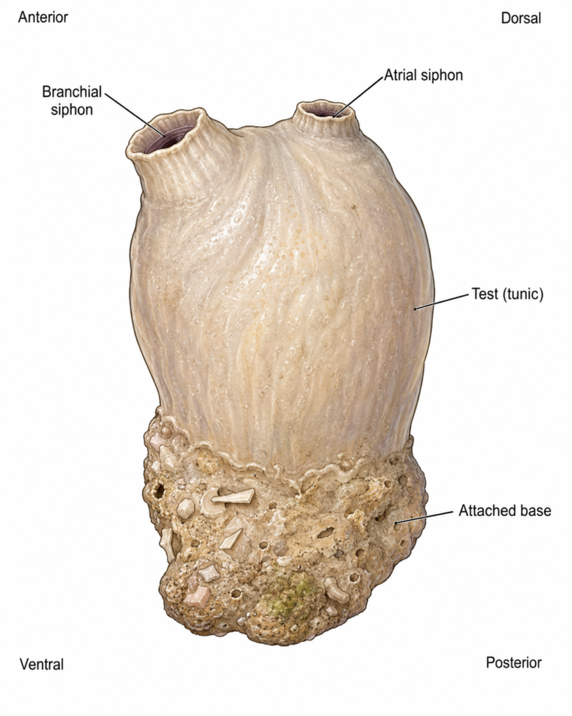
Body Form and Orientation
- The adult Herdmania has a sac-like, oval or oblong body. The exact shape is not same in all the species.
- The body can be roughly divided into the body proper and the basal attached region. The body proper is the free portion containing the soft parts of the animal.
- In classical description of Herdmania, the branchial aperture marks the anterior side. The opposite region represents the posterior side. The atrial aperture marks the dorsal side, and the opposite region is referred to as ventral side. This peculiar orientation is also found in the general adult ascidian body plan.
- The basal region helps in attachment with the substratum. In animals attached on a hard surface, the body may be fixed by a broad basal area and a distinct foot may not be formed.
- A foot-like extension is described in classical Herdmania specimens living over softer or sandy substratum. It is mainly formed by the test and helps in anchorage. Thus, foot should not be considered as an equally developed structure in every specimen.
- The size and colour differ considerably between the different species and even between populations. Therefore, a single exact body length or colour cannot be given as a general character of the genus. H. momus itself shows considerable variation in body size and colour in different geographical populations.
Branchial and Atrial Siphons
- Two siphons are present in the free region of the body. These are the branchial or oral siphon and atrial siphon.
- The branchial siphon is the incurrent siphon. Sea water enters through its branchial or oral aperture and passes towards the large pharyngeal region.
- The atrial siphon is the excurrent siphon. Filtered water from the atrial cavity is passed outside through this opening.
- The anus opens into the atrial cavity in ascidians. Thus, faecal material is also carried outside with the outgoing water through the atrial siphon.
- The reproductive ducts also discharge towards the atrial region. Gametes and wastes therefore pass out with the excurrent water current.
- The siphons are muscular structures and can contract. Their exact length, colour, prominence and external form vary according to the particular Herdmania species.
- Lobes or other fine siphonal characters should be described at the species level. These structures can form useful taxonomic characters and should not be assumed to be exactly same in all species of Herdmania.
Test or Tunic
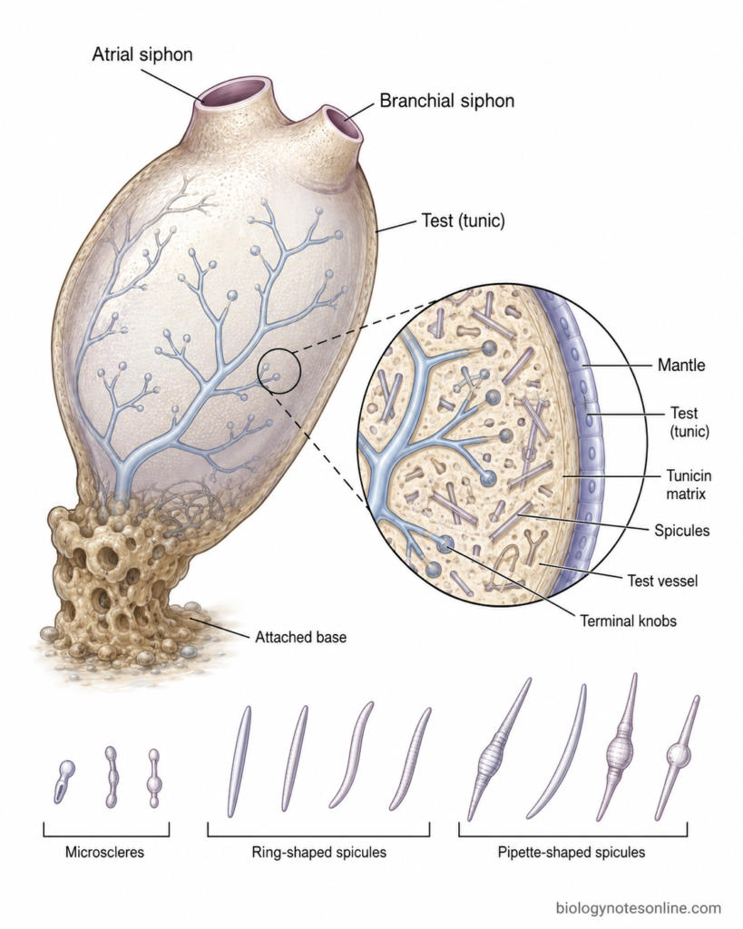
- The test or tunic is the external covering surrounding the body of Herdmania. It lies outside the true body wall or mantle.
- It is secreted in association with the epidermis and forms a characteristic extracellular covering of tunicates. The tunic is one of the important features from which the name Tunicata is derived.
- The matrix contains cellulose, traditionally referred to as tunicin in tunicates. Tunicates are unusual among animals because they can synthesize cellulose.
- The tunic mainly provides protection and mechanical covering to the soft body. In the basal region it also takes part in attachment of the animal with the substratum.
- The tunic and mantle are not the same structure. The mantle is the cellular body wall lying below the tunic, whereas the tunic forms the external extracellular matrix.
- Blood vessels can extend into the tunic in many solitary ascidians. In H. momus, tunic blood vessels are well demonstrated because the small tunic spicules are formed inside these vessels before passing into the tunic matrix.
- Thickness, transparency and toughness of the tunic can change according to species, age and specimen. It should not always be described as having one fixed thickness or appearance.
Calcareous Spicules
- Herdmania is characterized by the presence of calcareous spicules. These are mineralized structures occurring in different body tissues, while their distribution in the tunic also varies between species.
- The spicules are mainly made of calcium carbonate (CaCO₃). They form hard elements within the otherwise soft tissues of the ascidian.
- Body spicules can occur in the mantle, siphons and branchial basket. Their occurrence and arrangement have been studied particularly well in Herdmania momus.
- The spicules have a probable supporting and structural role, particularly those present within the mantle tissues. Tunic spicules may also have a protective role, although the exact ecological function is not completely established for every type.
- Shape, dimensions, number and exact distribution of the spicules are species-dependent characters. These should not be generalized from H. momus to every species of the genus.
- A special mineral condition is found in H. momus. Its body and tunic spicules are composed of vaterite, a polymorph of calcium carbonate. X-ray diffraction and later structural studies confirmed this condition.
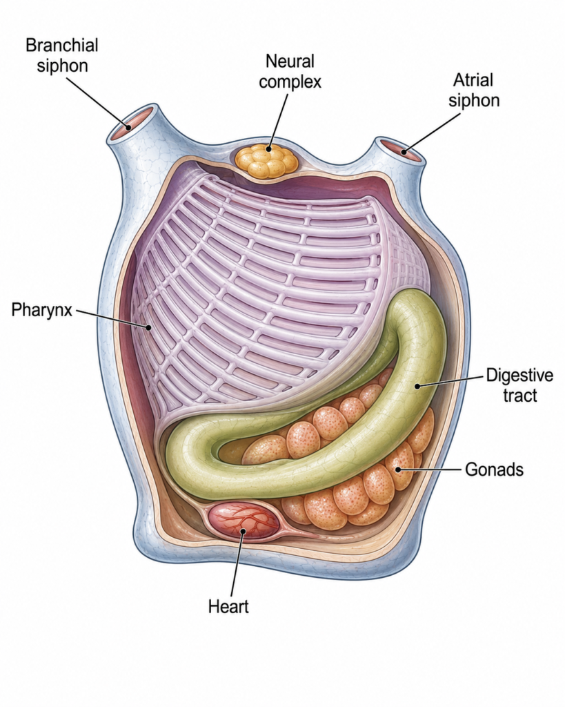
Body Wall and Internal Organization of Herdmania
The body wall of Herdmania lies below the external tunic or test. It is generally referred to as the mantle. Inside the mantle is the large pharyngeal region and the atrial or peribranchial cavity.
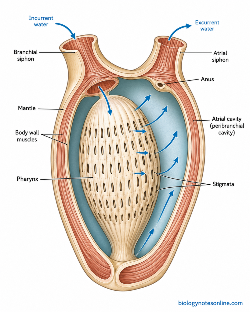
Mantle
- The mantle is the cellular body wall of Herdmania present just below the tunic. The tunic and mantle are therefore two different structures.
- It contains an outer epidermal component, connective tissue and well-developed muscle fibres. The muscles are mainly associated with the body wall and siphonal regions.
- The mantle muscles form bands over the body. Muscle fibres also extend into the branchial and atrial siphons.
- These muscles help in the contraction of body wall. They also take part in closing and retracting the siphons.
- When disturbed, the mantle can contract rapidly. Water present inside the branchial and atrial regions is forced outside through the siphons. This produces the characteristic squirting response of ascidians.
- The normal feeding water current is not produced by these muscular contractions. It is mainly maintained by the beating of cilia in the pharyngeal stigmata. The strong mantle contraction is used during withdrawal and squirting.
Atrial or Peribranchial Cavity
- The atrial cavity, also called peribranchial cavity, is a large cavity present outside the pharyngeal wall. It lies between much of the pharynx and the surrounding body wall.
- The large pharynx contains numerous small openings called stigmata or gill slits. These openings communicate with the atrial cavity.
- Sea water first enters the branchial siphon and reaches the pharyngeal cavity. From here it passes through the ciliated stigmata.
- After passing through the stigmata, the filtered water enters into the atrial or peribranchial cavity. Food particles are retained by the mucous feeding system of the pharynx.
- The atrial cavity communicates with the exterior through the atrial siphon. The water collected in this cavity is finally passed outside through this siphon.
- The anus also opens into the atrial cavity. Thus, faecal material can be carried outside with the outgoing water current through the atrial siphon.
- Therefore, the peribranchial cavity forms an important part of the water passage in Herdmania. The direction of water is mainly branchial siphon → pharynx → stigmata → atrial cavity → atrial siphon.
Digestive System of Herdmania
The digestive system of Herdmania is adapted for ciliary or filter feeding. It consists mainly of the alimentary canal and associated digestive glands. The alimentary canal is complete and forms a curved or U-shaped gut.
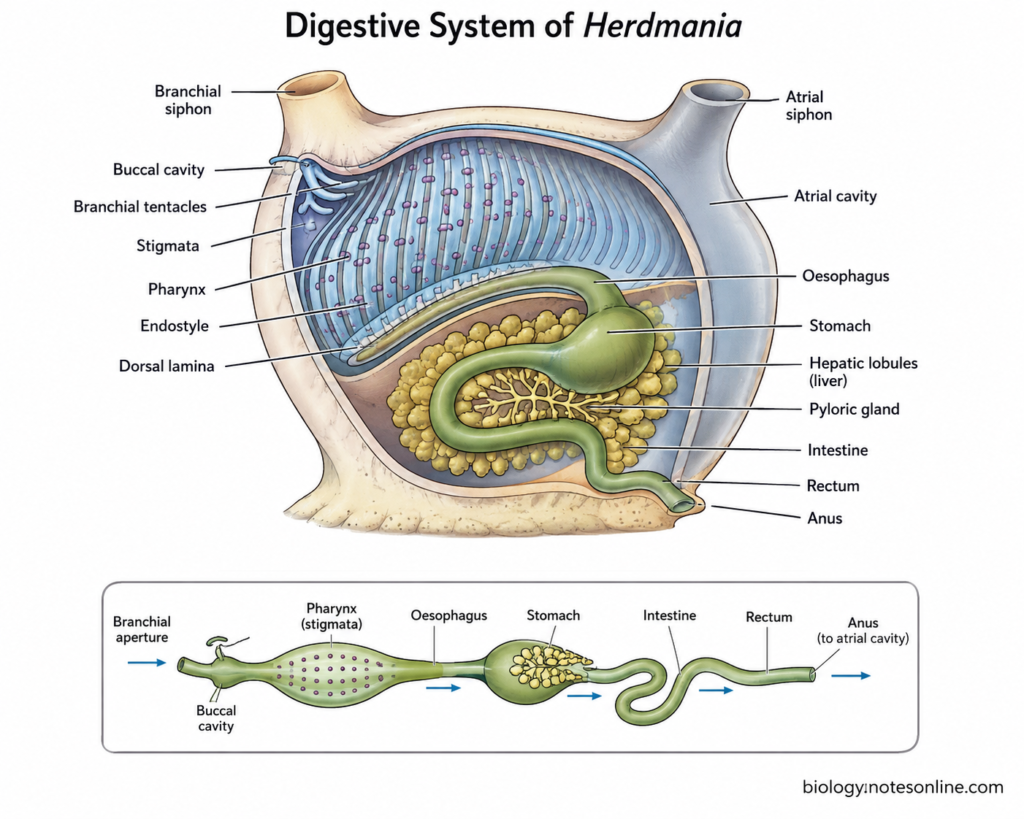
Alimentary Canal
The following are the main parts of alimentary canal-
- Branchial or oral aperture – It forms the entrance of the digestive system. The aperture is present at the end of branchial siphon and sea water containing food particles enters through it.
- Buccal cavity – The branchial siphon encloses a short cavity referred to as the buccal cavity or stomodaeum. A ring of branchial tentacles is present near its inner end. Their exact number and branching are different among species.
- Pharynx – The pharynx or branchial sac is the largest part. It occupies a major portion of body and its wall is perforated by numerous ciliated openings called stigmata. Water passes through these stigmata into the atrial cavity.
- Endostyle – It is a longitudinal glandular groove present along the ventral side of pharynx. The endostyle produces mucus which forms the food-trapping net. Food particles present in water are retained in this mucus.
- Dorsal lamina – It is present along the dorsal side of the pharynx. The mucus containing food particles is moved towards this region and finally passes towards the oesophagus.
- Oesophagus – It is a short tube arising from the posterior region of pharynx. The food cord from the pharynx enters into the oesophagus and then reaches the stomach.
- Stomach – The oesophagus opens into the stomach. It is a wider region of the alimentary canal and remains closely associated with hepatic tissue. In both H. momus and H. pallida, the stomach has been described as densely covered with hepatic lobules.
- Intestine – The stomach continues into the intestine. The intestine forms a broad loop and then turns towards the atrial region. A very wide intestinal loop is recorded in H. momus and H. pallida.
- Rectum – The terminal part of intestine forms a short rectum. It passes towards the atrial cavity.
- Anus – The rectum opens through the anus into the atrial or peribranchial cavity. Faecal matter is therefore carried outside with the outgoing water through atrial siphon. Shape and lobing of the anal margin can differ between Herdmania species.
Digestive Glands
The digestive glands associated with alimentary canal are as follows-
- Hepatic lobules or liver – A well-developed hepatic tissue is present around the stomach. In classical description it is referred to as the liver. It consists of numerous glandular lobules associated closely with the stomach. The arrangement and development can vary between different species.
- Pyloric gland – It is an extensively branched tubular gland associated with the stomach and intestine. The tubules form a network over the gut and communicate with the alimentary canal. It is considered an accessory digestive structure in classical descriptions of Herdmania.
Feeding Mechanism of Herdmania
Herdmania is a ciliary or filter feeder. The food mainly consists of microscopic particles suspended in sea water. The Herdmania feeding mechanism is carried out with the help of cilia, pharyngeal stigmata and mucus secreted by the endostyle. The process takes place in the following sequence-
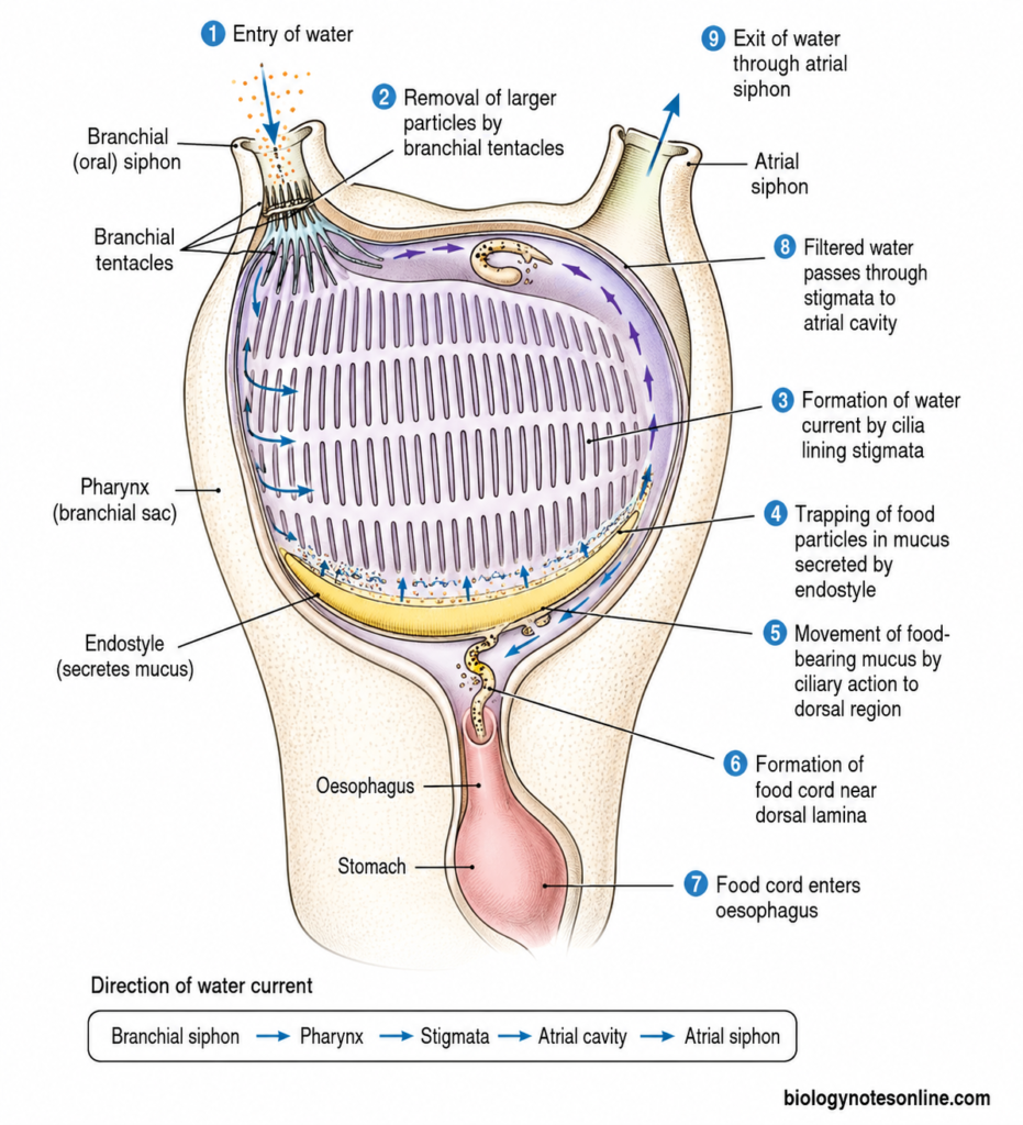
Step 1- Entry of water
Water containing suspended food particles enters through the branchial or oral siphon. From here, the water reaches the large pharynx or branchial sac.
Step 2- Removal of larger particles
The branchial tentacles present near the entrance act as a sieve. Larger particles are prevented from entering the pharynx, while smaller suspended particles pass with water.
Step 3- Formation of water current
The water current is mainly produced by beating of cilia lining the stigmata. Their continuous movement draws water through branchial siphon and across the pharyngeal wall. Thus, feeding in Herdmania is also referred to as ciliary feeding.
Step 4- Trapping of food particles
The endostyle is present along the ventral side of pharynx. It continuously secretes a fine mucous feeding net. Microscopic food particles become trapped in this mucus during filter feeding. This is the major endostyle function during feeding.
Step 5- Movement of food-bearing mucus
The mucus containing trapped food is moved over the inner surface of branchial sac by ciliary action. It gradually reaches the dorsal region of pharynx.
Step 6- Formation of food cord
Near the dorsal lamina, the food-bearing mucus is rolled into a mucus string or food cord. The food cord is then carried towards the oesophageal opening.
Step 7- Entry of food into oesophagus
The food cord now enters the oesophagus. From the oesophagus it is passed towards the stomach for further digestion.
Step 8- Passage of filtered water
The water is not passed into the alimentary canal. It moves through numerous ciliated stigmata present in the wall of branchial sac and enters the atrial or peribranchial cavity.
Step 9- Exit of water
The filtered water collected in the atrial cavity finally passes outside through the atrial siphon. Thus, the direction of water current is branchial siphon → pharynx → stigmata → atrial cavity → atrial siphon.
Respiration and Water Current of Herdmania
Herdmania carries out respiration mainly through the large pharyngeal or branchial surface. The branchial wall is thin and supplied with blood spaces or vessels. Sea water is continuously passed over this region by ciliary activity.
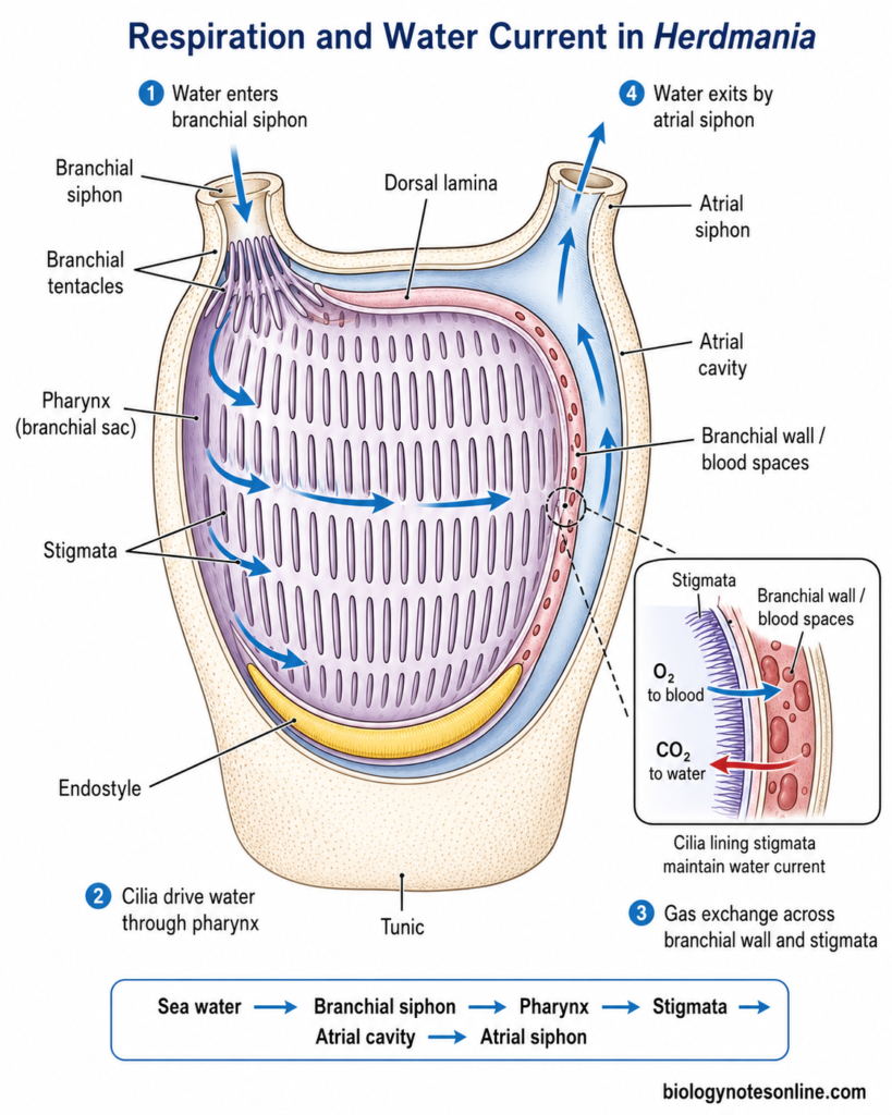
The direction of water current is as follows-
Sea water → Branchial siphon → Pharynx → Stigmata → Atrial cavity → Atrial siphon
- Sea water enters the body through the branchial or oral siphon. The water contains dissolved oxygen along with suspended food particles.
- From the branchial siphon, water passes into the large pharynx or branchial sac. It forms the major filtering as well as respiratory region.
- Numerous small openings called stigmata are present in the pharyngeal wall. The margins of stigmata contain ciliated cells. Continuous beating of these cilia maintains the water current.
- During this process, water passes through the stigmata. Blood flows through spaces in the thin branchial wall and occurs very close to the passing sea water.
- Respiratory exchange takes place mainly across this branchial surface. Oxygen from sea water diffuses towards the blood, while carbon dioxide passes in the opposite direction. The main gas transfer in ascidians is by diffusion.
- The blood of ascidians does not act as a highly specialized oxygen carrier like vertebrate blood. Its respiratory gas-carrying capacity is relatively low. Thus, continuous movement of oxygenated sea water through the branchial basket is important.
- After passing through the stigmata, water enters the atrial or peribranchial cavity. It does not return to the branchial chamber.
- Water collected in the atrial cavity finally passes outside through the atrial siphon. This completes the normal water current through the body.
- The tunic of many solitary ascidians also contains blood vessels. Tunic blood vessels are present in Herdmania momus, where tunic spicules are formed inside them.
- A vascular tunic may provide an additional surface for exchange in some ascidians. However, its respiratory importance is not established equally for all Herdmania species. The pharyngeal or branchial surface should be considered the major respiratory region.
- Thus, the same water current is used for both filter feeding and respiration. Cilia of the stigmata keep this current moving through the branchial basket.
Circulatory System of Herdmania
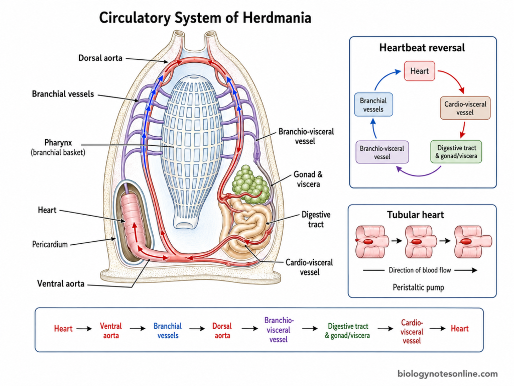
The circulatory system of Herdmania consists of a heart, blood or hemolymph and blood channels. In traditional zoology description, the circulation is referred to as open or lacunar type. The heart is simple and tubular. A characteristic feature is the periodic reversal of heartbeat.
Heart and Pericardium
- The heart of Herdmania is a simple elongated and tubular structure. Tunicate heart does not possess separate chambers like the vertebrate heart.
- It is present in the visceral region of the body. In most solitary ascidians, particularly stolidobranch ascidians, the heart develops towards the right side.
- The heart is enclosed within a pericardial sac or pericardium. Thus, the myocardial tube and its surrounding pericardium together form the heart-pericardial complex.
- The wall of the heart contains a single layer of contractile myocardial cells. It is surrounded externally by the non-muscular pericardial layer.
- The heart is a peristaltic pump. Contraction does not occur at the whole heart at once. A wave of contraction passes along the tubular heart and pushes the blood in one direction.
- Valves like those of vertebrate heart are absent. Movement of blood is mainly produced by the travelling peristaltic contraction of the heart tube.
Blood Vessels, Sinuses and Blood Cells
- Blood from the heart is distributed mainly towards the branchial basket, visceral organs, mantle and other body regions. A part of the circulation may also extend into the tunic.
- The branchial circulation is well developed. Blood passes through vascular channels in the wall of the large branchial basket, close to the numerous stigmata.
- Blood flowing through the branchial region is brought close to sea water passing through the stigmata. This arrangement also provides a large surface associated with gaseous exchange.
- The visceral circulation supplies the stomach, intestine, gonads and other internal organs. Blood then returns towards the heart through the vascular channels and spaces.
- Traditional descriptions of ascidians commonly refer to blood vessels, sinuses and lacunae and therefore describe the circulation as open. Modern observations show that the exact organization of these channels can differ between tunicates. In Corella, for example, much of the blood remains inside definite vessels and ducts.
- The tunic may also contain vascular extensions. In Herdmania momus, tunic blood vessels are definitely present and the small tunic spicules are actually formed inside these vessels before entering the tunic.
- The circulating fluid is generally referred to as blood or hemolymph. It contains plasma and different types of circulating cells called blood corpuscles or hemocytes.
- Different hemocytes occur in ascidians. These include undifferentiated cells, phagocytic cells, morula-type cells, pigment or storage cells and other forms. The exact cell types and terminology are not exactly same among different species.
Reversal of Heartbeat
- Reversal of heartbeat is one of the important characteristics of ascidian circulation. It is also classically described in Herdmania.
- In the beginning, a wave of contraction starts from one end of the tubular heart. The wave then travels along its length.
- During this period, blood is pumped mainly in one direction. Several successive contractions generally follow the same direction.
- After a period, the contractions stop briefly or change their activity. The new peristaltic waves now start from the opposite end of the heart.
- Thus, the direction of contraction becomes reversed. The direction of blood flow also reverses through the branchial and visceral circulation.
- After another period, the direction can change again. In this way, the heart alternately pumps blood towards one side and then towards the opposite side.
- This periodic reversal is not an abnormal condition. It is a characteristic feature of the ascidian heart and is found among different tunicates.
The direction can be represented simply as-
Heart → Branchial circulation → Visceral circulation → Heart
After reversal-
Heart → Visceral circulation → Branchial circulation → Heart
Excretion of Herdmania
Excretion in Herdmania is comparatively simple. A definite vertebrate-like kidney is absent. Nitrogenous wastes are removed or accumulated by different processes.
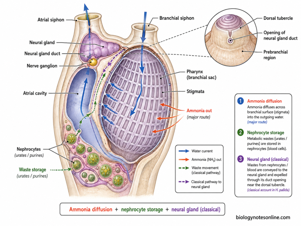
- Herdmania is mainly ammonotelic. Ammonia formed during metabolism can diffuse across the pharyngeal surface and passes outside with the outgoing water. Distinct excretory structures are absent in many tunicates.
- Some waste products are stored inside special blood cells called nephrocytes. These cells accumulate substances such as urates and other purine waste products. This type is referred to as storage excretion.
- In classical zoological description of Herdmania pallida, the neural gland or supraneural gland is also described as an excretory organ. This description is particularly associated with the studies of S. M. Das on H. pallida.
- The neural gland lies in the intersiphonal region, close above the cerebral or nerve ganglion. It is therefore a part of the neural gland complex.
- The gland consists of branching tubules which communicate with a longitudinal canal. Anteriorly, its duct passes towards the prebranchial region and opens near the base of the dorsal tubercle.
- According to the classical account of H. pallida, nephrocytes carrying accumulated waste were considered to be associated with this neural gland pathway and discharge towards the prebranchial region.
- The excretory function of the neural gland should be mentioned with some caution. The function of the ascidian neural gland complex has remained uncertain and several roles have been proposed for it.
- Modern studies on other ascidians, particularly Ciona, indicate that the neural gland complex may have an osmoregulatory and ionic homeostatic function. Endocrine-like functions have also been suggested. Thus, it should not be considered only as a kidney-like organ in all tunicates.
Excretion in Herdmania mainly involves diffusion of ammonia, storage of wastes in nephrocytes, and in the classical description of H. pallida, an excretory role is also assigned to the neural gland.
Nervous System and Sense Organs of Herdmania
The nervous system of adult Herdmania is simple and much reduced as compared to the larval nervous system. It mainly consists of a cerebral ganglion and nerves. The important parts are as follows-
Nervous System
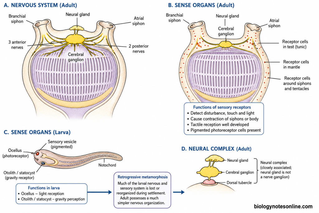
- The adult nervous system is represented mainly by a solid nerve ganglion or cerebral ganglion. It is also commonly referred to as the brain.
- The cerebral ganglion lies in the mantle on the dorsal side, between the branchial and atrial siphons. In the classical description of Herdmania, it lies below the neural gland.
- The ganglion contains nerve cells and nerve fibres. Bipolar and multipolar nerve cells have been described in the classical account of H. pallida.
- Nerves arise from the cerebral ganglion and pass towards the siphons and different parts of body. They take part in movements of the siphons, mantle and other reflex responses.
- In the classical description of H. pallida, three anterior nerves pass towards the branchial siphon and two posterior nerves towards the atrial siphon. This exact number should be considered a species-level classical description, rather than a fixed character for every Herdmania species.
- Adult ascidians also possess peripheral nerves supplying the siphons, body wall and visceral structures. Modern work on other ascidians has shown an extensive nervous supply from the cerebral ganglion to the siphons and peripheral organs.
- The cerebral ganglion, neural gland and dorsal tubercle are commonly grouped together as the neural complex. The neural gland is closely associated with the nervous system, but it is not simply another nerve ganglion.
Sense Organs
- Highly developed special sense organs are generally absent in the adult Herdmania. Different sensory receptor cells are present instead.
- Receptor cells occur in the test, mantle and siphonal regions. Classical studies of H. pallida demonstrated nerve cells, nerve fibres and receptor cells even in the test.
- Sensory cells around the siphons and tentacles respond to external stimulation. These regions help the animal to detect disturbance and produce contraction of the siphons or body.
- Tactile reception is well developed around exposed regions of the adult. Contact or mechanical stimulation can cause rapid withdrawal and closing of the siphons.
- Pigmented receptor cells associated with the test, mantle and siphonal region have also been described in ascidians. S. M. Das reported red pigmented cells acting as photoreceptors in tunicates.
- The dorsal tubercle has also been traditionally assigned a sensory role. Its exact function is not completely settled and it should not simply be described as a definite olfactory organ. The associated neural gland complex has been given several functions in different studies.
- The larval sensory system is much more prominent than that of adult. The ascidian tadpole larva possesses a sensory vesicle associated with pigmented sensory organs.
- In ascidian larvae, the ocellus is associated with light reception, while the otolith or statocyst-like organ is associated with gravity perception. These help in swimming and settlement behaviour.
- During retrogressive metamorphosis, much of the larval nervous and sensory system is lost or reorganized. The adult therefore possesses a much simpler nervous organization.
Reproductive System
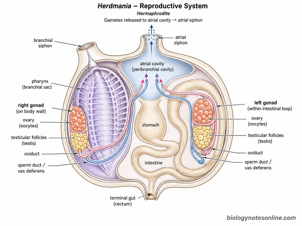
Herdmania is a hermaphrodite or bisexual ascidian. Both male and female reproductive organs are present in the same animal. In H. momus and H. pallida, a gonad is present on each side of the body. The detailed structure of gonads is not same in all species of Herdmania. In fact, gonadal characters are important for identification of species.
The following are the important parts of reproductive system-
- Gonads – Two elongated gonads, one on either side, are present in the commonly studied H. momus. The left gonad lies within the intestinal loop, while the right gonad is present on the right side of body wall. A similar one-gonad-per-side condition is also described in H. pallida.
- Ovary – Each gonad contains the female reproductive region or ovary. Developing ova or oocytes are present within it. The size and appearance of ovary change with the stage of reproductive maturity in H. momus.
- Testis – The male part is formed of testicular follicles associated with the ovarian region. These follicles produce spermatozoa. Small ducts from the testicular follicles finally communicate with the main sperm duct.
- Oviduct – The ovary is connected with an oviduct. It carries mature ova from the gonad towards the atrial region. The terminal structure and opening of the oviduct differ considerably between different Herdmania species.
- Sperm duct or vas deferens – The testicular part is connected with a sperm duct. In H. momus, sperm duct and oviduct are separate and run near each other towards the atrial cavity.
- Gonoduct openings – The oviduct and sperm duct open into the atrial or peribranchial cavity. In H. momus, their openings are present near the terminal region of the gut. Mature gametes are discharged into this cavity.
- The gametes finally pass outside through the atrial siphon with the outgoing water. Solitary ascidians generally release eggs and sperm into surrounding sea water.
- Fertilization is external in the commonly studied solitary ascidian condition. Development of H. momus from fertilized eggs has also been studied experimentally in sea water.
- The reproductive condition can change with season and geographical population. For example, H. momus populations from the Red Sea and Mediterranean show differences in reproductive period and gonadal activity.
- The exact number of gonadal lobes, arrangement of testicular follicles, form of sperm duct and structure around the oviduct opening should not be considered same for the whole genus. These are species-specific characters and are widely used in the taxonomy of Herdmania.
Reproduction Process of Herdmania
Herdmania reproduces mainly by sexual reproduction. The adult is hermaphroditic, having both male and female reproductive organs in the same individual. As it is a solitary ascidian, asexual budding is not the normal mode of reproduction. The reproduction process is as follows-
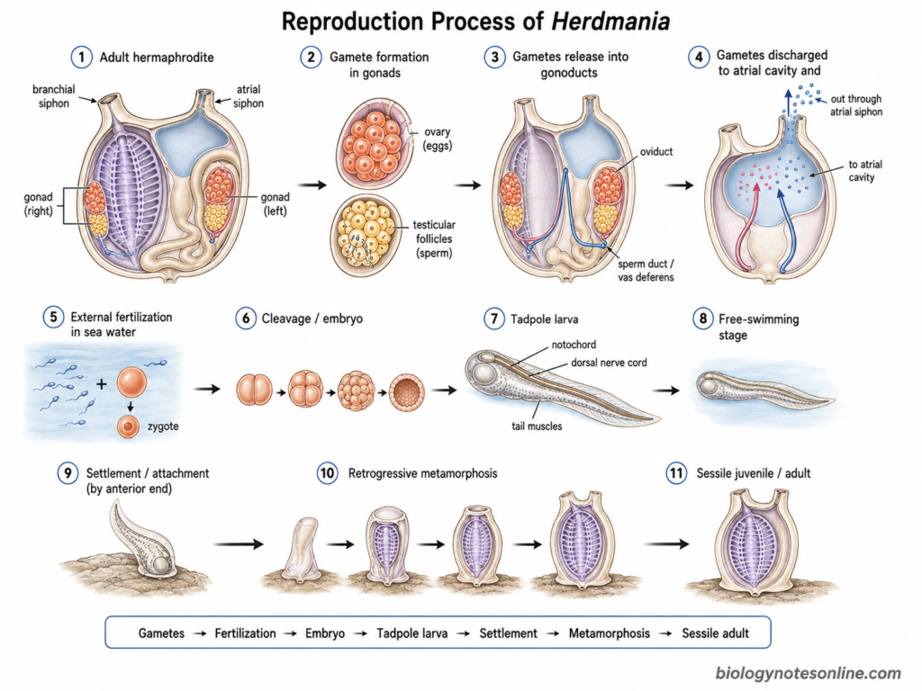
- Formation of gametes – The ovary produces ova or eggs, while the testicular follicles produce spermatozoa. Both reproductive parts occur in the same adult animal.
- Release into gonoducts – Mature ova enter the oviduct and spermatozoa pass into the sperm duct or vas deferens. The exact form of these ducts may vary between different Herdmania species. Gonadal structure itself is an important taxonomic character in the genus.
- Discharge of gametes – The reproductive ducts open towards the atrial cavity. Gametes are passed into this cavity and finally released outside with the outgoing water through the atrial siphon.
- Maturation of egg – The eggs become ready for fertilization after their release into sea water. In Herdmania pallida, oocyte maturation or germinal vesicle breakdown begins when the oocyte comes in contact with sea water. This observation is specifically demonstrated for H. pallida.
- Fertilization – Fertilization takes place externally in sea water, as occurs in solitary ascidians. The sperm fuses with the egg and a zygote is formed. Hermaphroditic condition does not necessarily mean that self-fertilization always occurs, because self-fertility differs among ascidian species.
- Cleavage and embryonic development – After fertilization, the zygote undergoes a series of cleavage divisions. The embryo gradually develops the characteristic ascidian body organization. The fertilized egg finally forms a tadpole-type larva.
- Hatching of larva – A free-swimming ascidian tadpole larva hatches from the egg. Development from fertilized eggs to hatching larvae has been directly studied in Herdmania momus.
- Free-swimming stage – The larva swims freely for a short period. It possesses a tail with notochord, dorsal nerve cord and muscles, showing prominent chordate characters.
- Settlement – After the swimming period, the larva selects a suitable substratum and becomes attached by its anterior region. In H. momus, settlement and larval attachment have been experimentally observed.
- Metamorphosis – After attachment, retrogressive metamorphosis takes place. The larval tail is resorbed and the notochord and much of the larval locomotory system disappear. Adult structures now develop further. H. momus larvae become competent for metamorphosis after hatching and undergo the typical ascidian transformation.
- The newly formed juvenile becomes sessile and develops gradually into the filter-feeding adult. Thus, the life cycle changes from a motile chordate-like larva into an attached adult ascidian.
The process can be represented as-
Gamete formation → Gamete release → External fertilization → Zygote → Cleavage → Embryo → Tadpole larva → Settlement → Retrogressive metamorphosis → Sessile adult
Development and Tadpole Larva of Herdmania
Development in Herdmania results in the formation of a free-swimming ascidian tadpole larva. The larva is quite different from the sessile adult and shows the chordate organization more clearly. Development of H. momus from egg to larval settlement has also been studied experimentally.
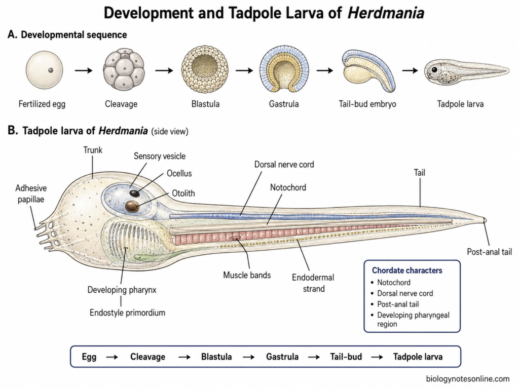
Early Development
- Development starts from the fertilized egg or zygote. After fertilization, repeated cleavage divisions are started.
- The cleavage is holoblastic and follows the characteristic ascidian pattern. The fertilized egg is divided into smaller blastomeres without increase in the total size of embryo.
- Continued cleavage results in the formation of the blastula stage. Different groups of blastomeres are now destined to form epidermis, nervous system, notochord, muscles, endoderm and other larval tissues.
- The blastula is followed by gastrulation. During this process the germ layers are rearranged and the basic body organization is established. Gastrulation in ascidians takes place when the embryo still contains relatively few cells.
- After gastrulation, neurulation and tail formation take place. The notochord becomes elongated, the neural tube develops and muscle cells are arranged on the sides of tail.
- The embryo gradually changes into a tail-bud stage and finally forms the tadpole larva. Thus, the main developmental sequence is-
Fertilized egg → Cleavage → Blastula → Gastrula → Tail-bud embryo → Tadpole larva
Structure of the Ascidian Tadpole Larva
- The larva of Herdmania is a small tadpole-like and free-swimming form. The body can be divided into a broad trunk and a long muscular tail.
- The trunk forms the anterior region. It contains the sensory and neural structures together with rudiments of different adult organs.
- Adhesive papillae are present at the anterior end of larva. They are used during attachment with a suitable substratum. In H. momus, the anterior papillary region is also important for initiation of settlement and metamorphosis.
- A large cerebral or sensory vesicle is present in the trunk. It forms the anterior expanded part of the larval nervous system.
- The sensory vesicle contains pigmented sensory structures. The ocellus is associated with reception of light, while the otolith has a role in gravity perception and orientation. The term statocyst is also commonly used in older zoology description for this gravity-sensing system.
- From the nervous region, a dorsal tubular nerve cord extends backwards into the tail. It lies dorsal to the notochord.
- The notochord forms the axial supporting structure of the tail. It gives rigidity to the larval tail and is one of the most important chordate characters.
- Bands of striated muscle cells occur on the lateral sides of notochord. Their contractions produce the swimming movement of the larva.
- The tail is therefore mainly formed of the notochord, dorsal nerve cord, muscle bands and endodermal strand, covered externally by epidermis and tunic.
- The trunk also contains a developing pharyngeal region, endostyle primordium and rudiments of the future digestive and branchial structures. These structures are not yet developed like those of the filter-feeding adult.
Chordate Characters of the Larva
The larval stage is important for showing the chordate nature of Herdmania. The major chordate characters are as follows-
- Notochord – A well-developed notochord is present in the larval tail. It forms the main axial supporting structure. During metamorphosis, the notochord is resorbed and is not retained in the adult.
- Dorsal hollow nerve cord – A dorsal neural tube or nerve cord is present above the notochord. Anteriorly it becomes enlarged into the cerebral or sensory vesicle.
- Pharyngeal slits – The pharyngeal apparatus is a chordate character of Herdmania. In the early swimming larva, the pharynx and branchial structures are mainly present as developing rudiments. The numerous functional stigmata or pharyngeal gill slits become conspicuous during post-larval development and in the adult branchial basket. Thus, it is not correct to describe a newly hatched ascidian tadpole as having the same extensive perforated pharynx as the adult.
- Post-anal tail – A long tail extends behind the trunk and contains the notochord, nerve cord and muscle cells. It is used for swimming during the short larval period.
These characters establish Herdmania as a chordate, although it is a non-vertebrate animal. The notochord, larval nerve cord and muscular tail are reduced or lost during metamorphosis. The pharyngeal apparatus, on the other hand, becomes highly developed in the sessile filter-feeding adult. This is the reason the chordate nature of Herdmania is particularly clear from its developmental history.
Retrogressive Metamorphosis in Herdmania
Retrogressive metamorphosis is the transformation of the free-swimming ascidian tadpole larva into a sessile adult. During this process, several important larval chordate characters are lost. At the same time, the structures required for the adult filter-feeding life are developed. This type of metamorphosis is characteristic of ascidians.
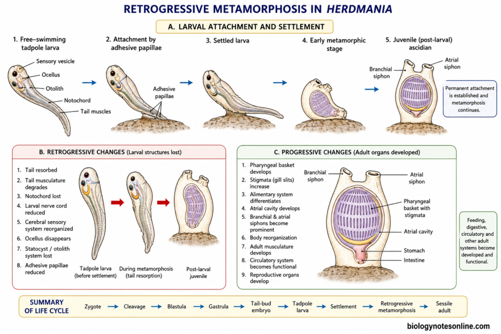
Larval Attachment
- The tadpole larva of Herdmania remains free-swimming for a short period. This stage mainly helps in dispersal and finding a suitable place for settlement.
- After attaining competence, the larva starts searching for a suitable substratum. Settlement behaviour occurs before the actual transformation of the body.
- The anterior end of larva bears adhesive papillae. These structures come in contact with the substratum and help in attachment. The papillae also have sensory and secretory functions in ascidians.
- In Herdmania momus, the anterior papillary region has an important role in starting metamorphosis. Experimental separation of this region prevents normal metamorphic changes in the remaining larval body.
- After attachment, the larva stops its free-swimming mode of life. The sessile phase is now started and metamorphic changes take place.
- Permanent attachment structures are gradually formed after settlement. The temporary larval adhesive structures are later reduced as the juvenile becomes fixed to the substratum.
Retrogressive Changes
The major retrogressive changes are as follows-
- Tail – The long larval tail is withdrawn and resorbed. Tail regression is one of the first conspicuous changes during ascidian metamorphosis. In H. momus, tail resorption is also used as an indication of successful settlement and metamorphosis.
- Tail musculature – The striated muscles which produce larval swimming are no longer required. The muscle fibres are degraded during metamorphosis. In H. momus, programmed degradation of larval myofibrils occurs during normal metamorphosis.
- Notochord – The larval notochord is completely lost. The notochord and other internal components of tail are withdrawn into the body and undergo degeneration. There is no evidence that the larval notochord is retained in the adult ascidian.
- Larval nerve cord – Much of the dorsal larval nervous system is reduced during metamorphosis. The elaborate nervous organization required for swimming is not retained in the same form in adult.
- Cerebral sensory system – The large larval cerebral or sensory vesicle undergoes extensive reorganization. Much of the larval sensory apparatus is lost with the change to sessile life.
- Ocellus – The larval ocellus, which is concerned with light reception, disappears during metamorphosis. A similar conspicuous larval visual organ is absent in the adult.
- Statocyst or otolith system – The larval gravity-sensing structure is also lost. Classical zoology commonly refers to it as a statocyst, while modern ascidian literature commonly describes the pigmented otolith within the sensory vesicle.
- Adhesive papillae – These larval structures function during substrate selection and initial attachment. After settlement their larval function ends and they are reduced or reorganized.
Thus, the important larval structures concerned mainly with swimming, orientation and dispersal become unnecessary after attachment and are lost.
Progressive Changes
Retrogressive metamorphosis does not mean that the complete animal undergoes degeneration. Several adult organs are formed, enlarged or reorganized during the same process. Adult organ formation is therefore an important progressive part of ascidian metamorphosis.
The major progressive changes are as follows-
- Pharyngeal basket – The pharyngeal region increases greatly and develops into the large branchial basket characteristic of adult ascidians. It becomes an important organ for filter feeding and respiration.
- Stigmata – Functional pharyngeal stigmata or gill slits develop and their number increases during juvenile growth. Water can now pass through the pharyngeal wall into the atrial region.
- Alimentary system – The digestive organs, which are not functional like those of adult during the swimming larval stage, become differentiated. The oesophagus, stomach and intestine develop as the animal changes towards active feeding.
- Atrial cavity – The atrial or peribranchial system becomes developed around the pharyngeal region. It receives water passing through the stigmata and communicates with the atrial siphon.
- Adult siphons – The branchial and atrial siphons become prominent. In normal post-larval development of H. momus, the branchial basket and siphons become clearly recognizable as adult morphology develops.
- Body orientation – The body undergoes considerable rearrangement after settlement. Visceral structures change their relative positions and the adult sessile body form is established.
- Adult musculature – New body-wall and siphonal muscles develop. These muscles are different in function from the larval tail muscles and are used for contraction of the adult body and siphons.
- Circulatory system – The adult heart, blood cells and circulatory arrangement develop and become functional during post-larval development. Heart precursor cells are already specified earlier, but the functional adult circulation is established with juvenile development.
- Reproductive organs – Gonadal rudiments and reproductive structures develop during post-larval growth. The gonads do not become fully functional immediately after tail loss. Sexual maturity is reached later as the juvenile grows into adult.
- Thus, larval locomotory structures are lost but feeding, digestive, circulatory and other adult systems become more developed. This is an important part of metamorphosis and should not be ignored.
Larva vs Adult
| Feature | Tadpole larva | Adult |
|---|---|---|
| Lifestyle | Free-swimming | Sessile |
| Notochord | Present in tail | Absent |
| Dorsal nerve cord | Well developed | Greatly reduced and reorganized |
| Tail | Present | Absent |
| Ocellus/statocyst | Present | Lost |
| Pharynx | Relatively less developed | Enlarged branchial basket |
| Feeding | Larval stage largely dispersive and non-feeding | Active filter feeder |
| Siphons | Developing or rudimentary | Prominent |
| Gonads | Undeveloped | Functional in sexually mature adult |
The tadpole larva mainly represents the dispersal stage, while the adult body is adapted for attachment, filter feeding and reproduction.
Why is it Called Retrogressive Metamorphosis?
It is called retrogressive metamorphosis because the active tadpole larva possesses conspicuous chordate locomotory and sensory structures, such as the notochord, dorsal nerve cord, muscular tail, ocellus and gravity-sensing organ, which are reduced or lost when the larva becomes a sessile adult.
However, the process is not complete degeneration. During the same period, the branchial basket, digestive system, siphons, circulation and other adult organs develop. Therefore, “retrogressive” mainly refers to the loss of prominent larval chordate and locomotory characters, not to the whole metamorphic process.
Affinities and Evolutionary Significance of Herdmania
Herdmania is an ascidian tunicate and shows clear affinities with other chordates. These characters are more prominent during the tadpole larval stage. The adult becomes sessile and several larval chordate structures are reduced during metamorphosis.
Affinities with Chordata
- Notochord – The tadpole larva possesses a well-developed notochord in the tail. It forms the axial supporting structure of larva and is one of the main chordate characters.
- Dorsal nerve cord – A dorsal tubular nerve cord occurs above the notochord. Anteriorly it becomes enlarged into the sensory or cerebral vesicle.
- Post-anal tail – The larva has a muscular post-anal tail. It is used for swimming during the short free-living stage.
- Pharyngeal apparatus – The pharyngeal region is another important chordate character. Rudimentary pharyngeal slits occur during development, while the adult develops a very large branchial pharynx with numerous stigmata.
- Endostyle – An endostyle is present along the ventral side of pharynx. It secretes mucus during filter feeding. The chordate endostyle is considered homologous with the vertebrate thyroid gland, supported by iodine-related functions and common developmental gene systems.
- The notochord, dorsal tubular nerve cord, pharyngeal apparatus and post-anal tail clearly establish the chordate nature of Herdmania. Most of these are much easier to recognize in larva than adult.
Affinities with Vertebrates
- Modern molecular studies place Tunicata as the sister group of Vertebrata. Tunicates and vertebrates together form the clade generally called Olfactores. Cephalochordates separated earlier from this lineage.
- Thus, Herdmania belongs to the tunicate lineage which represents the closest living invertebrate relatives of vertebrates. This does not mean that Herdmania itself is a direct ancestor of vertebrates.
- Ascidian embryos show several conserved developmental mechanisms with vertebrates. For example, Brachyury has an important role in notochord development across chordates. Most molecular experiments for these comparisons are from model ascidians such as Ciona, and not specifically from Herdmania.
- The relationship between endostyle and thyroid is another important affinity. Similar molecular regulators and iodine-related functions are present in these structures.
- The sessile adult condition should not be considered as the ancestral chordate condition. Modern evolutionary studies indicate that the highly modified adult ascidian body is a derived condition.
Evolutionary Significance of Herdmania
- Herdmania is important as a non-vertebrate chordate. Its larva retains the basic structural plan from which the chordate relationship can be clearly understood.
- The tadpole larva shows that a simple marine animal can possess a notochord, dorsal nerve cord and post-anal tail, although these are later reduced in adult.
- Retrogressive metamorphosis is particularly important. The swimming and sensory structures of larva disappear, while structures for sessile filter feeding become highly developed.
- This shows that evolution does not always involve an increase in structural complexity in every life stage. In ascidians, the adult body became specialized for a fixed and filter-feeding mode of life.
- Tunicates provide important material for studying the origin of chordate body plan and vertebrate characters. Development of notochord, nervous system, pharyngeal region and endostyle are particularly useful for such comparisons.
- Modern phylogeny gives tunicates special importance because they are more closely related to vertebrates than cephalochordates are. This position was different from the older morphology-based view in which amphioxus was commonly considered the closest vertebrate relative.
- Herdmania should therefore not be described as a “connecting link” or direct ancestor of vertebrates. It is a living, specialized ascidian belonging to the tunicate branch of chordates.
- Information obtained from Ciona and other model tunicates is useful for understanding the evolutionary position of Herdmania, but such molecular observations should not be presented as direct experimental findings from Herdmania unless specifically studied in this genus.
Biological and Ecological Importance of Herdmania
Some of the important biological and ecological importance of Herdmania are as follows-
- Chordate Study – Herdmania is important for the study of chordate organization. Its tadpole larva possesses notochord, dorsal nerve cord and muscular tail.
- Metamorphosis – It is used for understanding retrogressive metamorphosis. During this process, several larval chordate characters are lost and the sessile adult is formed.
- Filter Feeding – Herdmania is an important filter feeder of marine ecosystem. It removes phytoplankton, bacteria and other small suspended particles from sea water.
- Benthic Transfer – Filter feeding helps in transfer of organic matter from the water column to benthic region. In this way, ascidians take part in pelagic-benthic coupling.
- Nutrient Cycling – Herdmania takes part in nutrient turnover through feeding, metabolism and release of waste materials. Studies on H. momus have shown turnover of carbon, nitrogen and phosphorus.
- Benthic Community – The members form a part of marine benthic communities. They occur on rocks, reefs and other submerged surfaces.
- Biofouling – Some species can grow abundantly over artificial marine surfaces. H. momus is known from floating docks and other submerged structures, and can form part of fouling communities.
- Marine Invasion – H. momus is important in the study of marine biological invasion. It has spread from the Red Sea region into the Mediterranean Sea through the Suez Canal.
- Biomineralization – The calcareous spicules of H. momus are useful for studying biomineralization. These are hard mineral structures formed inside a soft-bodied animal.
- Vaterite Study – Spicules of H. momus contain vaterite, a form of calcium carbonate. Because of this, the species is also studied for formation and structure of biological calcium carbonate.
- Chordate Evolution – Herdmania has importance in understanding the evolution of chordates. Tunicates are the closest living invertebrate relatives of vertebrates, although Herdmania itself should not be considered a direct vertebrate ancestor.
- Larval Biology – The tadpole larva is useful for studying settlement and larval development. Experimental studies on H. momus have also examined larval attachment and beginning of metamorphosis.
Identification and Spotting Characters of Herdmania
Herdmania can be identified as a solitary marine ascidian or sea squirt. The following are the important spotting characters-
- Solitary Body – The animal is a solitary ascidian. Individuals do not form a common colonial body.
- Marine Habit – Herdmania is exclusively marine. The adult remains attached to rocks or other submerged substratum.
- Sessile Adult – The adult is sessile and fixed with the substratum. Only the tadpole larva is free-swimming for a short period.
- Body Form – The body is generally sac-like, oval or oblong. Exact shape and size may vary among different species.
- Tunic – The body is externally covered by a test or tunic. It is generally leathery in appearance, but thickness and toughness are not same in all Herdmania species.
- Two Siphons – Two prominent openings or siphons are present. These are the branchial siphon and atrial siphon.
- Branchial Siphon – It forms the incurrent aperture. Sea water enters through this siphon and reaches the pharynx.
- Atrial Siphon – It forms the excurrent aperture. Filtered water is passed outside through this opening.
- Body Colour – Colour can be useful during identification of a particular species. It should not be taken as one fixed character of the whole genus. H. momus, for example, may show characteristic red-pink and white bands on the siphons.
- Spicules – Calcareous spicules are an important character of Herdmania. Spicules occur in the body tissues and their form has taxonomic importance. Exact size and arrangement vary according to species.
- Branchial Basket – On internal examination, a very large pharynx or branchial basket occupies much of the body. Its wall contains numerous ciliated stigmata.
- Tadpole Larva – Developmental material shows an ascidian tadpole larva. It possesses a trunk and muscular tail and represents the free-swimming stage before settlement.
Practical Spotting Table
| Spotting Character | Identification |
|---|---|
| Habit | Solitary, marine and sessile ascidian |
| Body | Sac-like or oblong |
| Covering | Tunic or test present |
| Openings | Two siphons |
| Branchial siphon | Incurrent |
| Atrial siphon | Excurrent |
| Internal character | Large perforated branchial basket |
| Special character | Calcareous spicules |
| Larva | Ascidian tadpole larva |
Spotting identification: The given specimen can be identified as Herdmania due to its solitary ascidian body, tunic covering, two siphons, sessile marine habit and large branchial basket. Calcareous spicules provide another important character of the genus.
Branchial Siphon vs Atrial Siphon of Herdmania
| Character | Branchial Siphon | Atrial Siphon |
|---|---|---|
| Other name | Oral siphon | Cloacal siphon |
| Position | Present towards the anterior region | Present towards the dorsal region |
| Opening | Branchial or oral aperture | Atrial aperture |
| Nature | Incurrent siphon | Excurrent siphon |
| Water flow | Sea water enters through it | Water passes outside through it |
| Connection | Opens into the pharynx or branchial sac | Opens from the atrial or peribranchial cavity |
| Food particles | Water carrying food particles enters through it | Food particles normally do not pass out through it |
| Respiration | Brings oxygenated sea water into the body | Removes water after it passes through the stigmata |
| Waste removal | Not mainly concerned with removal of wastes | Faecal matter and other wastes pass outside with water |
| Gametes | Gametes are not normally discharged through it | Gametes pass outside through this siphon |
| Main function | Entry of sea water for filter feeding and respiration | Exit of filtered water, wastes and gametes |
The direction of water current is-
Sea water → Branchial siphon → Pharynx → Stigmata → Atrial cavity → Atrial siphon
Functions of Major Structures of Herdmania
| Structure | Major Function |
|---|---|
| Tunic or Test | Protects the soft body and also helps in attachment with the substratum. |
| Mantle | Forms the muscular body wall and helps in contraction of the body and siphons. |
| Branchial Siphon | Takes sea water into the pharynx for feeding and respiration. |
| Atrial Siphon | Passes filtered water, wastes and gametes outside. |
| Branchial Basket or Pharynx | Performs filter feeding and forms the major surface associated with respiration. |
| Stigmata | Allow water to pass from the pharynx into the atrial cavity. |
| Cilia | Produce the continuous water current through the branchial basket. |
| Endostyle | Secretes mucus which traps microscopic food particles. |
| Dorsal Lamina | Helps in carrying the food-bearing mucus towards the oesophagus. |
| Branchial Tentacles | Prevent larger particles from entering the pharyngeal region. |
| Oesophagus | Carries food from the pharynx towards the stomach. |
| Stomach | Takes part in digestion of food. |
| Intestine | Helps in further digestion and absorption of digested food. |
| Atrial Cavity | Receives water passing through the stigmata and directs it towards the atrial siphon. |
| Heart | Pumps blood through different regions of the body. |
| Cerebral Ganglion | Controls nervous activity and supplies nerves to the body and siphons. |
| Neural Gland | Has been traditionally associated with excretory and other physiological functions. |
| Gonads | Produce male and female gametes. |
| Calcareous Spicules | Provide structural support to different body tissues. |
| Adhesive Papillae | Help the tadpole larva in attachment with a suitable substratum. |
| Notochord | Provides axial support to the larval tail during swimming. |
| Larval Tail | Helps in locomotion during the free-swimming tadpole stage. |
Retrogressive vs Progressive Changes During Metamorphosis
During metamorphosis of ascidians, degeneration of larval structures and development of adult structures occur together. In Herdmania momus, tail resorption occurs together with development of adult structures such as the branchial basket and siphons.
| Structure/Feature | Retrogressive Changes | Progressive Changes |
|---|---|---|
| Tail | Larval tail is withdrawn and resorbed. | Sessile adult body form is established. |
| Tail Muscles | Larval striated muscles degenerate. | Adult body-wall and siphonal muscles develop. |
| Notochord | Notochord is completely lost during tail resorption. | No notochord is formed in the adult. |
| Nerve Cord | Much of the larval dorsal nerve cord is reduced or reorganized. | Adult cerebral ganglion and peripheral nerves become established. |
| Sensory Organs | Larval ocellus and otolith/statocyst are lost. | Adult develops a simpler sensory organization. |
| Adhesive Papillae | Larval adhesive papillae lose their function after settlement. | Permanent attachment with the substratum develops. |
| Pharynx | Larval pharyngeal region is relatively less developed. | Branchial basket becomes greatly enlarged. |
| Stigmata | Few or immature during early development. | Functional stigmata develop and increase during juvenile growth. |
| Digestive System | Larval stage is mainly dispersive and does not possess the functional adult feeding system. | Oesophagus, stomach and intestine become functional for feeding. |
| Atrial Cavity | Not developed like the adult condition. | Atrial or peribranchial cavity becomes well developed. |
| Siphons | Adult siphons are not prominent in the swimming larva. | Branchial and atrial siphons develop and become prominent. |
| Circulation | Larval circulatory condition is replaced during reorganization. | Adult heart and circulatory arrangement become functional. |
| Body Form | Free-swimming chordate-like larval form disappears. | Sac-like and sessile adult body is formed. |
The retrogressive changes mainly involve loss of structures required for larval swimming and sensory activity. The progressive changes include development of structures required for attachment, filter feeding and adult life. Thus, retrogressive metamorphosis does not mean degeneration of the complete animal. Adult organs develop at the same time.
Frequently Asked Questions (FAQs) about Herdmania
1. What is Herdmania?
Herdmania is a genus of solitary marine ascidians or sea squirts. It belongs to phylum Chordata, subphylum Tunicata and class Ascidiacea. The adult is sessile and filter feeding, while its tadpole larva shows prominent chordate characters.
2. What is the common name of Herdmania?
Herdmania is commonly referred to as a sea squirt. It is an ascidian tunicate. The name sea squirt is commonly used for members of class Ascidiacea.
3. Why is Herdmania called a sea squirt?
It is called a sea squirt because the muscular body and siphons contract when the animal is disturbed. During this contraction, water present inside the body is suddenly expelled through the siphonal openings.
4. Is Herdmania a chordate?
Yes. Herdmania belongs to Chordata. The chordate characters are more clearly present in its tadpole larva, which has a notochord, dorsal tubular nerve cord, pharyngeal structures and post-anal tail.
5. Is Herdmania a vertebrate or invertebrate chordate?
Herdmania is an invertebrate chordate. It belongs to Tunicata and does not possess a vertebral column or cranium. Its larval stage, however, shows the basic chordate body plan.
6. Which subphylum does Herdmania belong to?
Herdmania belongs to the subphylum Tunicata. Older zoology textbooks commonly use the name Urochordata for the same group. Modern WoRMS accepts Tunicata as the valid name.
7. Is Urochordata the same as Tunicata?
Yes. Urochordata is an older and still commonly used name for Tunicata. In current WoRMS classification, Urochordata is treated as an unaccepted junior synonym of Tunicata, not as a separate subphylum.
8. Is Herdmania solitary or colonial?
Herdmania is a solitary ascidian. Each adult forms an independent individual and does not produce a common colonial body. Taxonomic studies describe Herdmania as a pyurid solitary ascidian genus.
9. What is the tunic of Herdmania made of?
The tunic or test is an extracellular covering present outside the mantle. Like other tunicates, its matrix contains a large amount of cellulose, traditionally associated with the term tunicin. Tunicate cellulose is an unusual character among animals.
10. What are the two siphons of Herdmania?
Two siphons are present, the branchial or oral siphon and the atrial siphon. The branchial siphon takes sea water inside, while the atrial siphon passes filtered water and other discharged materials outside.
11. What is the function of the endostyle in Herdmania?
The endostyle is a glandular groove present along the ventral side of pharynx. It secretes mucus. Small suspended food particles become trapped in this mucus and are then carried towards the digestive tract.
12. How does Herdmania obtain food?
Herdmania is a filter feeder or ciliary feeder. Sea water enters through the branchial siphon. Microscopic food particles are trapped in mucus produced by the endostyle, while filtered water passes through the stigmata into the atrial cavity.
13. How does respiration occur in Herdmania?
Respiration takes place mainly across the large branchial or pharyngeal surface. Water continuously passes through the pharynx and its stigmata. Gaseous exchange takes place between the passing sea water and blood present in the branchial vascular system.
14. Why does the heart of Herdmania reverse its beating?
The ascidian heart is a tubular peristaltic heart. Its contraction waves move in one direction for a period and later start from the opposite end. Thus, the direction of blood flow also reverses. The exact physiological advantage of this reversal is still not completely established.
15. What is the excretory organ of Herdmania?
A vertebrate-like kidney is absent. Classical descriptions of H. pallida assign an excretory role to the neural or supraneural gland and nephrocytes. Its function should be mentioned cautiously, because modern studies suggest that the ascidian neural gland complex may also take part in ionic or osmotic regulation.
16. Is Herdmania hermaphrodite?
Yes. Commonly studied Herdmania species are hermaphroditic, with male and female reproductive tissues present in the same individual. The detailed arrangement of the gonads and gonoducts varies between species and forms an important taxonomic character.
17. What is the larval form of Herdmania?
The larval form is an ascidian tadpole larva. It is a short-lived, free-swimming stage having an anterior trunk and muscular tail. The larval stage mainly helps in dispersal and selection of a suitable substratum.
18. Which chordate characters are present in the tadpole larva?
The larva shows a notochord, dorsal tubular nerve cord, post-anal muscular tail and developing pharyngeal apparatus. These characters clearly show why Herdmania is placed under Chordata. Several of them are later lost during metamorphosis.
19. What is retrogressive metamorphosis in Herdmania?
Retrogressive metamorphosis is the change of the active tadpole larva into a sessile adult. The tail, notochord, larval muscles and much of larval nervous system are lost. At the same time, adult structures such as the branchial basket and digestive organs develop.
20. What is the difference between Herdmania momus and Herdmania pallida?
H. momus and H. pallida are two separate species of Herdmania. They differ in several taxonomic characters, particularly gonadal structure, gonoducts, body-wall muscles, test characters and branchial structures. Therefore, measurements and fine anatomical characters of one species should not be directly applied to the other.
References
- Gilbert, S. F. (2000). Developmental biology (6th ed.). Sinauer Associates.
- Kotpal, R. L. (2010). Modern text book of zoology: Vertebrates. Rastogi Publications.
- Vanni, V., Ballarin, L., Gasparini, F., Peronato, A., & Manni, L. (2022). Studying regeneration in ascidians: An historical overview. In S. Blanchoud & B. Galliot (Eds.), Whole-body regeneration: Methods and protocols (Methods in Molecular Biology, Vol. 2450, pp. 27–48). Humana. https://doi.org/10.1007/978-1-0716-2172-1_2
- Young, C. M., Swalla, B. J., & Cloney, R. A. (2025). Phylum Chordata: Tunicata. In M. J. Boyle, C. M. Young, & M. A. Sewell (Eds.), Atlas of marine invertebrate larvae (2nd ed., pp. 755–791). Academic Press. https://doi.org/10.1016/B978-0-08-102871-1.00027-0
- Anderson, H. E., & Christiaen, L. (2016). Ciona as a simple chordate model for heart development and regeneration. Journal of Cardiovascular Development and Disease, 3(3), 25. https://doi.org/10.3390/jcdd3030025
- Baldwin, D., McCabe, M., & Thomas, F. (1984). The respiratory gas carrying capacity of ascidian blood. Comparative Biochemistry and Physiology Part A: Physiology, 79(3), 479–482. https://doi.org/10.1016/0300-9629(84)90549-8
- Bingham, B. L., & Walters, L. J. (1989). Solitary ascidians as predators of invertebrate larvae: Evidence from gut analyses and plankton samples. Journal of Experimental Marine Biology and Ecology, 131(2), 147–159. https://doi.org/10.1016/0022-0981(89)90004-X
- Bone, Q., Carré, C., & Chang, P. (2003). Tunicate feeding filters. Journal of the Marine Biological Association of the United Kingdom, 83(5), 907–919. https://doi.org/10.1017/S002531540300804Xh
- Braun, K., Leubner, F., & Stach, T. (2020). Phylogenetic analysis of phenotypic characters of Tunicata supports basal Appendicularia and monophyletic Ascidiacea. Cladistics, 36(3), 259–300. https://doi.org/10.1111/cla.12405
- Cahill, P. L., Atalah, J., Selwood, A. I., & Kuhajek, J. M. (2016). Metamorphosis of the invasive ascidian Ciona savignyi: Environmental variables and chemical exposure. PeerJ, 4, e1739. https://doi.org/10.7717/peerj.1739
- Cameron, C. B., Garey, J. R., & Swalla, B. J. (2000). Evolution of the chordate body plan: New insights from phylogenetic analyses of deuterostome phyla. Proceedings of the National Academy of Sciences of the United States of America, 97(9), 4469–4474. https://doi.org/10.1073/pnas.97.9.4469
- Cima, F., Ballarin, L., Gasparini, F., & Burighel, P. (2006). External amebocytes guard the pharynx entry in a tunicate (Ascidiacea). Developmental & Comparative Immunology, 30(5), 463–472. https://doi.org/10.1016/j.dci.2005.07.004
- Cloney, R. A. (1982). Ascidian larvae and the events of metamorphosis. American Zoologist, 22(4), 817–826. https://doi.org/10.1093/icb/22.4.817
- Das, S. M. (1948). Receptor organs in Tunicata. Nature, 162, 1000. https://doi.org/10.1038/1621000a0
- Degnan, B. M., Souter, D., Degnan, S. M., & Long, S. C. (1997). Induction of metamorphosis with potassium ions requires development of competence and an anterior signalling centre in the ascidian Herdmania momus. Development Genes and Evolution, 206(6), 370–376. https://doi.org/10.1007/s004270050066
- Delsuc, F., Brinkmann, H., Chourrout, D., & Philippe, H. (2006). Tunicates and not cephalochordates are the closest living relatives of vertebrates. Nature, 439(7079), 965–968. https://doi.org/10.1038/nature04336
- Deyts, C., Casane, D., Vernier, P., Bourrat, F., & Joly, J.-S. (2006). Morphological and gene expression similarities suggest that the ascidian neural gland may be osmoregulatory and homologous to vertebrate peri-ventricular organs. European Journal of Neuroscience, 24(8), 2299–2308. https://doi.org/10.1111/j.1460-9568.2006.05073.x
- El-Sayed, A. A. M., El-Damhogy, K. A., Hanafy, M. H., & Gad El-Kareem, A. F. (2018). Reproductive biology of the solitary ascidian, Herdmania momus (Ascidiacea: Hemichordta) from Hurghada coasts, Red Sea, Egypt. Egyptian Academic Journal of Biological Sciences, B. Zoology, 10(1), 47–60. https://doi.org/10.21608/eajbsz.2018.13429
- Evans, J., Borg, J. A., & Schembri, P. J. (2013). First record of Herdmania momus (Ascidiacea: Pyuridae) from the central Mediterranean Sea. Marine Biodiversity Records, 6, e134. https://doi.org/10.1017/S1755267213001127
- Franchi, N., & Ballarin, L. (2017). Immunity in protochordates: The tunicate perspective. Frontiers in Immunology, 8, 674. https://doi.org/10.3389/fimmu.2017.00674
- Hatcher, A. (1991). Effect of temperature on carbon, nitrogen and phosphorus turnover by the solitary ascidian Herdmania momus (Savigny). Journal of Experimental Marine Biology and Ecology, 152(1), 15–31. https://doi.org/10.1016/0022-0981(91)90132-G
- Herdman, W. A. (1885). On the evolution of the blood-vessels of the test in the Tunicata. Nature, 31, 247–249. https://doi.org/10.1038/031247a0
- Hotta, K., Dauga, D., & Manni, L. (2020). The ontology of the anatomy and development of the solitary ascidian Ciona: The swimming larva and its metamorphosis. Scientific Reports, 10, 17916. https://doi.org/10.1038/s41598-020-73544-9
- Hwang, C.-H., Lee, S. J., Seok, H. J., Kim, H.-J., Hwang, I., Kang, M. G., & Park, J. M. (2024). The distribution of the red-throated ascidian Herdmania momus shifts northwards in association with ocean warming in the Korean Peninsula. Journal of the Marine Biological Association of the United Kingdom, 104, e44. https://doi.org/10.1017/S0025315424000365
- Inoue, J., Nakashima, K., & Satoh, N. (2019). ORTHOSCOPE analysis reveals the presence of the cellulose synthase gene in all tunicate genomes but not in other animal genomes. Genes, 10(4), 294. https://doi.org/10.3390/genes10040294
- Jacobi, Y., Yahel, G., & Shenkar, N. (2018). Efficient filtration of micron and submicron particles by ascidians from oligotrophic waters. Limnology and Oceanography, 63(S1), S267–S279. https://doi.org/10.1002/lno.10736
- Jacobs, M. W., Degnan, S. M., Woods, R., Williams, E., Roper, K. E., Green, K., & Degnan, B. M. (2006). The effect of larval age on morphology and gene expression during ascidian metamorphosis. Integrative and Comparative Biology, 46(6), 760–776. https://doi.org/10.1093/icb/icl015
- Jeffery, W. R. (2002). Ascidian gene-expression profiles. Genome Biology, 3(10), reviews1030.1–reviews1030.4. https://doi.org/10.1186/gb-2002-3-10-reviews1030
- Jiang, D., & Smith, W. C. (2007). Ascidian notochord morphogenesis. Developmental Dynamics, 236(7), 1748–1757. https://doi.org/10.1002/dvdy.21184
- Kabalah-Amitai, L., Mayzel, B., Zaslansky, P., Kauffmann, Y., Cloetens, P., & Pokroy, B. (2013). Unique crystallographic pattern in the macro to atomic structure of Herdmania momus vateritic spicules. Journal of Structural Biology, 183(2), 191–198. https://doi.org/10.1016/j.jsb.2013.05.002
- Knott, N. A., Underwood, A. J., Chapman, M. G., & Glasby, T. M. (2004). Epibiota on vertical and on horizontal surfaces on natural reefs and on artificial structures. Journal of the Marine Biological Association of the United Kingdom, 84(6), 1117–1130. https://doi.org/10.1017/S0025315404010550h
- Konrad, M. W. (2016). Blood circulation in the ascidian tunicate Corella inflata (Corellidae). PeerJ, 4, e2771. https://doi.org/10.7717/peerj.2771
- Koplovitz, G., Shmuel, Y., & Shenkar, N. (2016). Floating docks in tropical environments—A reservoir for the opportunistic ascidian Herdmania momus. Management of Biological Invasions, 7(1), 43–50. https://doi.org/10.3391/mbi.2016.7.1.06
- Kott, P. (2002). The genus Herdmania Lahille, 1888 (Tunicata, Ascidiacea) in Australian waters. Zoological Journal of the Linnean Society, 134(3), 359–374. https://doi.org/10.1046/j.1096-3642.2002.00009.x
- Lambert, C. C. (2005). Signaling pathways in ascidian oocyte maturation: Effects of various inhibitors and activators on germinal vesicle breakdown. Development, Growth & Differentiation, 47(4), 265–272. https://doi.org/10.1111/j.1440-169X.2005.00796.x
- Lambert, G., & Lambert, C. C. (1987). Spicule formation in the solitary ascidian, Herdmania momus. Journal of Morphology, 192(2), 145–159. https://doi.org/10.1002/jmor.1051920206
- Lee, S. S.-C., Teo, S. L.-M., & Lambert, G. (2013). New records of solitary ascidians on artificial structures in Singapore waters. Marine Biodiversity Records, 6, e93. https://doi.org/10.1017/S1755267213000638
- Lemaire, P., & Piette, J. (2015). Tunicates: Exploring the sea shores and roaming the open ocean. A tribute to Thomas Huxley. Open Biology, 5(6), 150053. https://doi.org/10.1098/rsob.150053
- Lemaire, P., Smith, W. C., & Nishida, H. (2008). Ascidians and the plasticity of the chordate developmental program. Current Biology, 18(14), R620–R631. https://doi.org/10.1016/j.cub.2008.05.039
- Lowenstam, H. A., & Abbott, D. P. (1975). Vaterite: A mineralization product of the hard tissues of a marine organism (Ascidiacea). Science, 188(4186), 363–365. https://doi.org/10.1126/science.1118730
- Marques, L., Calado, R., & Lillebø, A. I. (2022). Potential of ascidians as extractive species and their added value in marine integrated multitrophic aquaculture systems—From pests to valuable blue bioresources. Frontiers in Marine Science, 9, 849870. https://doi.org/10.3389/fmars.2022.849870
- Martinucci, G. B., Dallai, R., Burighel, P., & Casagrande, L. (1992). Ciliary specializations in branchial stigmatal cells of protochordates. Tissue and Cell, 24(2), 229–241. https://doi.org/10.1016/0040-8166(92)90096-P
- Nishikawa, T. (2002). Revision of the ascidian genus Herdmania (Urochordata: Ascidiacea) inhabiting Japanese waters. Species Diversity, 7(3), 217–250. https://doi.org/10.12782/specdiv.7.217
- Olivo, P., Palladino, A., Ristoratore, F., & Spagnuolo, A. (2021). Brain sensory organs of the ascidian Ciona robusta: Structure, function and developmental mechanisms. Frontiers in Cell and Developmental Biology, 9, 701779. https://doi.org/10.3389/fcell.2021.701779
- Osugi, T., Sasakura, Y., & Satake, H. (2017). The nervous system of the adult ascidian Ciona intestinalis type A (Ciona robusta): Insights from transgenic animal models. PLOS ONE, 12(6), e0180227. https://doi.org/10.1371/journal.pone.0180227
- Park, J., Lee, Y.-S., Kim, D., & Shin, S. (2020). Effects of temperature and salinity on egg development and larval settlement of an invasive ascidian species, Herdmania momus (Savigny, 1816). Korean Journal of Environmental Biology, 38(4), 625–633. https://doi.org/10.11626/KJEB.2020.38.4.625
- Pennati, R., & Rothbächer, U. (2015). Bioadhesion in ascidians: A developmental and functional genomics perspective. Interface Focus, 5(1), 20140061. https://doi.org/10.1098/rsfs.2014.0061
- Petersen, J. K. (2007). Ascidian suspension feeding. Journal of Experimental Marine Biology and Ecology, 342(1), 127–137. https://doi.org/10.1016/j.jembe.2006.10.023
- Razy-Krajka, F., & Stolfi, A. (2019). Regulation and evolution of muscle development in tunicates. EvoDevo, 10, 13. https://doi.org/10.1186/s13227-019-0125-6
- Riisgård, H. U., & Larsen, P. S. (2025). Filtration and respiration of filter-feeding marine invertebrates are linked through allometric power-law functions. Biology Open, 14(6), bio062024. https://doi.org/10.1242/bio.062024
- Rius, M., & Shenkar, N. (2012). Ascidian introductions through the Suez Canal: The case study of an Indo-Pacific species. Marine Pollution Bulletin, 64(10), 2060–2068. https://doi.org/10.1016/j.marpolbul.2012.06.029
- Sasakura, Y., & Hozumi, A. (2018). Formation of adult organs through metamorphosis in ascidians. WIREs Developmental Biology, 7(2), e304. https://doi.org/10.1002/wdev.304
- Satoh, N. (2003). The ascidian tadpole larva: Comparative molecular development and genomics. Nature Reviews Genetics, 4(4), 285–295. https://doi.org/10.1038/nrg1042
- Satoh, N., Makabe, K. W., Katsuyama, Y., Wada, S., & Saiga, H. (1996). The ascidian embryo: An experimental system for studying genetic circuitry for embryonic cell specification and morphogenesis. Development, Growth & Differentiation, 38(4), 325–340. https://doi.org/10.1046/j.1440-169X.1996.t01-3-00001.x
- Sawada, H., & Saito, T. (2022). Mechanisms of sperm–egg interactions: What ascidian fertilization research has taught us. Cells, 11(13), 2096. https://doi.org/10.3390/cells11132096
- Schreiber, L., Kjeldsen, K. U., Funch, P., Jensen, J., Obst, M., López-Legentil, S., & Schramm, A. (2016). Endozoicomonas are specific, facultative symbionts of sea squirts. Frontiers in Microbiology, 7, 1042. https://doi.org/10.3389/fmicb.2016.01042
- Shenkar, N., & Loya, Y. (2008). The solitary ascidian Herdmania momus: Native (Red Sea) versus non-indigenous (Mediterranean) populations. Biological Invasions, 10(8), 1431–1439. https://doi.org/10.1007/s10530-008-9217-2
- Šolc, D. (2007). The heart and heart conducting system in the kingdom of animals: A comparative approach to its evolution. Experimental & Clinical Cardiology, 12(3), 113–118.
- Thompson, J. M., & Di Gregorio, A. (2015). Insulin-like genes in ascidians: Findings in Ciona and hypotheses on the evolutionary origins of the pancreas. Genesis, 53(1), 82–104. https://doi.org/10.1002/dvg.22832
- Tsuda, M., Sakurai, D., & Goda, M. (2003). Direct evidence for the role of pigment cells in the brain of ascidian larvae by laser ablation. Journal of Experimental Biology, 206(8), 1409–1417. https://doi.org/10.1242/jeb.00235
- Turon, X., Holan, J. R., & Davis, A. R. (2018). Defence behind the ramparts: Spicule armament against specialist predators in a subtidal habitat-forming ascidian. Journal of Experimental Marine Biology and Ecology, 507, 31–38. https://doi.org/10.1016/j.jembe.2018.07.008
- Ueda, N., & Degnan, S. M. (2013). Nitric oxide acts as a positive regulator to induce metamorphosis of the ascidian Herdmania momus. PLOS ONE, 8(9), e72797. https://doi.org/10.1371/journal.pone.0072797
- Yamagishi, M., Huang, T., Hozumi, A., Onuma, T. A., Sasakura, Y., & Ogasawara, M. (2022). Differentiation of endostyle cells by Nkx2-1 and FoxE in the ascidian Ciona intestinalis type A: Insights into shared gene regulation in glandular- and thyroid-equivalent elements of the chordate endostyle. Cell and Tissue Research, 390(2), 189–205. https://doi.org/10.1007/s00441-022-03679-w
- Zamora-Mendoza, L., Valdiviezo-Cuenca, Y. J., Agurto-Rodríguez, G., Guillén, P. O., López, F., Guanoliquin, L. V., Zambrano, C., Zamora-Ledezma, C., Rodríguez, J., Gómez-Guerra, G., & Alexis, F. (2026). Ascidians as a sustainable source of cellulose: Physicochemical characterization, degradability, and relevance for bioplastic applications. Biopolymers, 117(3), e70099. https://doi.org/10.1002/bip.70099
pharnx daigram of herdmania