

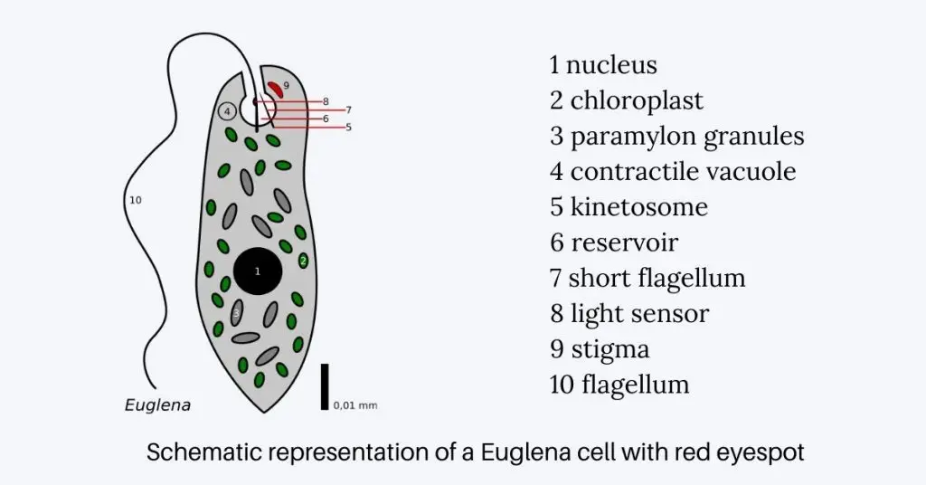
Euglena eyespot is a small pigmented light sensitive structure present at the anterior end of Euglena. It is also called stigma or eyespot apparatus. It appears as red or orange spot under microscope due to presence of carotenoid pigment containing lipid granules.
The eyespot is not used for image formation. It helps the organism to detect the direction and intensity of light. The pigment granules mainly contain zeaxanthin, which absorbs light and works as a shading body.
The complete eyespot apparatus has two main parts. These are the carotenoid-rich pigment granules and the paraflagellar body. The paraflagellar body is present near the base of flagellum and contains the real light-sensitive proteins.
During swimming, Euglena rotates around its own axis. In this movement the eyespot produces periodic shadow on the paraflagellar photoreceptor. This shadowing helps the cell to identify from which side light is coming.
Blue light activates a special enzyme called photoactivated adenylyl cyclase (PAC). This produces a chemical signal inside the cell. This signal changes the beating pattern of the flagellum and helps in movement towards or away from light.
This movement in response to light is called phototaxis. By this process Euglena moves towards suitable light for photosynthesis. It can also avoid very strong light which may damage the cell. Thus eyespot acts as a light detecting and guiding structure in Euglena.
Definition of Eyespot
An eyespot is a specialized, light-sensitive organelle found in certain unicellular organisms, enabling them to detect light direction and intensity, thereby facilitating phototactic behaviors.
Location of Eyespot Apparatus in Euglena
- Eyespot apparatus is present in the anterior region of Euglena cell.
- It is situated near the reservoir or flagellar pocket.
- The reservoir is an inward fold of the outer covering of the cell and it is present near the base of flagellum.
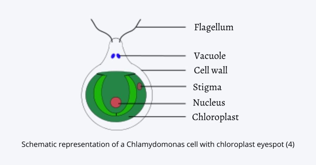
- In Euglena, the eyespot is extraplastidial. It means it is not embedded inside the chloroplast.
- It remains free in the cytoplasm and present close to the flagellar pocket.
- The eyespot is found near the base of the long emergent flagellum.
- It is closely associated with the paraflagellar body, which acts as actual photoreceptor.
- Due to this position, the eyespot can cast shadow on the paraflagellar body during rotation of Euglena.
- Thus, the eyespot apparatus is located at the anterior cytoplasmic region, close to reservoir, flagellar base and paraflagellar body.
Eyespot Structure
Eyespot Detail Structure
- Eyespot apparatus of Euglena is made up of two main parts. These are pigmented lipid granules and paraflagellar body (PFB).
- The pigmented lipid granules are called stigma or eyespot proper. These granules give red or orange colour to the eyespot.
- These granules are extraplastidial in nature. It means they are not present inside the chloroplast.
- The pigment granules remain freely in the cytoplasm. They are loosely and irregularly arranged near the flagellar pocket or reservoir.
- The granules are mainly lipid droplets which contain isoprenoid pigments. These pigments include β-carotene, diadinoxanthin, diatoxanthin and zeaxanthin.
- Zeaxanthin has important role in maintaining the packet like structure of granules. It helps to keep the structural arrangement of the eyespot.
- The second part is paraflagellar body (PFB). It is the actual light sensing part of the eyespot apparatus.
- The PFB is an ovoid swelling present on the basal region of the large emergent flagellum.
- The paraflagellar body has crystalline lamellar structure. It is made up of protein rods arranged in tight helical form.
- This crystalline region contains large number of chromophore bearing proteins. These proteins are arranged in dense and ordered manner.
- The important light sensing protein in this region is photoactivated adenylyl cyclase (PAC). It is a flavoprotein which responds mainly to blue light.
- Along with PAC, the eyespot apparatus also has structural proteins, metabolic enzymes and signalling molecules. These help in receiving and transferring light signal.
- Botanically, Euglena eyespot is generally considered as Type C eyespot. It is because it has independent cluster of granules without its own membrane covering.
- Thus, the eyespot of Euglena is formed by pigment granules and paraflagellar body. The pigment part acts as shading body and the paraflagellar body acts as photoreceptive part.
Different Eyespot Proteins
- PAC
Photoactivated Adenylyl Cyclase (PAC) is the main blue light receptor of Euglena eyespot. It changes ATP into cAMP after light activation. - PACα
PACα is the triggering subunit of PAC. It catches light efficiently and starts light induced change in the protein. - PACβ
PACβ is the resetting subunit of PAC. It resets quickly and helps Euglena to detect changing light during rotation. - PKA
Protein Kinase A (PKA / PK.4) is activated by cAMP. It changes flagellar motor proteins and helps in changing the beating of flagellum. - Structural proteins
These proteins maintain the crystalline structure of paraflagellar body (PFB). They keep the light sensitive proteins in ordered arrangement. - Metabolic enzymes
These enzymes work with photoreceptor proteins. They help in processing the light signal inside the cell. - Accessory molecules
Accessory signalling molecules help to transfer the signal from eyespot region to flagellum. They coordinate the light response. - CaM2
Calmodulin 2 (CaM2) is mainly related with gravity sensing. It also interacts with the same signalling pathway used in eyespot response. - EgPCDUF4201
EgPCDUF4201 also works in gravity related response. It may show connection with adenylyl cyclase and PKA pathway. - Rhodopsin-like proteins
Rhodopsin-like proteins were considered as possible photoreceptors. But PAC is the main protein for phototactic movement in Euglena.
The Photoreception and signal transduction
- The photoreception mechanism in Euglena starts when blue light falls on the Photoactivated Adenylyl Cyclase (PAC) present near the base of the flagellum. This PAC is the main photoreceptor protein of the eyespot apparatus.
- During swimming, Euglena rotates around its own axis and the red-orange pigment granules of the eyespot work as a shading body. These granules cast shadow on the paraflagellar body again and again.
- Due to this alternate shading, the photoreceptor receives light and dark condition in a repeated manner. This produces an oscillating light signal which helps the cell to know the direction of light.
- The FAD chromophore present in PAC absorbs blue light. After absorption, proton coupled electron transfer takes place inside the photoreceptor protein.
- This light absorption causes change in the structure of PAC molecule. The protein changes into active form and this is the important step for starting signal transduction.
- The active PAC opens its adenylyl cyclase catalytic region. In this step the enzyme becomes functional and begins to convert ATP into cyclic AMP (cAMP).
- The reaction is as follows-
ATP → cAMP - The cAMP formed in this reaction acts as a second messenger. It carries the light signal from the eyespot region to the flagellar region of the cell.
- The cAMP then activates Protein Kinase A (PKA / PK.4) present near the reservoir and flagellar pocket. This is a signalling protein which works after receiving the chemical signal.
- Activated PKA phosphorylates the motor proteins present inside the flagellum. In this process phosphate group is added to the flagellar proteins.
- After phosphorylation, the sliding rate of microtubules inside the flagellum is changed. Due to this, the normal beating pattern and speed of the flagellum is altered.
- The changed flagellar beating helps Euglena to turn its body in proper direction. It may move towards the light or may move away from the light depending on light intensity.
- Movement towards suitable light is called positive phototaxis. This helps Euglena to get proper light for photosynthesis.
- Movement away from very strong light is called negative phototaxis. This protects the cell from damage caused by excess light.
- Thus, the eyespot apparatus first receives blue light by PAC, then forms cAMP as chemical messenger and finally controls the flagellar movement by PKA. This whole process helps Euglena in phototactic movement.
Types of Eyespot
- Type A
Type A eyespot is present in the chloroplast stroma. It is located below the outer chloroplast envelope and lies between the thylakoid sheets. It has no association with the flagella. This type is found in Chlorophyceae and Cryptophyceae. - Type B
Type B eyespot is also present in the chloroplast stroma. It is associated with the swollen flagellar base and remains arranged with the flagellar pocket. This type is found in Chrysophyceae, Xanthophyceae and Phaeophyceae. - Type C
Type C eyespot is independent from the chloroplast. It is present as cytoplasmic cluster of granules without any separate membrane covering. It is close to the flagellar base but remains structurally separate. This type is found in Euglenophyceae like Euglena and Eustigmatophyceae. - Type D
Type D eyespot is separated from the chloroplast and closely related with the flagellar base. It is enclosed in a special triple membraned sac which has no pores. This type is found in some members of Dinophyceae. - Type E
Type E eyespot is a highly complex independent organelle. It has lens like hyalosome, cornea like mitochondria, iris rings and thylakoid derived retinal body. This complex eyespot is called ocellus and found in Warnowiaceae family of Dinophyceae. - Euglena type
In Euglena, the eyespot is generally Type C eyespot. It is present outside chloroplast as pigment granules in cytoplasm and works near the flagellar apparatus.
Mechanism of Eyespot-Mediated Light Perception
- The mechanism starts when blue light photons fall on the paraflagellar body of Euglena. This body contains Photoactivated Adenylyl Cyclase (PAC), which works as the main photoreceptor protein.
- During swimming, Euglena rotates continuously on its longitudinal axis. In this condition the red coloured eyespot comes in front of the photoreceptor at regular interval.
- The eyespot contains carotenoid-rich pigment granules. These granules act as a shading body and cast shadow over the photoreceptor.
- Due to this repeated shadowing, the photoreceptor receives alternate light and dark condition. This condition helps the cell to understand the direction of light.
- When blue light is absorbed by the chromophore present in PAC, rapid molecular change takes place in the protein. Proton coupled electron transfer and changes in hydrogen bond occurs.
- This light induced change alters the structure of PAC molecule. The protein becomes active and it can now begin the chemical signalling process.
- The active PAC opens its adenylyl cyclase catalytic region. Then the enzyme converts ATP into cyclic AMP (cAMP).
- The reaction is as follows-
ATP → cAMP - The cAMP acts as a second messenger in the cell. It carries the light signal from the eyespot region towards the flagellar motor region.
- The cAMP then binds with Protein Kinase A (PKA / PK.4), which is present near the flagellar pocket. After this binding, the PKA becomes active.
- Activated PKA phosphorylates the motor proteins present inside the flagellum. In this process, phosphate group is added to the flagellar motor proteins.
- Due to phosphorylation, the sliding rate of microtubules inside the flagellum is changed. This also changes the normal beating pattern and speed of the flagellum.
- The changed beating of flagellum helps Euglena to turn its body in proper direction. The flagellum works as the main moving structure of the cell.
- The movement of Euglena in response to light is called phototaxis. It may move towards suitable light or away from very strong light.
- In suitable light condition, Euglena shows positive phototaxis for photosynthesis. In excess light condition, it shows negative phototaxis to protect the cell from damage.
- Thus eyespot mediated light perception includes light reception, repeated shading, PAC activation, cAMP formation, PKA activation and change in flagellar beating. This whole process finally produces directed movement of Euglena.
Functions of Eyespot Apparatus
- Eyespot apparatus is used for detecting light in Euglena. It detects the intensity and direction of light coming from outside.
- It acts as a small photoreceptive structure of the cell. It does not form any image, but only helps in sensing light.
- The carotenoid-rich pigment granules work as light absorbing shield. These granules absorb and block light during movement of the cell.
- During swimming, Euglena rotates on its own axis. At this time the eyespot gives shadow on the paraflagellar body again and again.
- This repeated shading helps the cell to know the direction of light source. So the eyespot works as a screening body for light perception.
- The eyespot apparatus helps in phototaxis. It means movement of Euglena in response to light.
- In suitable light condition, Euglena moves towards light. This is called positive phototaxis and it helps in photosynthesis.
- In very strong light condition, Euglena moves away from light. This is called negative phototaxis and it protects the cell from light injury.
- The eyespot apparatus also controls the beating of flagellum. After receiving light, it produces chemical signal which changes the flagellar movement.
- The changed beating pattern of flagellum helps the cell to turn in proper direction. Thus light detection is converted into actual movement.
- It helps Euglena to remain in proper light condition. The cell can avoid too dim and too bright region by the help of eyespot.
- The main importance of eyespot apparatus is that it helps in better photosynthesis. By selecting suitable light, the organism can produce energy and also protect its cellular parts from excess light.
Differences between Type A, Type B, Type C, Type D and Type E Eyespot
| Type | Position | Flagellar relation | Membrane character | Found in |
|---|---|---|---|---|
| Type A | It is present inside the chloroplast stroma. It lies between thylakoid sheets below the outer chloroplast envelope. | It has no structural association with flagella. | It has no separate special membrane. It remains within chloroplast region. | Chlorophyceae and Cryptophyceae. |
| Type B | It is also present inside the chloroplast stroma. | It is associated with swollen base of flagellum and arranged with flagellar pocket. | It has no separate special membrane. It is chloroplast associated. | Chrysophyceae, Xanthophyceae and Phaeophyceae. |
| Type C | It is present outside chloroplast as cytosolic cluster of granules. | It is close to flagellar base but structurally separate. | It has no independent bounding membrane. | Euglenophyceae like Euglena and Eustigmatophyceae. |
| Type D | It is physically independent from chloroplast. | It is highly coordinated with flagellar base. | It is enclosed in a special triple-membraned sac and the sac lacks pores. | Some members of Dinophyceae. |
| Type E | It is completely independent and very complex organelle. | It is integrated with a larger sensory network. | It forms complex ocellus with lens like hyalosome, cornea like mitochondria, iris rings and retinal body. | Warnowiaceae family of Dinophyceae. |
The main difference is based on their relation with chloroplast, flagellar apparatus and membrane covering. Type A and Type B are chloroplast associated. Type C, Type D and Type E are independent from chloroplast. Type C has no membrane covering, Type D has triple membrane sac and Type E is the most complex ocellus like structure.
References
- Investigation of gravitaxis and phototaxis in Euglena gracilis. (n.d.). ResearchGate.
- Richter, P. R., Graf, J., Haag, F. W. M., Scudlo, V., Wiesmeth, S., Hauslage, J., Richter, M., Geißler, D., & Lebert, M. (2026). Acceleration and light-induced changes in cytosolic cAMP concentration in Euglena gracilis. Biomolecules, 16(3), 451.
- Algal eyespot. (n.d.). SlideShare.
- Biophysical characterization, evolutionary lineage, and signal transduction of the Euglena eyespot apparatus. (n.d.).
- Carotenoids biosynthesis, accumulation, and applications of a model microalga Euglena gracilis. (n.d.). PMC.
- Carotenoids in the eyespot apparatus are required for triggering phototaxis in Euglena gracilis. (n.d.). PMC.
- Comparison of composition and contents of carotenoids among strains of Euglena. (n.d.).
- Discovery and characterization of photoactivated adenylyl cyclase (PAC), a novel blue-light receptor flavoprotein, from Euglena gracilis. (n.d.). ResearchGate.
- Liu, P., Wang, J., Hong, Y., Chen, Z., Liu, X., Chen, H., Zeng, G., & Pan, X. (2026). Environmental concentrations of PFOS selectively accumulate in the Euglena eyespot and directly inhibit chloroplast ATP synthase: A dual impairment of phototaxis and photosynthetic light reactions without inhibiting photosynthesis. Preprints.org.
- Euglena. (n.d.).
- Euglena. (2026, March 13). In Wikipedia. https://en.wikipedia.org/wiki/Euglena
- BYJU’S. (n.d.). Euglena classification.
- Guiry, M. D., & Guiry, G. M. (2026). Euglena Ehrenberg, 1830. AlgaeBase.
- Supriya, N. (n.d.). Euglena eyespot – Definition, location, functions & mechanism. Biology Reader.
- Biology Notes Online. (n.d.). Euglena eyespot – Definition, function, types, structure, proteins [Video]. YouTube.
- Noble, R. (2021, December 1). Euglena eye and nature’s invention. Understanding Living Systems.
- Häder, D.-P., & Hemmersbach, R. (2022). Euglena, a gravitactic flagellate of multiple usages. Life, 12, 1522.
- Euglena: The photoreceptor system for phototaxis. (n.d.). PubMed.
- Leander, B. S., Lax, G., Karnkowska, A., & Simpson, A. G. B. (2017). Euglenida. In J. M. Archibald et al. (Eds.), Handbook of the protists (pp. 1-42). Springer International Publishing AG.
- Eyespot apparatus. (2026, January 4). In Wikipedia.
- Eyespot apparatus. (n.d.). Bionity.com.
- Carolina Staff. (2023, January 30). From Euglena to zebra fish: An overview of the diversity of eyes. Carolina Knowledge Center.
- Identification of key residues in allosteric signaling of photoactivated adenylyl cyclase. (n.d.). bioRxiv.
- Light and the evolution of vision. (n.d.). PMC.
- Looser, J., Schröder-Lang, S., Hegemann, P., & Nagel, G. (2009). Mechanistic insights in light-induced cAMP production by photoactivated adenylyl cyclase alpha (PACalpha). Biological Chemistry, 390(11), 1105-1111.
- Nasir, A., Richter, P. R., Le Bail, A., Daiker, V., Stoltze, J., Prasad, B., Strauch, S. M., & Lebert, M. (2022). Molecular cross-talk between gravity- and light-sensing mechanisms in Euglena gracilis. International Journal of Molecular Sciences, 23(5), 2776.
- Ohki, M., Sato-Tomita, A., Matsunaga, S., Iseki, M., Tame, J. R. H., Shibayama, N., & Park, S.-Y. (2017). Molecular mechanism of photoactivation of a light-regulated adenylate cyclase. Proceedings of the National Academy of Sciences, 114(32), 8562-8567.
- Morphological investigations of the Euglena gracilis isolated paraflagellar body. (n.d.). ResearchGate.
- Piccinni, E., & Mammi, M. (1978). Motor apparatus of Euglena gracilis: Ultrastructure of the basal portion of the flagellum and the paraflagellar body. Boll Zoologia, 45, 405-414.
- Peculiar proteome of dark-cultivated Euglena gracilis. (n.d.). PMC.
- Photoactivated adenylyl cyclase controls phototaxis in the flagellate Euglena gracilis. (n.d.). PubMed.
- Ntefidou, M., & Häder, D. P. (2005). Photoactivated adenylyl cyclase (PAC) genes in the flagellate Euglena gracilis mutant strains. Photochemical & Photobiological Sciences, 4(9), 732-739.
- Photoactivated adenylyl cyclase. (2025, November 18). In Wikipedia.
- Photoactivated adenylyl cyclase controls phototaxis in the flagellate Euglena gracilis. (n.d.). PubMed. (Duplicate title in source list)
- Häder, D. P., & Iseki, M. (2017). Photomovement in Euglena. Advances in Experimental Medicine and Biology, 979, 207-235.
- Horiguchi, T., Kawai, H., Kubota, M., Takahashi, T., & Watanabe, M. (1999). Phototactic responses of four marine dinoflagellates with different types of eyespot and chloroplast. Phycological Research, 47, 101-107.
- Diehn, B. (1973). Phototaxis and sensory transduction in Euglena. Science, 181(4104), 1009-1015.
- Tamaki, S., Ozasa, K., Nomura, T., Ishikawa, M., Yamada, K., Suzuki, K., & Mochida, K. (2023, January 30). Pigments responsible for Euglena’s eyespot apparatus identified. RIKEN Center for Sustainable Resource Science.
- Hontani, Y., Mehlhorn, J., Domratcheva, T., Beck, S., Kloz, M., Hegemann, P., Mathes, T., & Kennis, J. T. M. (2023). Spectroscopic and computational observation of glutamine tautomerization in the blue light sensing using flavin domain photoreaction. Journal of the American Chemical Society, 145(2), 1040-1052.
- Dodge, J. D. (1984). The functional and phylogenetic significance of dinoflagellate eyespots. Biosystems, 16, 259-267.
- The involvement of a protein kinase in phototaxis and gravitaxis of Euglena gracilis. (n.d.). ResearchGate.
- The involvement of a protein kinase in phototaxis and gravitaxis of Euglena gracilis. (n.d.). PubMed. (Duplicate title in source list)
- Taguchi, M., Sakuraba, S., Chan, J., & Kono, H. (2024). Unveiling the photoactivation mechanism of BLUF photoreceptor protein through hybrid quantum mechanics/molecular mechanics free-energy calculation. ACS Physical Chemistry Au, 4(6), 647-659.
- Konupková, A., Peña-Diaz, P., & Hampl, V. (2025). Visualisation of Euglena gracilis organelles and cytoskeleton using expansion microscopy. Life Science Alliance, 8(4), e202403110.
- Konupková, A., Peña-Diaz, P., & Hampl, V. (2025). Visualisation of Euglena gracilis organelles and cytoskeleton using expansion microscopy. Life Science Alliance, 8(4), e202403110. (Duplicate file in source list)
- Tamaki, S., Ozasa, K., Nomura, T., Ishikawa, M., Yamada, K., Suzuki, K., & Mochida, K. (2023). Zeaxanthin is required for eyespot formation and phototaxis in Euglena gracilis. Plant Physiology, 191(4), 2414-2426.
- Koumura, Y., Suzuki, T., Yoshikawa, S., Watanabe, M., & Iseki, M. (2004). The origin of photoactivated adenylyl cyclase (PAC), the Euglena blue-light receptor: Phylogenetic analysis of orthologues of PAC subunits from several euglenoids and trypanosome-type adenylyl cyclases from Euglena gracilis. Photochemical & Photobiological Sciences, 3(6), 580-586.
What organisms have eyespots?
Eyespots, or ocelli, are eye-like markings found in a diversity of organisms including lepidopterans (butterflies, moths, and skippers), reptiles, fish, birds, and cats.