

The extrinsic pathway of apoptosis is a type of programmed cell death pathway which starts after receiving death signals from outside the cell. It is also called the death receptor pathway.
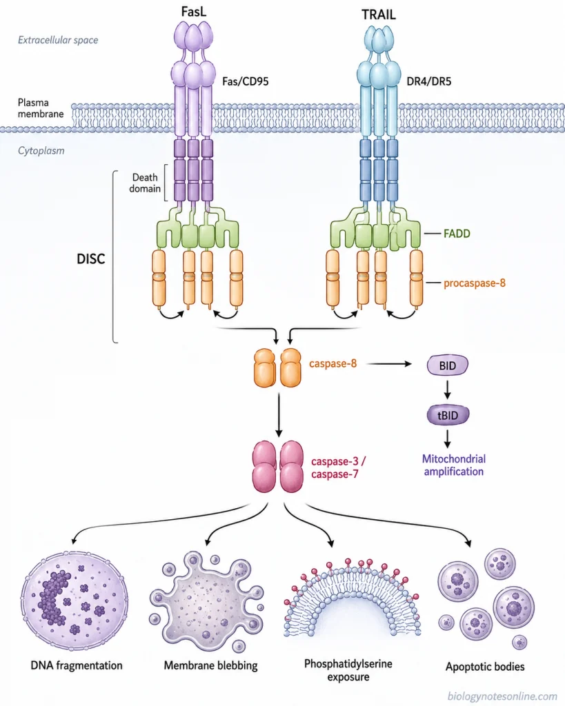
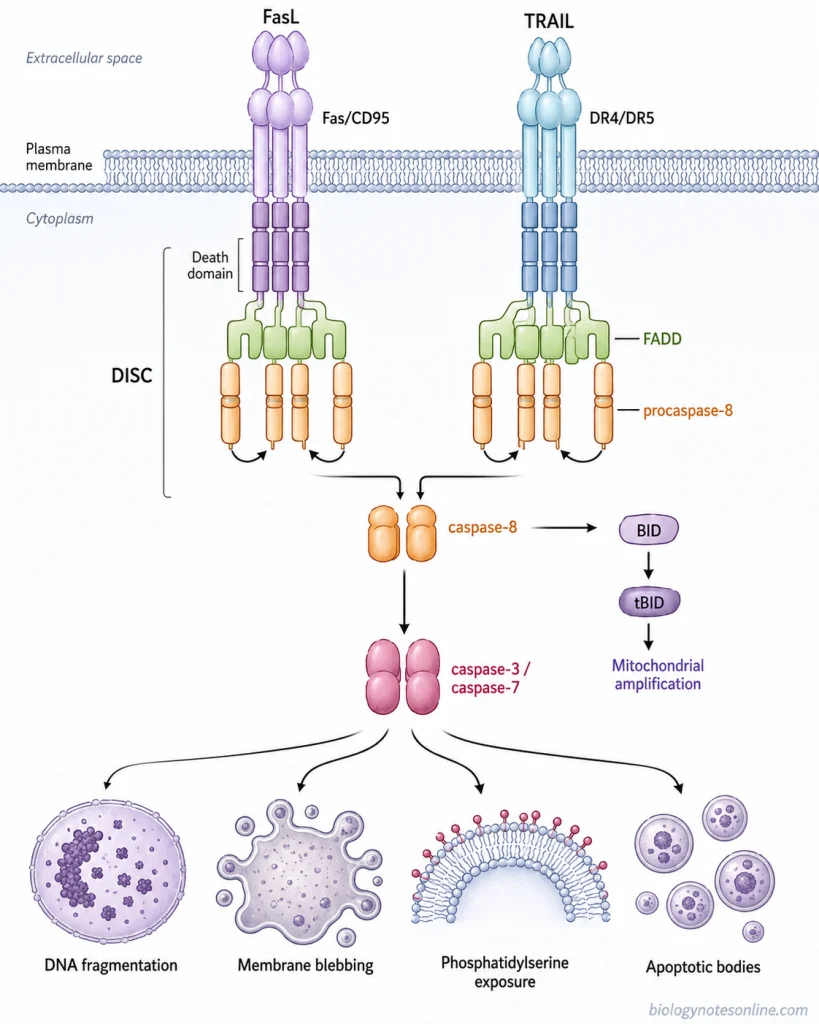
Fas ligand (FasL) and TRAIL are the death ligands involved in this pathway, which bind with their specific receptors present on the plasma membrane such as Fas (CD95), DR4, and DR5. These receptors contain an intracellular death domain.
After binding of the ligand, the adaptor protein FADD is recruited to this region and procaspase-8 is then recruited by FADD. A protein complex is formed by these components. This is referred to as the death-inducing signaling complex (DISC).
During this process, procaspase-8 molecules come close together and are activated into caspase-8. The activated caspase-8 further activates caspase-3 and caspase-7, which are executioner caspases and carry out the cellular changes during apoptosis. In some cells, caspase-8 also acts on BID and cleaves it to form truncated BID (tBID). This links the extrinsic pathway with the mitochondrial pathway and increases the apoptotic signal inside the cell.
Components of the Extrinsic Apoptosis Pathway
The following are the important components involved in the extrinsic apoptosis pathway–
- Death ligands– These are extracellular signaling proteins which start the death receptor-mediated pathway. Fas ligand (FasL/CD95L), TNF and TRAIL are the major death ligands. They bind with their respective receptors present on the cell surface.
- Death receptors– Death receptors are members of the tumor necrosis factor receptor (TNFR) superfamily having an intracellular death domain. Some of the important death receptors are Fas (CD95/APO-1), TNFR1, DR4 (TRAIL-R1) and DR5 (TRAIL-R2). The death domain is used for recruitment of proteins required for transmission of apoptotic signal.
- FADD (Fas-associated death domain protein)– It is an adaptor protein. FADD binds with the death domain of activated receptors and also contains a death effector domain (DED) for recruitment of procaspase-8. In Fas and TRAIL receptor signaling, it is recruited directly to the activated receptor.
- TRADD (TNFR1-associated death domain protein)– This adaptor protein is mainly involved with TNFR1 signaling. After activation of TNFR1, TRADD is recruited first and other proteins including FADD can then become associated with the signaling complex.
- Death-inducing signaling complex (DISC)– It is a multiprotein complex formed after activation of several death receptors. FADD and procaspase-8 are brought together in this complex. This is referred to as the DISC and it acts as the site for activation of initiator caspase-8.
- Procaspase-8 and caspase-8– Procaspase-8 is an inactive initiator caspase recruited to the DISC through FADD. During this process, procaspase-8 molecules are brought close together and active caspase-8 is formed. Caspase-10 can also participate in some death receptor signaling systems.
- c-FLIP– It is a regulatory protein which can be recruited to the DISC. c-FLIP regulates caspase-8 activation and can prevent the apoptotic signal depending upon its form and the cellular condition.
- Executioner caspases– Caspase-3 and caspase-7 are the important executioner caspases activated downstream of caspase-8. These act on different cellular proteins, producing the changes of apoptotic cell death.
- BID– BID is a BH3-only protein which provides a link between extrinsic and mitochondrial apoptosis. Caspase-8 cleaves BID and truncated BID (tBID) is formed. It then acts through the mitochondrial pathway for amplification of the death signal in cells where this additional signal is required.
Step-by-Step Mechanism of the Extrinsic Pathway
The following are the steps involved in the extrinsic pathway of apoptosis–
Step 1- Binding of death ligand
The first step is the binding of a death ligand with its specific receptor present on the plasma membrane. Fas ligand (FasL) binds to Fas (CD95), while TRAIL binds with DR4 or DR5. TNF can bind to TNFR1. After binding, the receptor undergoes changes in its intracellular region and the death domain becomes available for further signaling.
Step 2- Recruitment of adaptor protein
The activated Fas and TRAIL receptors recruit FADD (Fas-associated death domain protein) through interaction of their death domains. FADD contains another region called the death effector domain (DED). In case of TNFR1, FADD is not recruited directly. TRADD is first associated with TNFR1 and FADD can be recruited during formation of the apoptotic signaling complex.
Step 3- Recruitment of procaspase-8
FADD now provides the site for binding of procaspase-8. The death effector domain of FADD interacts with the DED regions present in procaspase-8 and several procaspase-8 molecules are brought close together. In humans, procaspase-10 can also be recruited in some of these receptor complexes.
Step 4- Formation of DISC
A multiprotein signaling complex is formed at the activated death receptor. This is referred to as the death-inducing signaling complex (DISC). In Fas signaling, it mainly contains the ligand-bound receptor, FADD and procaspase-8. The DISC acts as a platform for activation of the initiator caspase.
Step 5- Activation of caspase-8
During this process, procaspase-8 molecules present in the signaling complex are brought into close proximity and become activated. Active caspase-8 is then produced. Its activity can also be controlled by proteins such as c-FLIP, which can become associated with the DISC and regulate caspase-8 activation.
Step 6- Activation of executioner caspases
The activated caspase-8 acts on downstream executioner caspases. Caspase-3 and caspase-7 are activated and the caspase cascade is now started. These enzymes act on different cellular proteins responsible for maintaining normal structure and functions of the cell.
Step 7- Mitochondrial amplification in some cells
In some cell types, direct activation of executioner caspases is not sufficient. Caspase-8 then cleaves the BH3-only protein BID, forming truncated BID (tBID). tBID acts on the mitochondrial apoptotic machinery through BAX and BAK, causing mitochondrial outer membrane permeabilization and amplification of the death signal. This pathway is particularly required in the type II form of death receptor-mediated apoptosis.
Step 8- Apoptotic changes
After sufficient activation of executioner caspases, different cellular proteins are cleaved. Changes such as membrane blebbing, DNA fragmentation, phosphatidylserine exposure and formation of apoptotic bodies are produced during this process.
How Does the Extrinsic Pathway Connect to the Intrinsic Pathway?
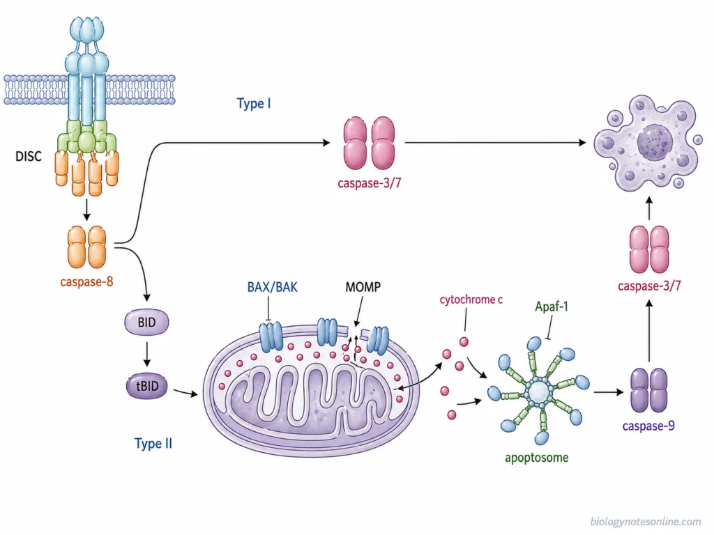
The major connection between extrinsic pathway and intrinsic pathway is through BID protein. BID is a pro-apoptotic BH3-only protein of BCL-2 family. During death receptor-mediated apoptosis, caspase-8 is activated in the death-inducing signaling complex (DISC) and it also acts on BID. BID is cleaved during this process, producing a shorter active form called truncated BID (tBID).
The tBID formed then moves towards the outer mitochondrial membrane. Here, it acts on the pro-apoptotic proteins BAX and BAK and mitochondrial outer membrane becomes permeable. This process is referred to as mitochondrial outer membrane permeabilization (MOMP). During this process, cytochrome c is released from mitochondria into the cytoplasm.
The released cytochrome c combines with Apaf-1 and procaspase-9. An apoptosome is now formed. Caspase-9 is activated in this complex which further activates caspase-3 and caspase-7.
In type II cells, direct activation of executioner caspases by caspase-8 is not sufficient. BID-mediated mitochondrial pathway is therefore required for amplification of apoptotic signal. In type I cells, stronger activation of caspase-8 occurs and the executioner caspases can be activated directly. The mitochondrial amplification generally is not required in these cells.
Major Death-Receptor Signaling Routes
The death receptor pathway occurs through different receptors of TNF receptor family. Some of the important signaling routes are as follows-
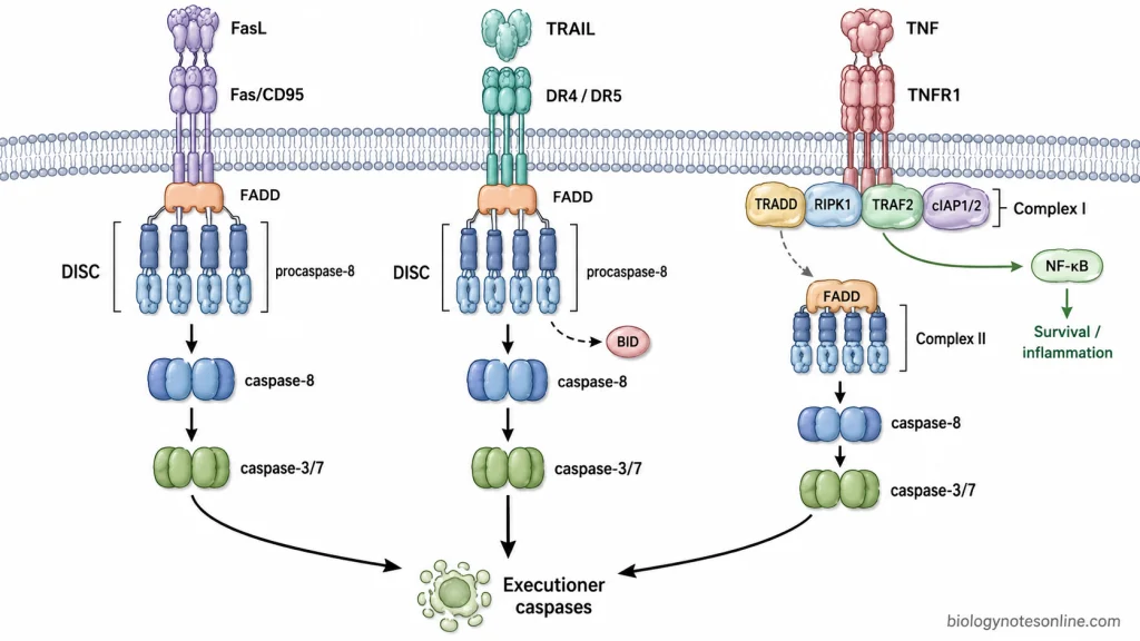
- Fas/FasL signaling- Fas (CD95/APO-1) is a death receptor present on cell surface. Fas ligand (FasL/CD95L) binds with Fas and the receptor becomes activated. After this binding, FADD is recruited to the intracellular death domain of Fas. Procaspase-8 also binds with FADD and these proteins together form a complex. This is referred to as death-inducing signaling complex (DISC). In this complex procaspase-8 molecules are brought close together and caspase-8 is formed by their activation. The activated caspase-8 then acts on executioner caspases.
- TRAIL receptor signaling- TRAIL (TNF-related apoptosis-inducing ligand) is another death ligand involved in extrinsic apoptosis. It acts mainly through DR4 (TRAIL-R1) and DR5 (TRAIL-R2). Both are death receptors having an intracellular death domain. After binding of TRAIL, FADD is recruited. Procaspase-8 is also recruited and DISC formation takes place. During this process, caspase-8 becomes activated and the executioner caspases can be activated by it. In some cells, caspase-8 also cleaves BID. The mitochondrial pathway becomes involved in such cells.
- TNF/TNFR1 signaling- TNF binds with TNFR1 on the cell membrane. The signaling through this receptor is somewhat different because more than one protein complex can be formed. In the first step, TRADD, RIPK1, TRAF2 and cIAP proteins are recruited to the activated receptor and a membrane-associated complex I is formed.Complex I is mainly involved in inflammatory and cell-survival signaling. For apoptotic pathway, a second complex can be formed in the cytoplasm. This is called complex II. FADD and procaspase-8 are present in this complex, where activation of procaspase-8 takes place. The caspase-8 formed further activates the executioner caspases.
Regulation of the Extrinsic Apoptosis Pathway
The extrinsic apoptosis pathway is regulated at different steps after binding of death ligand. Regulation may occur at receptor level, during formation of DISC and also after activation of caspases. Some of the important regulatory mechanisms are as follows-
- Death receptor and decoy receptor level- The amount of functional death receptor present on the cell surface can affect the response towards a death ligand. In TRAIL pathway, DR4 and DR5 transmit the apoptotic signal. DcR1 and DcR2 also bind with TRAIL, but they do not contain a functional death domain for producing the normal apoptotic signal. These are referred to as decoy receptors. Higher decoy receptor activity can therefore reduce signaling through DR4 and DR5.
- Regulation by c-FLIP- c-FLIP is an important regulatory protein present at the death-inducing signaling complex (DISC). It is structurally related to procaspase-8 and contains death effector domains by which it can become associated with FADD. The short form, c-FLIP(_S), prevents proper activation of procaspase-8. c-FLIP(_L) has a more variable action and its effect depends on its amount and the composition of DISC. Thus, the ratio of c-FLIP and procaspase-8 is one of the factors controlling whether sufficient caspase-8 activity is produced.
- TNFR1 complex and NF-κB signaling- In TNFR1 signaling, binding of TNF does not always result in apoptosis. At first, TRADD, RIPK1, TRAF2 and cIAP1/cIAP2 become associated and complex I is formed. The cIAP proteins add ubiquitin chains to RIPK1. During this process proteins involved in NF-κB activation are recruited, and NF-κB induces different cell-survival genes including FLIP. When this survival signaling is reduced, the TNFR1 complex can change towards a cytoplasmic death complex where FADD and caspase-8 are recruited.
- Inhibitor of apoptosis proteins (IAPs)- XIAP is a member of inhibitor of apoptosis protein family which acts at a later stage of the pathway. It inhibits caspase-9 and the executioner caspases caspase-3 and caspase-7. Hence, activation of caspase-8 alone may not always be sufficient to produce strong executioner caspase activity. During mitochondrial amplification, Smac/DIABLO and Omi are released and they reduce the inhibitory action of XIAP.
- Regulation through mitochondrial pathway- This regulation is especially important in type II cells. The amount of caspase-8 produced after death-receptor activation is not sufficient in these cells for direct activation of executioner caspases. Caspase-8 cleaves BID and forms truncated BID (tBID), which acts on BAX and BAK at mitochondria. The mitochondrial signal is therefore used for amplification of death receptor signal. Anti-apoptotic proteins such as BCL-2 and BCL-xL can oppose BAX/BAK-mediated mitochondrial permeabilization and reduce this amplification.
Biological Significance of Extrinsic Apoptosis
Extrinsic apoptosis is a mechanism by which cells are removed after receiving death signals from outside the cell. It has an important role especially in immune system. Some of the important biological significance are as follows-
- Maintenance of immune homeostasis- During an immune response, large number of lymphocytes are produced after antigen stimulation. These cells are not required continuously. Fas/FasL-mediated apoptosis removes many of the activated T lymphocytes after the immune response and the lymphocyte population is brought back towards normal level. It is an important mechanism for maintenance of immune homeostasis.
- Removal of self-reactive lymphocytes- Fas-mediated activation-induced cell death (AICD) is involved in maintaining tolerance towards self-antigens. Self-reactive or repeatedly activated lymphocytes can be eliminated by this process. When the Fas/FasL pathway becomes defective, these lymphocytes may survive for longer period and autoimmunity can develop.
- Killing of infected cells- Cytotoxic T lymphocytes and NK cells can use death-receptor signaling for killing of target cells. FasL present on the cytotoxic lymphocyte binds with Fas on the target cell and apoptosis is produced. TRAIL-mediated signaling also takes part in antiviral response. During viral infection, death receptor-mediated apoptosis helps in elimination of some virus-infected cells.
- Tumour immune surveillance- TRAIL is expressed by different cells of immune system including NK cells and T lymphocytes. It can bind with DR4 and DR5 on susceptible tumour cells and causes apoptosis. The TRAIL pathway has an important role in recognition and removal of some transformed cells during tumour immune surveillance.
- Maintenance of immune-privileged sites- Fas/FasL signaling is also associated with immune privilege in some tissues. In the eye, FasL can induce apoptosis of inflammatory cells entering the tissue. During this process, excessive inflammatory cell accumulation is restricted and damage to the immune-privileged tissue can be reduced.
Extrinsic Apoptosis in Disease
A change in death receptor-mediated apoptosis is associated with different disease conditions. In some diseases this pathway becomes defective, while excessive activation of the same pathway can also produce tissue injury. Some of the important are as follows-
- Cancer- Defective extrinsic apoptosis is commonly associated with cancer. Different alterations can occur in Fas/FasL, TRAIL receptors, FADD, caspase-8 and other proteins of the pathway. Reduced death receptor signaling or increased amount of inhibitory proteins such as c-FLIP can make the tumour cells resistant to apoptosis. TRAIL resistance is also found in different tumour cells. The cells which should normally undergo apoptosis can remain alive during this condition.
- Autoimmune lymphoproliferative syndrome (ALPS)- It is a disorder in which normal removal of lymphocytes by apoptosis becomes defective. The major defect is associated with Fas-mediated apoptosis, and mutation of FAS is found in many patients. Defects involving FASL or CASP10 can also occur. As a result, abnormal accumulation of lymphocytes takes place and lymphadenopathy, splenomegaly and autoimmune manifestations may be produced. Increased risk of lymphoma is also present in these patients.
- Liver diseases- Death receptors such as Fas, TNFR1, DR4 and DR5 are expressed in liver cells. Because of this, excessive death receptor-mediated apoptosis can take part in liver injury. Fas and TNFR1 signaling have been associated with different conditions including viral hepatitis, fulminant hepatic failure, cholestatic liver disease, alcoholic hepatitis, NAFLD/NASH and ischemia-reperfusion injury. In viral hepatitis, FasL present on cytotoxic T lymphocytes can bind with Fas of infected hepatocytes and apoptosis occurs. Hepatocyte apoptosis can also contribute to progression of hepatic fibrosis.
- Viral infections- Virus infection can affect extrinsic apoptosis in different ways. Apoptosis of virus-infected cells is one of the mechanisms used by host for removal of infected cells. Fas, TRAIL receptors and TNFR1 can be involved during this process. On the other hand, many viruses produce proteins which interfere with death receptors, DISC formation or caspase activity and the infected cell escapes from apoptosis for some period. Some viruses can also use apoptosis during release and spread of viral progeny.
References
- Arakaki, R., Yamada, A., Kudo, Y., Hayashi, Y., & Ishimaru, N. (2014). Mechanism of activation-induced cell death of T cells and regulation of FasL expression. Critical Reviews in Immunology, 34(4), 301–314. https://doi.org/10.1615/critrevimmunol.2014009988
- Brunner, T., Wasem, C., Torgler, R., Cima, I., Jakob, S., & Corazza, N. (2003). Fas (CD95/Apo-1) ligand regulation in T cell homeostasis, cell-mediated cytotoxicity and immune pathology. Seminars in Immunology, 15(3), 167–176. https://doi.org/10.1016/S1044-5323(03)00035-6
- Ferguson, T. A., & Griffith, T. S. (2006). A vision of cell death: Fas ligand and immune privilege 10 years later. Immunological Reviews, 213(1), 228–238. https://doi.org/10.1111/j.1600-065X.2006.00430.x
- Franklin, J. L. (2011). Redox regulation of the intrinsic pathway in neuronal apoptosis. Antioxidants & Redox Signaling, 14(8), 1437–1448. https://doi.org/10.1089/ars.2010.3596
- Galluzzi, L., Vitale, I., Aaronson, S. A., Abrams, J. M., Adam, D., Agostinis, P., Alnemri, E. S., Altucci, L., Amelio, I., Andrews, D. W., Annicchiarico-Petruzzelli, M., Antonov, A. V., Arama, E., Baehrecke, E. H., Barlev, N. A., Bazan, N. G., Bernassola, F., Bertrand, M. J. M., Bianchi, K., … Kroemer, G. (2018). Molecular mechanisms of cell death: Recommendations of the Nomenclature Committee on Cell Death 2018. Cell Death & Differentiation, 25(3), 486–541. https://doi.org/10.1038/s41418-017-0012-4
- Green, D. R. (2022). The death receptor pathway of apoptosis. Cold Spring Harbor Perspectives in Biology, 14(2), a041053. https://doi.org/10.1101/cshperspect.a041053
- Guicciardi, M. E., & Gores, G. J. (2010). Apoptosis as a mechanism for liver disease progression. Seminars in Liver Disease, 30(4), 402–410. https://doi.org/10.1055/s-0030-1267540
- Jeng, P. S., Inoue-Yamauchi, A., Hsieh, J. J., & Cheng, E. H. (2018). BH3-dependent and independent activation of BAX and BAK in mitochondrial apoptosis. Current Opinion in Physiology, 3, 71–81. https://doi.org/10.1016/j.cophys.2018.03.005
- Kaufmann, T., Strasser, A., & Jost, P. J. (2012). Fas death receptor signalling: Roles of Bid and XIAP. Cell Death & Differentiation, 19(1), 42–50. https://doi.org/10.1038/cdd.2011.121
- Lamb, H. M. (2020). Double agents of cell death: Novel emerging functions of apoptotic regulators. The FEBS Journal, 287(13), 2647–2663. https://doi.org/10.1111/febs.15308
- MacFarlane, M., & Williams, A. C. (2004). Apoptosis and disease: A life or death decision. EMBO Reports, 5(7), 674–678. https://doi.org/10.1038/sj.embor.7400191
- Mérino, D., Lalaoui, N., Morizot, A., Solary, E., & Micheau, O. (2007). TRAIL in cancer therapy: Present and future challenges. Expert Opinion on Therapeutic Targets, 11(10), 1299–1314. https://doi.org/10.1517/14728222.11.10.1299
- O’Brien, M. A., & Kirby, R. (2008). Apoptosis: A review of pro-apoptotic and anti-apoptotic pathways and dysregulation in disease. Journal of Veterinary Emergency and Critical Care, 18(6), 572–585. https://doi.org/10.1111/j.1476-4431.2008.00363.x
- Pimentel, J. M., Zhou, J.-Y., & Wu, G. S. (2023). The role of TRAIL in apoptosis and immunosurveillance in cancer. Cancers, 15(10), 2752. https://doi.org/10.3390/cancers15102752
- Pistritto, G., Trisciuoglio, D., Ceci, C., Garufi, A., & D’Orazi, G. (2016). Apoptosis as anticancer mechanism: Function and dysfunction of its modulators and targeted therapeutic strategies. Aging, 8(4), 603–619. https://doi.org/10.18632/aging.100934
- Plati, J., Bucur, O., & Khosravi-Far, R. (2011). Apoptotic cell signaling in cancer progression and therapy. Integrative Biology, 3(4), 279–296. https://doi.org/10.1039/C0IB00144A
- Ramaswamy, M., Cleland, S. Y., Cruz, A. C., & Siegel, R. M. (2009). Many checkpoints on the road to cell death: Regulation of Fas-FasL interactions and Fas signaling in peripheral immune responses. In H. Kalthoff (Ed.), Death receptors and cognate ligands in cancer (Results and Problems in Cell Differentiation, Vol. 49, pp. 17–47). Springer. https://doi.org/10.1007/400_2008_24
- Refaat, A., Abd-Rabou, A., & Reda, A. (2014). TRAIL combinations: The new ‘trail’ for cancer therapy (Review). Oncology Letters, 7(5), 1327–1332. https://doi.org/10.3892/ol.2014.1922
- Shah, S., Wu, E., Rao, V. K., & Tarrant, T. K. (2014). Autoimmune lymphoproliferative syndrome: An update and review of the literature. Current Allergy and Asthma Reports, 14(9), 462. https://doi.org/10.1007/s11882-014-0462-4
- Tian, X., Srinivasan, P. R., Tajiknia, V., Sanchez Sevilla Uruchurtu, A. F., Seyhan, A. A., Carneiro, B. A., De La Cruz, A., Pinho-Schwermann, M., George, A., Zhao, S., Strandberg, J., Di Cristofano, F., Zhang, S., Zhou, L., Raufi, A. G., Navaraj, A., Zhang, Y., Verovkina, N., Ghandali, M., … El-Deiry, W. S. (2024). Targeting apoptotic pathways for cancer therapy. The Journal of Clinical Investigation, 134(14), e179570. https://doi.org/10.1172/JCI179570
- Tummers, B., & Green, D. R. (2017). Caspase-8: Regulating life and death. Immunological Reviews, 277(1), 76–89. https://doi.org/10.1111/imr.12541
- van Loo, G., & Bertrand, M. J. M. (2023). Death by TNF: A road to inflammation. Nature Reviews Immunology, 23(5), 289–303. https://doi.org/10.1038/s41577-022-00792-3
- Van Opdenbosch, N., & Lamkanfi, M. (2019). Caspases in cell death, inflammation, and disease. Immunity, 50(6), 1352–1364. https://doi.org/10.1016/j.immuni.2019.05.020
- Wang, C., & Youle, R. J. (2009). The role of mitochondria in apoptosis. Annual Review of Genetics, 43, 95–118. https://doi.org/10.1146/annurev-genet-102108-134850
- Waring, P., & Müllbacher, A. (1999). Cell death induced by the Fas/Fas ligand pathway and its role in pathology. Immunology & Cell Biology, 77(4), 312–317. https://doi.org/10.1046/j.1440-1711.1999.00837.x
- Zhou, X., Jiang, W., Liu, Z., Liu, S., & Liang, X. (2017). Virus infection and death receptor-mediated apoptosis. Viruses, 9(11), 316. https://doi.org/10.3390/v9110316