

What is Muscle?
Muscle is a specialized contractile tissue of the body. It is made up of muscle cells or muscle fibres. These cells have the ability to contract, shorten and produce pulling force. Due to this, movement of body and internal organs takes place.
Muscle cells are excitable in nature. They respond to electrical or chemical stimulus. The contraction of muscle mainly occurs by the interaction of actin and myosin proteins. During this process, these proteins slide over one another and shortening of muscle fibre takes place.
There are three types of muscle tissues in human body. These are skeletal muscle, cardiac muscle and smooth muscle.
Skeletal muscles are attached with bones. These muscles are responsible for voluntary movement such as walking and movement of body parts. It also helps in maintaining posture and production of body heat.
Cardiac muscle is present only in the wall of heart. It contracts rhythmically and involuntarily. Due to this contraction, blood is continuously pumped throughout the body.
Smooth muscle is present in the wall of hollow internal organs such as stomach, intestine and blood vessels. It is involuntary in nature. It helps in movement of food, air and blood through different organs.
Characteristics of Muscle
- Excitability (Irritability or Responsiveness) – It is the ability of muscle cell to receive and respond to electrical, chemical or mechanical stimulus. During this, an action potential is generated in the muscle cell.
- Contractility – It is the special ability of muscle to shorten after stimulation. Due to this shortening, pulling force is produced.
- Extensibility – It is the ability of muscle cells to stretch or extend without any damage. The muscle fibre can increase its length during this condition.
- Elasticity – It is the ability of muscle tissue to return back to its original resting length. This occurs after the muscle is stretched or deformed.
Location of Muscle
- Skeletal Muscle – It is mainly attached with bones by the tendons. It helps in movement of different body parts. Skeletal muscle is also present in the skin of face, tongue, diaphragm, eye socket and upper part of oesophagus. It is also found around the openings of mouth and anus.
- Cardiac Muscle – It is present only in the wall of heart. The muscular wall of heart is called myocardium.
- Smooth Muscle – It is present in the walls of hollow internal organs. These include stomach, intestine, urinary bladder, uterus and other reproductive tracts. Smooth muscle is also found in blood vessels, respiratory tract, skin and eyes. In skin, it forms the erector pili muscle, while in eyes it is present in iris and ciliary muscle.
Structure of Muscle
- Whole Muscle – The complete skeletal muscle is covered by an outer connective tissue layer called epimysium. It gives protection and support to the whole muscle.
- Fascicles – Inside the muscle, the muscle fibres are arranged in bundles called fascicles. Each fascicle is surrounded by a connective tissue layer called perimysium.
- Muscle Fibres (Myocytes) – Muscle fibres are long and cylindrical muscle cells present inside each fascicle. Each muscle fibre is covered by a thin connective tissue layer called endomysium.
- Sarcolemma – It is the plasma membrane of muscle fibre. The sarcolemma forms deep tube-like inward extensions called T-tubules. These tubules carry electrical signal into the inner part of muscle fibre.
- Sarcoplasm – It is the cytoplasm present inside the muscle fibre. It contains myofibrils, mitochondria, glycogen and other cellular materials.
- Sarcoplasmic Reticulum (SR) – It is a specialized membranous network present around the myofibrils. It stores and releases calcium ions required for muscle contraction. After contraction, calcium ions are again taken back into it.
- Myofibrils – These are long rod-like structures present inside the muscle fibre. They extend through the whole length of fibre. Each myofibril is formed by repeated units called sarcomeres.
- Sarcomere – It is the basic structural and functional unit of muscle contraction. A sarcomere is present between two Z-discs or Z-lines. Many sarcomeres are arranged one after another to form a myofibril.
- Myofilaments – These are very thin protein filaments present inside each sarcomere. Their sliding causes shortening of sarcomere and muscle contraction.
- Thick Filaments – Thick filaments are present mainly in the central region of sarcomere. These are made up of myosin protein.
- Thin Filaments – Thin filaments are attached with the Z-discs. These are mainly made up of actin protein. The regulatory proteins troponin and tropomyosin are also present with actin and control muscle contraction.

Types of Muscle
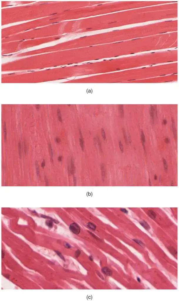
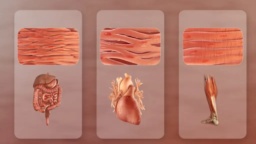
There are three types of muscles in human body. These are skeletal muscle, cardiac muscle and smooth muscle.
- Skeletal Muscle – It is also known as voluntary muscle. It is attached with bones and helps in movement of body. These muscles also maintain body posture. Muscle fibres are long, cylindrical and unbranched. Many nuclei are present at the peripheral region. Light and dark bands are present, therefore it is striated in appearance. The bands are formed by arrangement of actin and myosin.
- Cardiac Muscle – It is found only in the wall of heart. These muscles are involuntary and contract rhythmically. By this contraction, blood is pumped to different body parts. The cells are short, branched and striated. Generally one nucleus is present in the central region. The cells are connected by intercalated discs. These discs help in the combined contraction of cardiac muscle cells.
- Smooth Muscle – It is also called visceral muscle. It is present in the walls of stomach, intestine, urinary bladder and uterus. These are also present in blood vessels and respiratory tract. Smooth muscles are involuntary in nature. They help in movement of food, air and blood. The cells are spindle-shaped and have tapering ends. One nucleus is present at the centre. Striations are absent.
Development of Muscle
The development of muscle takes place by the following steps-
- Skeletal muscles mainly develop from the embryonic mesoderm. Most of the muscles arise from the paraxial mesoderm.
- The paraxial mesoderm divides from head region towards tail region. Small segments called somitomeres are formed. Seven somitomeres form muscles of head and neck. The remaining region forms 35 pairs of somites in the trunk.
- The somitomeres undergo epithelialization. During this process, groups of epithelial cells are formed.
- The mesodermal cells are changed into myogenic cells. These cells divide by mitosis and form postmitotic myoblasts.
- The myoblasts increase in number during the early stage. Each myoblast contains one nucleus. Hundreds of myoblasts come close and fuse with each other.
- The fusion of myoblasts forms long and multinucleated structures called myotubes. These myotubes later develop into myocytes or muscle fibres.
- The newly formed myotubes start synthesis of structural and contractile proteins. These include actin, myosin, troponin and tropomyosin.
- The thick myosin filaments and thin actin filaments become arranged along the long axis of muscle cell. These filaments aggregate near the cell membrane in regular arrays.
- The arranged thick and thin filaments combine to form myofibrils. This process is referred to as myogenesis. The myofibrils form the contractile structures inside the mature muscle fibre.
Functions of Muscle
- Movement and Locomotion – Muscles produce pulling force and move the bones. It helps in walking, running, grasping objects and movement of different body parts. Facial expressions are also formed by muscle movement.
- Maintenance of Posture – Skeletal muscles maintain the upright position of body against gravity. Small contractions continuously take place in these muscles. It also stabilizes the joints and prevents unwanted movement.
- Production of Heat – During muscle contraction, ATP is broken down and heat is released. This heat helps in maintaining normal body temperature. During shivering, rapid muscle contractions produce more heat.
- Pumping of Blood – Cardiac muscle contracts continuously and rhythmically. Due to this contraction, blood is pumped from heart to different body parts. It also helps in maintaining blood pressure.
- Control of Blood Flow – Smooth muscle is present in the walls of blood vessels. Its contraction causes vasoconstriction, while relaxation causes vasodilation. By this process, blood flow and blood pressure are regulated.
- Movement of Food and Other Materials – Smooth muscles move food through the digestive tract by wave-like contraction called peristalsis. It also helps in movement of air, secretions and other substances inside the body.
- Control of Body Openings – Muscles are present around the openings of different internal tracts. These muscles control swallowing, urination and defecation. Their action may be voluntary or involuntary.
- Protection of Internal Organs – Skeletal muscles form a supporting layer around internal organs. It supports the organs of abdominal and pelvic region. It also protects these organs from external injury.
Examples of Muscle
The following are some examples of muscles in human body-
Skeletal Muscles
- Biceps brachii – It is present at the front side of upper arm. It helps in flexion of elbow.
- Triceps brachii – It is present at the back side of upper arm. It helps in extension of elbow.
- Gastrocnemius and Soleus – These are calf muscles present in the lower leg. They help in movement of foot.
- Quadriceps femoris – It is present at the front region of thigh. It extends the leg at knee joint.
- Gluteus maximus – It is a large muscle present in the buttock region. It helps in movement of hip.
- Sartorius – It is a long muscle present in the thigh. It helps in movement of hip and knee.
- Latissimus dorsi – It is a broad muscle present in the lower back. It helps in movement of arm.
- Pectoralis major – It is present in the chest region. It helps in flexion and adduction of arm.
- Trapezius – It is present in upper back and neck. It raises and rotates the shoulder blade.
- Deltoid – It is present around the shoulder. It helps in moving the arm away from body.
- Sternocleidomastoid – It is present in the neck region. It bends the neck and rotates the head.
- External oblique – It is present in abdominal region. It helps in bending and rotation of vertebral column.
- Diaphragm – It is present below the lungs. It helps in voluntary and involuntary breathing.
Cardiac Muscle
- Myocardium – It is the muscular tissue present in the wall of heart. It contracts rhythmically and pumps blood throughout the body.
Smooth Muscles
- Muscles of stomach and intestine – These muscles are present in the wall of digestive tract. They move food by peristalsis.
- Muscles of blood vessels – These are present in the walls of arteries and veins. They regulate blood flow and blood pressure.
- Muscle of urinary bladder – It expands during storage of urine and contracts during urination.
- Muscles of bronchioles – These muscles are present in the respiratory tract. They control the passage of air into lungs.
- Muscle of uterus – It is present in the wall of uterus. It contracts strongly during childbirth and helps in movement of fetus outside the uterus.
Difference between Skeletal, Cardiac and Smooth Muscle
| Characteristics | Skeletal Muscle | Cardiac Muscle | Smooth Muscle |
|---|---|---|---|
| Control | It is voluntary muscle. It is controlled consciously by nervous system. | It is involuntary muscle. It contracts automatically without conscious control. | It is involuntary muscle. Its activity is regulated automatically by body. |
| Appearance | It is striated in appearance. Light and dark bands are present. | It is also striated in appearance. Light and dark bands are present. | It is non-striated in appearance. Regular bands are absent. |
| Cell shape | Cells are long, cylindrical and unbranched. | Cells are short and branched. | Cells are short and spindle-shaped with tapering ends. |
| Nucleus | Many nuclei are present. These are located at peripheral region of cell. | One or two nuclei are generally present. These are located at central region. | One nucleus is present at the centre of cell. |
| Location | It is attached with bones and skin. It is also present around mouth and anus. | It is present only in the wall of heart or myocardium. | It is present in walls of stomach, intestine, urinary bladder, blood vessels and respiratory tract. |
| Speed of contraction | Contraction is fast. | Contraction is moderate and rhythmic. | Contraction is slow. |
| Automaticity | It cannot contract by itself. Signal from motor neuron is required. | It shows automaticity. Pacemaker cells produce its own rhythm. | Some smooth muscles show automaticity. |
| Cellular connection | Gap junctions are absent between muscle fibres. | Cells are connected by intercalated discs and gap junctions. These help in combined contraction. | Gap junctions are present in some types. These help in wave-like contraction. |
| Calcium-binding protein | Troponin binds with calcium and starts contraction. | Troponin binds with calcium and helps in contraction. | Troponin is absent. Calmodulin binds with calcium for contraction. |
References
- Betts, J. G., Young, K. A., Wise, J. A., Johnson, E., Poe, B., Kruse, D. H., Korol, O., Johnson, J. E., Womble, M., & DeSaix, P. (2022). 10.2 Skeletal muscle. Anatomy and Physiology 2e. OpenStax.
- Oregon State University. (n.d.). 10.5 Types of muscle fibers – Anatomy & physiology 2e.
- Morgan, K. (n.d.). 16 Muscle contraction The sliding filament theory.
- Fowler, S., Roush, R., & Wise, J. (2013). 16.5 Musculoskeletal system. Concepts of Biology. OpenStax.
- Zedalis, J., & Eggebrecht, J. (2018). 29.4 Muscle contraction and locomotion. Biology for AP® Courses. OpenStax.
- Clark, M. A., Douglas, M., & Choi, J. (2018). 38.4 Muscle contraction and locomotion. Biology 2e. OpenStax.
- Betts, J. G., Young, K. A., Wise, J. A., Johnson, E., Poe, B., Kruse, D. H., Korol, O., Johnson, J. E., Womble, M., & DeSaix, P. (2022). 4.4 Muscle tissue and motion. Anatomy and Physiology 2e. OpenStax.
- PubMed. (n.d.). A physiological role for titin and nebulin in skeletal muscle. National Institutes of Health.
- Lumen Learning. (n.d.). ATP and muscle contraction | Biology for majors II.
- Cooper, G. M. (2000). Actin, myosin, and cell movement. The Cell: A Molecular Approach (2nd ed.). Sinauer Associates; NCBI Bookshelf.
- Pearson. (n.d.). Anatomy & physiology study guide: Skeletal muscle fiber.
- PubMed Central (PMC). (n.d.). Biomechanical aspects of the muscle-bone interaction. National Institutes of Health.
- PubMed Central (PMC). (n.d.). Calcium-induced calcium release in smooth muscle. National Institutes of Health.
- Wikipedia contributors. (2025). Calcium-induced calcium release. Wikipedia, The Free Encyclopedia.
- Klabunde, R. E. (2021). Cardiac excitation-contraction coupling. Cardiovascular Physiology Concepts (3rd ed.).
- LOUIS Pressbooks. (n.d.). Chapter 12. Biomechanics – Human anatomy and physiology I.
- Pearson. (n.d.). Compare and contrast skeletal, cardiac, and smooth muscle – Marieb, Hoehn 7th edition ch 4 problem 19.
- PubMed. (n.d.). Dihydropyridine receptor-ryanodine receptor interactions in skeletal muscle excitation-contraction coupling. National Institutes of Health.
- Bolaños, P., & Calderón, J. C. (2022). Excitation-contraction coupling in mammalian skeletal muscle: Blending old and last-decade research. Frontiers in Physiology, 13.
- Meyer, L. C., & Wright, N. T. (2013). Frontiers | Structure of giant muscle proteins. Frontiers in Physiology, 4(368).
- Knya. (n.d.). Golgi tendon organ vs muscle spindle | Key differences explained!
- Martin, M. (2017). Golgi tendon organs & muscle spindles explained | ACE exam prep blog. ACE Fitness.
- Author Unknown. (n.d.). Hierarchical architecture and physiological dynamics of human muscular systems: A comprehensive review.
- Nutrabio. (n.d.). How do muscles contract: The sliding filament theory explained.
- Institute of Biophysics, Chinese Academy of Sciences. (n.d.). In situ structural insights into the excitation-contraction coupling mechanism of skeletal muscle.
- PubMed Central (PMC). (n.d.). In situ structural insights into the excitation-contraction coupling mechanism of skeletal muscle. National Institutes of Health.
- Human Kinetics. (n.d.). Levers work to create movement in the human body.
- PubMed Central (PMC). (n.d.). Molecular mechanisms underlying the impact of muscle fiber types on meat quality in livestock and poultry. National Institutes of Health.
- Biophysical Society. (n.d.). Muscle contraction & sliding filaments.
- OpenEd CUNY. (n.d.). Muscle contraction and locomotion.
- Agriculture Notes by Agriculture.Institute. (n.d.). Muscle fiber types: Differences between red, white, and intermediate fibers.
- Jack Westin. (n.d.). Muscle structure and control of contraction – Muscle system – MCAT content.
- Taylor, T. (2026). Muscle tissue: Exploring 3 types – skeletal, cardiac & smooth. Innerbody Research.
- Wikipedia contributors. (2026). Myofibril. Wikipedia, The Free Encyclopedia.
- Khan Academy. (n.d.). Myosin and actin (video) | Muscles.
- Visible Body. (n.d.). Neuromuscular interactions: How to move a muscle.
- Healthdirect. (2025). Neuromuscular system and diseases.
- PubMed Central (PMC). (n.d.). New functions for the proprioceptive system in skeletal biology. National Institutes of Health.
- Henderson, C. A., Gomez, C. G., Novak, S. M., Mi-Mi, L., & Gregorio, C. C. (2017). Overview of the muscle cytoskeleton. Comprehensive Physiology, 7(3), 891–944.
- Noto, R. E., Leavitt, L., & Edens, M. A. (2023). Physiology, muscle. StatPearls. NCBI Bookshelf.
- McCuller, C., Jessu, R., & Callahan, A. L. (2023). Physiology, skeletal muscle. StatPearls. NCBI Bookshelf.
- McCuller, C., Jessu, R., & Callahan, A. L. (2023). Physiology, skeletal muscle. StatPearls. NCBI Bookshelf.
- Hafen, B. B., & Burns, B. (2023). Physiology, smooth muscle. StatPearls. NCBI Bookshelf.
- Study.com. (n.d.). Proprioceptors & muscle spindles | Definitions & function – Lesson.
- PubMed Central (PMC). (n.d.). Roles of titin in the structure and elasticity of the sarcomere. National Institutes of Health.
- Marzuca-Nassr, G. N., Vitzel, K. F., Mancilla-Solorza, E., & Márquez, J. L. (2018). Sarcomere structure: The importance of desmin protein in muscle atrophy. International Journal of Morphology, 36(2), 576-583.
- Nutrabio. (n.d.). Skeletal muscle: The difference between the 3 fiber types.
- Taylor, T. (2026). Skeletal muscles & levers: Types, roles & examples. Innerbody Research.
- Study.com. (n.d.). Skeletal muscles: Interactions, fascicle arrangement & lever systems.
- Lumen Learning. (n.d.). Sliding filament model of contraction | Biology for majors II.
- Wikipedia contributors. (2026). Sliding filament theory. Wikipedia, The Free Encyclopedia.
- EdTech Books. (n.d.). Smooth muscle contraction.
- Introduction to Neurobiology. (n.d.). Spinal motor control and proprioception.
- Meyer, L. C., & Wright, N. T. (2013). Structure of giant muscle proteins. Frontiers in Physiology, 4(368).
- PubMed Central (PMC). (n.d.). Structure of giant muscle proteins. National Institutes of Health.
- PubMed Central (PMC). (n.d.). The latch-bridge hypothesis of smooth muscle contraction. National Institutes of Health.
- Purves, D., Augustine, G. J., Fitzpatrick, D., et al. (2001). The motor unit. Neuroscience (2nd ed.). Sinauer Associates; NCBI Bookshelf.
- PubMed Central (PMC). (n.d.). The excitation–contraction coupling mechanism in skeletal muscle. National Institutes of Health.
- PubMed. (n.d.). The latch-bridge hypothesis of smooth muscle contraction. National Institutes of Health.
- Khan Academy. (n.d.). Three types of muscle (video).
- MedlinePlus. (n.d.). Types of muscle tissue: MedlinePlus medical encyclopedia image.