

Lamina propria is a thin layer of loose or areolar connective tissue located directly beneath the epithelium of a mucous membrane. It contains blood vessels, lymphatic vessels, connective-tissue fibres and different immune cells. Its major functions are supporting and nourishing the epithelium, providing immune defence and connecting the mucosa with deeper tissues.
What is Lamina Propria?
Lamina propria is a thin layer of loose or areolar connective tissue present directly below the epithelium and basement membrane of a mucous membrane. It forms the main supporting connective tissue part of the mucosa.
The epithelial lining does not contain blood vessels of its own. Blood capillaries present in the lamina propria supply oxygen and nutrients to the epithelial cells. Removal of metabolic wastes is also carried out through these capillaries.
Blood vessels, lymphatic vessels, nerve endings, collagen fibres and different types of cells are present in this layer. Fibroblasts are the major resident cells and take part in formation and maintenance of the connective tissue matrix.
Different immune cells such as lymphocytes, macrophages, mast cells and plasma cells are also found. The loose arrangement of the tissue allows movement of these cells. Protection of the mucosal surface against entering pathogens is carried out by these immune cells.
Characteristics of Lamina Propria
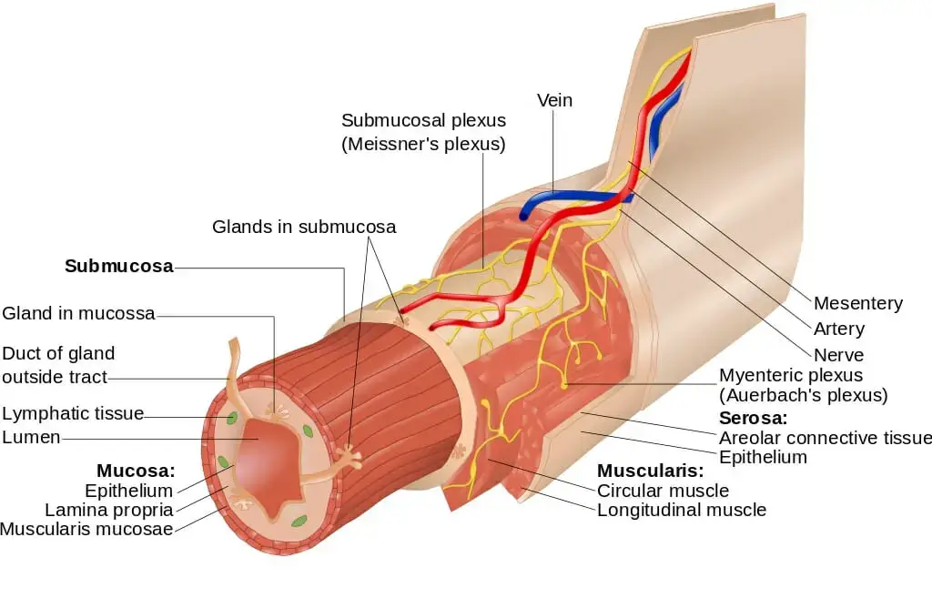
- Location– Lamina propria is present directly below the epithelial lining and basement membrane. The epithelium and lamina propria together form the mucous membrane or mucosa.
- Tissue type– It is made up of loose or areolar connective tissue. It contains more ground substance and less amount of protein fibres. Due to this, the tissue is flexible, elastic and compressible.
- Matrix– The extracellular matrix contains hydrated gel, glycoproteins and proteoglycans. Collagen fibres mainly type I and type III and elastic fibres are also present. These fibres are arranged in loose and wavy form. In some regions, it is divided into superficial papillary layer and deeper reticular layer.
- Vascularity– The epithelial lining does not have blood vessels of its own. The major blood supply is provided by the capillaries present in lamina propria. Lymphatic vessels such as lacteals are also found in the intestine. These vessels supply oxygen and nutrients and also removes metabolic wastes.
- Nerve supply– It contains afferent and efferent nerve endings. These nerve endings help in sensing mechanical stretch. It also controls different local physiological responses.
- Stromal cells– Fibroblasts are the major resident cells present in lamina propria. Other cells such as myofibroblasts, telocytes and interstitial cells of Cajal are also found. These cells take part in tissue support, maintenance and signalling.
- Immune cells– The loose arrangement of this tissue allows free movement of immune cells. Some of the important cells present are lymphocytes, macrophages, mast cells, dendritic cells and eosinophils. These cells take part in protection against the entering pathogens.
- Specialization– The structure of lamina propria changes according to the organ where it is present. It forms the central core of intestinal villi. Mucosal glands are found in the cervix and oesophagus. In urinary bladder, it provides flexibility and tensile strength.
Location of Lamina Propria
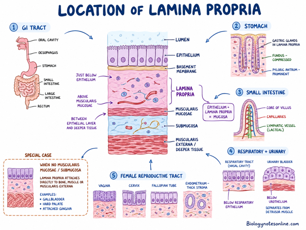
- Lamina propria is present just below the epithelial lining and basement membrane. The epithelium together with lamina propria forms the mucosa or mucous membrane.
- It is located above the muscularis mucosae and submucosa. It remains between the epithelial layer and deeper tissue of the organ.
- In some regions, muscularis mucosae or submucosa is not present. Here, the lamina propria becomes attached directly with the underlying bone, muscle or muscularis externa. It is found in gallbladder, hard palate and attached gingiva.
- In gastrointestinal tract, it is present throughout the digestive tube. It is found in oral cavity, oesophagus, stomach, small intestine, large intestine and rectum.
- In stomach, it is compressed in the fundus region. It is more prominent in pyloric antrum. The gastric glands are present within this layer.
- In small intestine, it forms the central core of intestinal villi. Blood capillaries, lymphatic vessels and different cells are present in this core.
- In respiratory tract, it is present below the epithelial lining of nasal cavity and trachea. It is found in both respiratory region and olfactory region of nasal cavity.
- In urinary bladder, lamina propria is found below the urothelium. It separates the urothelium from underlying detrusor muscle or muscularis propria.
- It is also present in female reproductive tract. It is found below the epithelial layer of vagina, cervix and fallopian tubes. In endometrium, it forms a thick connective tissue stroma.
Structure and Histology of Lamina Propria
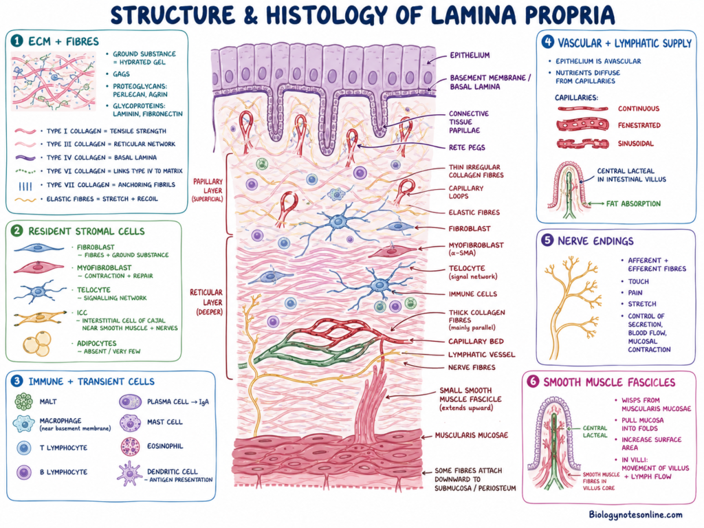
Lamina propria is a layer of loose or areolar connective tissue found immediately below the lining epithelium and basement membrane. It contains more ground substance and loosely arranged fibres. This type of arrangement provides flexibility, diffusion of nutrients and movement of immune cells.
Extracellular Matrix and Fibrous Framework
- The extracellular matrix is present in the form of hydrated gel. It contains glycosaminoglycans (GAGs), proteoglycans and glycoproteins.
- Perlecan and agrin are some of the proteoglycans found in the matrix. Laminin and fibronectin are the major glycoproteins present.
- Different collagen fibres remain scattered within the ground substance. Type I collagen provides tensile strength and is mainly found in the superficial stromal region.
- Type III collagen forms a thin and wavy reticular network. Supporting the cells and blood vessels is carried out by this network.
- Type IV collagen forms the basal lamina. Type VI collagen connects Type IV collagen with the surrounding matrix.
- Type VII collagen forms anchoring fibrils. These fibrils attach the basal lamina with the underlying reticular tissue.
- Elastic fibres are also present between collagen fibres. These allow stretching and recoil of the tissue, mainly in urinary bladder and respiratory tract.
Structural Sublayers
- In some tissues, lamina propria is divided into papillary and reticular layers. This type of division is mainly found in the oral mucosa.
- The papillary layer is the superficial part. It contains thin and irregularly arranged collagen fibres with many capillary loops.
- This layer forms connective tissue papillae which enter into the epithelial layer. The epithelial rete pegs also extend downward between these papillae.
- The reticular layer is the deeper part. It contains thick collagen fibres mainly arranged parallel with the epithelial surface.
- Some fibres of reticular layer pass downward and attach with the submucosa or periosteum.
Resident Stromal Cells
- Fibroblasts are the major resident cells present in lamina propria. Formation and renewal of fibres and ground substance are carried out by these cells.
- Myofibroblasts are also found in this layer. They contain both synthetic property of fibroblasts and contractile property of smooth muscle cells.
- Alpha-smooth muscle actin (α-SMA) is present in myofibroblasts. These cells take part in contraction and tissue repair.
- Telocytes are specialised interstitial cells having very long processes called telopodes. These processes form three-dimensional signalling network in the tissue.
- Telopodes contain thin portions called podomeres and dilated portions called podoms. Mitochondria, caveolae and endoplasmic reticulum are present in podoms.
- Interstitial cells of Cajal (ICCs) are branched or stellate cells. They are generally found close with smooth muscle fibres and nerve bundles.
- Fat cells are generally absent or present in very less number. Some adipocytes may be seen in regions of the urogenital tract.
Immune and Transient Cells
- A large number of immune cells are present in lamina propria. It forms an important site of mucosa-associated lymphoid tissue (MALT).
- Macrophages, T lymphocytes, B lymphocytes, plasma cells, mast cells, eosinophils and dendritic cells are commonly found.
- Macrophages are generally present close to the basement membrane. Removal of microorganisms, dead cells and tissue debris is carried out by these cells.
- Plasma cells mainly produce immunoglobulin A (IgA). This antibody provides protection to the mucosal surface.
- Dendritic cells take part in detection and presentation of foreign antigens. Other immune cells are involved in local inflammatory and defensive reactions.
Vascular and Lymphatic Supply
- The epithelial layer does not contain blood vessels of its own. Therefore, rich capillary beds are present in lamina propria.
- Supplying oxygen and nutrients to the epithelial cells is carried out by these capillaries. The nutrients reach epithelial cells by diffusion.
- Depending upon the organ, capillaries may be continuous, fenestrated or sinusoidal. Fenestrated capillaries are mainly found in regions of rapid absorption.
- Lymphatic vessels are also present in this layer. In intestinal villi, a blind-ended lymphatic vessel called central lacteal is found.
- Absorption and transportation of dietary fats are carried out through the central lacteal.
Nerve Endings
- Lamina propria is richly supplied with afferent and efferent nerve fibres. These fibres remain associated with stromal cells and blood vessels.
- Detection of touch, pain and mechanical stretching is carried out through these nerve endings.
- They also take part in controlling local secretion, blood flow and contraction of mucosal tissue.
Smooth Muscle Fascicles
- Small bundles or wisps of smooth muscle fibres may extend into lamina propria from the underlying muscularis mucosae.
- These muscle fibres pull the mucosa into folds. Increasing the surface area is carried out by this type of movement.
- In intestinal villi, the smooth muscle fibres extend into the villus core. Their contraction helps in movement of villi and lymph through the central lacteal.
Cells of Lamina Propria
The cells of lamina propria are divided into resident stromal cells and transient immune cells.
Resident Stromal and Structural Cells
- Fibroblasts are the major resident cells found in lamina propria. Formation, renewal and remodelling of collagen fibres, elastic fibres and ground substance are carried out by these cells.
- Myofibroblasts are modified fibroblast-like cells having both synthetic and contractile properties. They contain alpha-smooth muscle actin (α-SMA). Taking part in tissue contraction, wound healing and sensing of mechanical stretch are related with these cells.
- Telocytes are specialised interstitial cells having very long and thin processes called telopodes. The thin parts are called podomeres and dilated portions are called podoms.
- Forming a three-dimensional supporting network in the tissue is carried out by telocytes. These cells also take part in cell signalling, tissue repair and maintenance of stem cell regions such as intestinal crypts.
- Interstitial cells of Cajal (ICCs) are branched or stellate cells found near smooth muscle fibres and nerve endings. Producing electrical slow waves and helping in transmission of nerve signals are the major activities of these cells.
- Adipocytes are generally absent or found in very less number in lamina propria. Some fat cells may be present in certain regions such as urinary bladder.
Transient and Immune Cells
- Macrophages are phagocytic cells present mainly close to the basement membrane. Removal of microorganisms, dead cells, foreign materials and tissue debris are carried out by these cells.
- Macrophages also take part in tissue repair. During injury, they release different factors which stimulate fibroblast multiplication and formation of new connective tissue.
- T lymphocytes and B lymphocytes are scattered throughout the lamina propria. These cells form an important part of mucosa-associated lymphoid tissue (MALT).
- B lymphocytes may change into plasma cells. Production and secretion of immunoglobulin A (IgA) are carried out by plasma cells, providing local protection to the mucosal surface.
- Dendritic cells are important antigen-presenting cells. They collect foreign antigens from the mucosal surface and present them to T lymphocytes, initiating the immune response.
- Some dendritic cells extend their processes between the epithelial cells. Antigens may also be received from specialised M cells present in the epithelium.
- Mast cells contain cytoplasmic granules filled with histamine and heparin. Releasing these substances produces vascular changes during inflammation and allergic reactions.
- Eosinophils are commonly present in mucosal connective tissue. They take part in allergic reactions, inflammatory responses and defence against parasites.
- Neutrophils are normally present in very low number. During active infection or inflammation, a large number of neutrophils may enter into the lamina propria.
Lamina Propria in Different Organs
Oral Lamina Propria
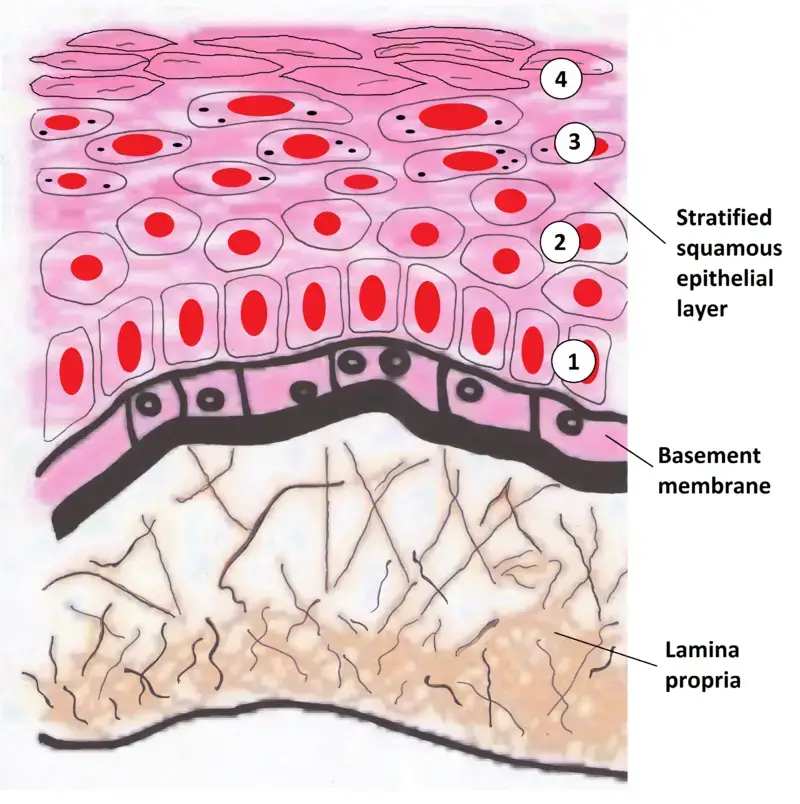
- Oral lamina propria is present below the stratified squamous epithelium of oral cavity. Providing support against friction, pressure and mechanical forces of chewing are the major activities of this layer.
- It is divided into papillary layer and reticular layer. The papillary layer forms the superficial part, while reticular layer is present deeper.
- The papillary layer contains thin and irregularly arranged collagen fibres. A large number of capillary loops are also present within this region.
- Forming connective tissue papillae which extend toward the epithelium is carried out by the papillary layer. The epithelial rete pegs pass downward between these papillae, producing a strong interlocking arrangement.
- This irregular interface increases the area of attachment between epithelium and lamina propria. Supplying nutrients to the avascular epithelial cells is also carried out through the capillary loops present here.
- The reticular layer contains thick collagen fibres arranged mainly parallel to the epithelial surface. Providing tensile strength and deeper mechanical support are related with these fibres.
- In hard palate and attached gingiva, submucosa is absent. Here, fibres of lamina propria become attached directly with the underlying bone.
- This direct attachment forms the mucoperiosteum. Resisting shear force and compression during mastication is carried out by this rigid arrangement.
Bladder Lamina Propria
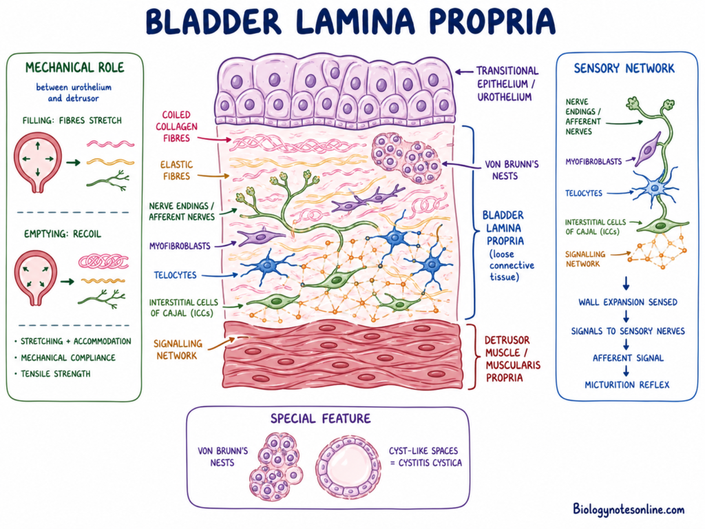
- In urinary bladder, lamina propria is present between the transitional epithelium or urothelium and the detrusor muscle or muscularis propria.
- It forms an important mechanical layer of bladder wall. Allowing stretching and accommodation of large amount of urine without tissue damage is carried out by this region.
- Collagen fibres and elastic fibres are present in a coiled and irregular arrangement. During filling of bladder, these fibres become stretched and during emptying they return toward the earlier arrangement.
- Providing mechanical compliance and tensile strength at the same time are the main properties of this fibrous framework.
- A large number of nerve endings are present in bladder lamina propria. Myofibroblasts, telocytes and interstitial cells of Cajal (ICCs) are also found close with these nerves.
- These cells form a connected signalling network. Sensing the expansion of bladder wall and passing the signals toward sensory nerves are related with this cellular system.
- The afferent signals produced during stretching take part in micturition reflex. This produces the sensation and response related with urination.
- Small islands of urothelial cells may be found within the lamina propria. These are called Von Brunn’s nests.
- In some conditions, these cell nests may form cyst-like spaces. This change is referred to as cystitis cystica.
Intestinal Lamina Propria
- Intestinal lamina propria is mainly adapted for nutrient absorption and mucosal immune defence. It is highly developed in small intestine.
- It extends upward into each intestinal villus and forms its central connective tissue core. It also fills the spaces present between the intestinal glands or crypts of Lieberkühn.
- A rich network of fenestrated blood capillaries is found inside the villus core. Absorption of digested carbohydrates and amino acids is carried out through these capillaries.
- A blind-ended lymphatic vessel called central lacteal is present within each villus. Absorption and transportation of dietary lipids are carried out by this vessel.
- Small smooth muscle fibres extend from the underlying muscularis mucosae into the lamina propria of villi.
- Rhythmic contraction of these muscle fibres produces movement of intestinal villi. It also helps in pushing lymph from the lacteal toward the deeper lymphatic vessels.
- A large number of immune cells are present in intestinal lamina propria. Macrophages, dendritic cells, lymphocytes and IgA-secreting plasma cells are commonly found.
- Detecting, trapping and destroying microorganisms entering with food are carried out by these immune cells. The layer forms an important part of gut-associated lymphoid tissue (GALT).
- In ileum, immune cells form large organised lymphoid nodules called Peyer’s patches. These may extend from lamina propria into the submucosa.
Functions of Lamina Propria
The functions of lamina propria include:
- Support– Providing the epithelial lining with structural and mechanical support, attaching the epithelium with the submucosa, muscle or bone found below it.
- Exchange– It contains a rich supply of blood capillaries while the epithelium does not have blood vessels. Supplying oxygen and nutrients to epithelial cells and removal of metabolic wastes are carried out through these capillaries.
- Defence– It also provided a major region for mucosal defence. Different immune cells including lymphocytes, macrophages, dendritic cells, mast cells and plasma cells are present here, detecting and destroying the microorganisms entering through mucosa.
- Flexibility– The loose and elastic nature of lamina propria allows stretching and returning of the mucosal tissue. This is used in organs having continuous change in volume such as stomach, intestine and urinary bladder.
- Sensation– Housing different sensory receptors and nerve endings for the detection of touch, pain, temperature and mechanical stretching. In urinary bladder, stretching of the tissue produces signals taking part in urination reflex.
- Absorption– In small intestine, it forms the main connective tissue core of intestinal villi. The blood capillaries take up digested carbohydrates and amino acids, while the lacteals are used for absorption and transportation of fats.
- Repair– Fibroblasts present in this layer are involved in producing collagen fibres and other extracellular matrix materials. Myofibroblasts also take part in contraction, tissue repair and the formation of new tissue after injury.
- Lubrication– It gives space for different mucosal glands present in organs such as oesophagus and cervix. The secretion produced by these glands and fluid coming from the blood vessels keeps the mucosal surface moist and lubricated.
Clinical Significance of Lamina Propria
Lamina propria is an important region during inflammation, cancer invasion and different mucosal diseases. Having many blood vessels, lymphatic vessels and immune cells makes this layer highly involved in pathological changes.
Cancer Invasion and Metastasis
- Epithelial tumours initially remain above the basement membrane. At this stage, direct access to blood and lymphatic vessels is generally absent.
- After breaking the basement membrane, tumour cells enter into lamina propria. Reaching the capillary and lymphatic network becomes possible in this stage, increasing the chance of metastasis.
- In urinary bladder, invasion of tumour into lamina propria is referred to as T1 bladder cancer. The tumour has crossed the basement membrane but has not entered into the detrusor muscle.
- In oesophageal and gastric cancer, invasion limited to lamina propria is included under early T1a stage. Presence of lymphatic channels in this region may allow early spread toward regional lymph nodes.
- In Barrett’s oesophagus, duplication of muscularis mucosae may be present. Distinguishing the newly formed muscular layer from original muscularis mucosae is necessary for correct measurement of tumour invasion.
Microscopic Colitis
- Lamina propria is the major region examined during diagnosis of microscopic colitis. The colon may appear normal during colonoscopy, but characteristic changes are seen in biopsy.
- Collagenous colitis shows deposition of a thick collagen band below the epithelial lining. The thickness is generally more than 10 micrometres.
- A large number of lymphocytes and plasma cells are found in superficial lamina propria. The inflammatory cells remain crowded near the thick collagen band.
- Lymphocytic colitis does not show a thick collagen band. Increased intraepithelial lymphocytes and mononuclear inflammatory cells in lamina propria are mainly present.
Celiac Disease
- In celiac disease, gluten produces an immune reaction mainly in the intestinal lamina propria.
- A large number of T lymphocytes, plasma cells, macrophages and neutrophils enter into this tissue. Continuous inflammation becomes present below the intestinal epithelium.
- During this process, the intestinal villi become short, blunt or completely flattened. Absorption of nutrients is reduced due to loss of normal villus structure.
Dystrophic Epidermolysis Bullosa
- The basement membrane is attached with the upper part of lamina propria through anchoring fibrils. These fibrils are mainly formed of Type VII collagen.
- Mutation in the COL7A1 gene causes defective or absent Type VII collagen. This condition is called dystrophic epidermolysis bullosa (DEB).
- Due to loss of anchoring fibrils, the epithelium becomes easily separated from the underlying connective tissue. Severe blistering, wound formation and tissue scarring are commonly produced.
Systemic Amyloidosis
- In systemic AL amyloidosis, abnormal protein material called amyloid becomes deposited in extracellular tissue.
- Amyloid deposits may be found in the interstitial region and walls of blood vessels present in gastrointestinal lamina propria.
- Rectal mucosal biopsy is commonly used for detecting this deposition. Staining with Congo red helps in identification of amyloid present in lamina propria and submucosa.
Von Brunn’s Nests and Cystitis Cystica
- In urinary bladder, small groups of urothelial cells may become embedded within lamina propria. These epithelial groups are called Von Brunn’s nests.
- Most of these nests are benign and do not produce serious changes. In some cases, the central part undergoes cystic change.
- Formation of fluid-filled cysts from Von Brunn’s nests is referred to as cystitis cystica.
Lamina Propria vs Basement Membrane
| Basis | Lamina Propria | Basement Membrane |
|---|---|---|
| Location | It is present below the basement membrane. Forms the deeper connective tissue part of mucosa. | It is found immediately below the epithelial cells. Forms the boundary between epithelium and connective tissue. |
| Thickness | Comparatively thick layer. | Very thin layer. |
| Tissue nature | It is made up of loose or areolar connective tissue. | It is a thin non-cellular sheet-like layer. |
| Parts | It is not generally divided into basal lamina and reticular lamina. In some regions, papillary and reticular parts may be present. | Basal lamina and lamina reticularis together form the basement membrane. |
| Origin | Mainly formed and maintained by fibroblasts and other connective tissue cells. | Basal lamina is produced by epithelial cells. Lamina reticularis is formed from underlying connective tissue. |
| Major fibres | Type I collagen, Type III collagen and elastic fibres are mainly present. | Type IV collagen is the major collagen. Type VII collagen forms anchoring fibrils. |
| Ground substance | Contains glycosaminoglycans, proteoglycans and glycoproteins in hydrated ground substance. | Contains laminin, fibronectin and other basement membrane glycoproteins. |
| Cells | Highly cellular layer. Fibroblasts, myofibroblasts, immune cells, telocytes and other cells are found. | Cells are not present within this layer. |
| Blood vessels | Contains a rich network of blood capillaries and lymphatic vessels. | Blood vessels are absent. |
| Nerve supply | Different nerve endings are present, taking part in sensation and local responses. | Nerve endings are not present within the membrane. |
| Nutrient supply | Supplying oxygen and nutrients to epithelial cells is carried out by the capillaries present here. | Nutrients pass across this membrane from lamina propria to the epithelium. |
| Immune role | A large number of immune cells are present. It forms an important region for mucosal defence. | It mainly acts as a physical supporting and separating layer. |
| Main function | Providing support, flexibility, immune defence, tissue repair and nutrition to epithelium. | Providing attachment, filtration and separation of epithelium from connective tissue. |
| Connection | Its Type III collagen fibres are attached with anchoring fibrils coming from basement membrane. | Type VII collagen anchoring fibrils connect it with the lamina propria. |
| Part of mucosa | Forms the main connective tissue part of mucosa. | Forms the thin layer between epithelium and lamina propria. |
Lamina Propria vs Submucosa
Lamina propria and submucosa are two connective tissue layers found in the wall of mucous membrane. Both layers provide support, blood supply and nerve supply, but their position, tissue arrangement and components are different.
| Basis | Lamina Propria | Submucosa |
|---|---|---|
| Location | It is present immediately below the epithelial lining and basement membrane. | It is present below the mucosa and lies deeper in the wall of organ. |
| Part of | The epithelium, lamina propria and muscularis mucosae together form the mucosa. | It is not a part of mucosa. It is found between mucosa and muscular layer. |
| Boundary | It is separated from submucosa by the muscularis mucosae. | It begins below the muscularis mucosae. |
| Absence | In regions where submucosa is absent, lamina propria becomes directly attached with the underlying bone or muscle. This is found in hard palate and attached gingiva. | It is absent in some regions such as hard palate and attached gingiva. |
| Tissue type | It is made up of loose or areolar connective tissue. Having more ground substance and loosely arranged fibres gives it a soft and flexible nature. | It is mainly made up of dense irregular connective tissue. Thick fibrocollagenous bundles are present in this layer. |
| Density | Comparatively loose, thin and highly cellular layer. | Comparatively broad, thick and dense layer. |
| Flexibility | Its loose arrangement allows movement of cells, diffusion of nutrients and stretching of mucosa. | It provides stronger mechanical support and allows the mucosa to move over the deeper muscular layer. |
| Fat cells | Fat cells are generally absent or present in very less number. | Adipose tissue is commonly found in many regions of submucosa. |
| Blood vessels | It contains small blood capillaries supplying oxygen and nutrients to the avascular epithelium. | Larger blood vessels are present and pass through this layer before giving smaller branches to the mucosa. |
| Lymphatics | Small lymphatic vessels are found. In intestinal villi, lacteals are present within this layer. | Larger lymphatic vessels are carried through the submucosa. |
| Nerve supply | Small nerve endings are present, taking part in sensation and local mucosal responses. | It contains larger nerve bundles and the submucosal plexus (Meissner’s plexus). |
| Nerve function | Detection of touch, pain and mechanical stretching is carried out by its nerve endings. | Controlling local secretion and contraction of muscularis mucosae is carried out by the submucosal plexus. |
| Glands | Mucosal glands may be present, such as cardiac and pyloric glands of stomach. | Deeper glands such as Brunner’s glands of duodenum and minor salivary glands may be present. |
| Immune cells | A large number of immune cells are present. It forms an important part of mucosa-associated lymphoid tissue (MALT). | Large lymphoid collections may extend into this layer from lamina propria. |
| Lymphoid tissue | Lymphocytes, macrophages, plasma cells and other immune cells are commonly scattered throughout the layer. | Large lymphoid structures such as Peyer’s patches may extend deeply into submucosa. |
| Fibroblasts | Fibroblasts are present and involved in formation and renewal of the extracellular matrix. | Fibroblasts produce the thick collagen bundles forming the dense connective tissue framework. |
| Telocytes | Telocytes mainly form a two-dimensional network below the epithelium. Maintaining epithelial barrier and intestinal stem cell region are related with these cells. | Telocytes form a dense three-dimensional supporting network around vessels, glands and nerve ganglia. |
| Main function | Providing nutrition to epithelium, immune defence, tissue repair and flexible support are its major functions. | Providing strong support and carrying large vessels, nerves, glands and lymphatic structures are the major functions. |
FAQs on Lamina Propria
What type of tissue is lamina propria?
Lamina propria is made up of loose or areolar connective tissue. Having more ground substance and loosely arranged fibres gives this tissue its flexible nature.
What is lamina propria made of?
It is made up of extracellular matrix, collagen fibres, elastic fibres, blood vessels, lymphatic vessels, nerve endings and different types of cells. Type I collagen and Type III collagen are mainly present in its fibrous framework.
Is lamina propria part of the mucosa?
Yes, lamina propria is a part of mucosa. The epithelium, lamina propria and muscularis mucosae together form the mucosa in most organs.
How to identify lamina propria in histology?
Lamina propria is identified as the loose connective tissue region present just below the epithelial lining and basement membrane. Loosely arranged collagen fibres, many cells, small blood vessels and more ground substance are commonly seen in this region. In intestinal villi, it forms the central connective tissue core.
What is lamina propria?
Lamina propria is a thin layer of loose connective tissue found below the epithelial lining. It forms the main supporting connective tissue part of mucosa.
Where is the lamina propria located?
It is located immediately below the epithelium and its basement membrane. In most organs, it remains above the muscularis mucosae.
What cells are found in the lamina propria?
Different stromal and immune cells are present. Fibroblasts, myofibroblasts, telocytes, macrophages, lymphocytes, plasma cells, mast cells, eosinophils and dendritic cells are commonly found.
Does the lamina propria contain blood vessels?
Yes, many small blood capillaries and lymphatic vessels are present. Supplying oxygen and nutrients to the avascular epithelium and removal of metabolic wastes are carried out through these vessels.
What is the difference between lamina propria and submucosa?
Lamina propria is a loose connective tissue layer present just below the epithelium. Submucosa is a deeper and denser connective tissue layer, generally found below the muscularis mucosae. Larger vessels, nerves and deeper glands are commonly present in submucosa.
What is the immune function of lamina propria?
It forms an important site for mucosal immune defence. A large number of lymphocytes, macrophages, plasma cells and dendritic cells are present, detecting and destroying microorganisms entering through the mucosal surface. It also forms a major part of mucosa-associated lymphoid tissue (MALT).
References
- Andersson, K.-E., & McCloskey, K. D. (2014). Lamina propria: The functional center of the bladder? Neurourology and Urodynamics.
- Telocytes, a distinct type of cell among the stromal cells present in the lamina propria of jejunum. (n.d.). ResearchGate.
- Amyloid in biopsies of the gastrointestinal tract—a retrospective observational study on 542 patients. (n.d.). PMC.
- Anatomy and physiology, energy, maintenance, and environmental exchange, the digestive system. (n.d.). OERTX.
- Collins, J. T., Nguyen, A., Omole, A. E., & Badireddy, M. (2025). Anatomy, abdomen and pelvis, small intestine. In StatPearls. StatPearls Publishing.
- Naini, B. V., Souza, R. F., & Odze, R. D. (2016). Barrett’s esophagus: A comprehensive and contemporary review for pathologists. The American Journal of Surgical Pathology, 40(5), e45–e66.
- Basement membranes in development and disease. (n.d.). PMC – NIH.
- Binding of laminin to type IV collagen: A morphological study. (n.d.). PMC – NIH.
- Zukauskas, R. (2024). Capillary (human physiology). In Health and Medicine | Research Starters. EBSCO.
- Capillary endothelium with fenestrations: Structure & function. (n.d.). King of the Curve.
- Clinical and endoscopic manifestations of gastrointestinal amyloidosis: A case series. (n.d.). PMC.
- Hempel, K. A., & Sharma, A. V. (2022). Collagenous and lymphocytic colitis. In StatPearls. StatPearls Publishing.
- Collagenous and lymphocytic colitis. (n.d.). Johns Hopkins Medicine.
- Colonoscopy leading to the diagnosis of AL amyloidosis in the gastrointestinal tract mimicking an acute ulcerative colitis flare. (n.d.). PMC.
- Combination of two T1 substaging systems (T1a/b/c and T1m/e) better predicts tumor outcomes in patients with T1 high grade bladder cancer. (n.d.). PMC.
- Turan, T., Efiloğlu, Ö., Günaydin, B., Özkanli, Ş., Nikerel, E., Atiş, G., Çaşkurlu, T., & Yildirim, A. (2018). Comparative differences between T1a/b and T1e/m as substages in T1 urothelial carcinoma of the bladder. International Braz J Urol, 44(2), 267-272.
- Comparative differences between T1a/b and T1e/m as substages in T1 urothelial carcinoma of the bladder. (n.d.). PMC.
- Saremian, J., & Yeh, M. M. (2008). DIAGNOSIS: Collagenous colitis. Discussion. OU Health Campus.
- Diagnosis of celiac disease. (n.d.). Celiac Disease Foundation.
- Emerging diverse roles of telocytes. (n.d.). PMC.
- Female genital tract. (n.d.). WebPath.
- GI tract. (2024). Duke Histology.
- Gastrointestinal system – histology education. (n.d.). GW Blogs – The George Washington University.
- Grading and staging. (n.d.). Action Bladder Cancer UK.
- Bolla, S. R., Odeluga, N., Amraei, R., & Jetti, R. (2023). Histology, bladder. In StatPearls. StatPearls Publishing.
- Godwin, L., Tariq, M. A., & Crane, J. S. (2023). Histology, capillary. In StatPearls. StatPearls Publishing.
- Brizuela, M., & Winters, R. (2023). Histology, oral mucosa. In StatPearls. StatPearls Publishing.
- Kia’i, N., & Bajaj, T. (2023). Histology, respiratory epithelium. In StatPearls. StatPearls Publishing.
- Histopathology. (n.d.). PMC – NIH.
- Inhibition of laminin self-assembly and interaction with type IV collagen by antibodies to the terminal domain of the long arm. (n.d.). PMC.
- Radenkovic, G., Petrovic, V., Zivanovic, D., Stoiljkovic, N., Sokolovic, D., Zivkovic, N., Radenkovic, D., Velickov, A., & Jovanovic, J. (2021). Interstitial cells of Cajal and neural structures in the human fetal appendix. Journal of Neurogastroenterology and Motility, 27(1), 127-133.
- Interstitial cells of Cajal and telocytes in the gut: Twins, related or simply neighbor cells? (n.d.). PubMed.
- Localization of type IV collagen, laminin, heparan sulfate proteoglycan, and fibronectin to the basal lamina of basement membranes. (n.d.). PMC.
- Microscopic colitis: Is it a spectrum of inflammatory bowel disease? (n.d.). PMC – NIH.
- Mucosa-associated lymphoid tissue (MALT). (2025). Cleveland Clinic.
- Mucosal immune response in health & disease – mini-review. (n.d.). Bio-Rad Antibodies.
- Mucosal immunity of the intestine. (n.d.). R&D Systems.
- Hall, E. J. (2007). Mucosal immunology–Why it’s important. WSAVA2007, Veterinary Information Network.
- Noninfectious colitides: Collagenous colitis, lymphocytic colitis, diversion colitis, and chemically induced colitis. (n.d.). PMC.
- Organization, barrier function and antimicrobial lipids of the oral mucosa. (n.d.). PMC.
- Oral: Four layers of the GI tract. (n.d.). Digestive: The Histology Guide – University of Leeds.
- Sweet, T., Abraham, C. M., & Rich, A. (2024). Origin and development of interstitial cells of Cajal. The International Journal of Developmental Biology, 68, 93-102.
- Overview of the digestive system. (n.d.). Anatomy and Physiology II – Lumen Learning.
- Tinguria, M. (2024). Pathology of celiac disease: An insight about etiopathogenesis, pathophysiology and histologic diagnosis. Medical Research Archives, 12(9).
- Hill, M. A. (2026). Respiratory system – histology. UNSW Embryology.
- Staging & grade – bladder cancer. (n.d.). Johns Hopkins Pathology.
- Telocytes and interstitial cells of Cajal in the biliary system. (n.d.). PMC – NIH.
- Telocytes are reduced during fibrotic remodelling of the colonic wall in ulcerative colitis. (n.d.). PMC.
- The charming world of the extracellular matrix: A dynamic and protective network of the intestinal wall. (n.d.). PMC.
- The lamina propria: A comprehensive research report on the structural, functional, and pathological dynamics of the mucosal connective tissue. (n.d.).
- Vannucchi, M. G. (2020). The telocytes: Ten years after their introduction in the scientific literature. An update on their morphology, distribution, and potential roles in the gut. International Journal of Molecular Sciences, 21(12), 4478.
- Janeway, C. A. Jr., Travers, P., Walport, M., et al. (2001). The mucosal immune system. In Immunobiology: The immune system in health and disease (5th ed.). Garland Science.
- Type VII collagen: The anchoring fibril protein at fault in dystrophic epidermolysis bullosa. (2018). Plastic Surgery Key.
- Type VII collagen: The anchoring fibril protein at fault in dystrophic epidermolysis bullosa. (n.d.).
- Type VII collagen forms an extended network of anchoring fibrils. (n.d.). PMC – NIH.
- Understanding your pathology report: Barrett’s esophagus and dysplasia. (2025). American Cancer Society.
- Li, T., Huang, X., Cheng, S., Zhao, L., Ren, G., Chen, W., Wang, Q., Zeng, C., & Liu, Z. (2017). Utility of abdominal skin plus subcutaneous fat and rectal mucosal biopsy in the diagnosis of AL amyloidosis with renal involvement. PLoS ONE, 12(9), e0185078.