

Voluntary muscles are skeletal muscles that can be deliberately activated, whereas involuntary muscles work without conscious control and include smooth muscle and cardiac muscle. The main differences involve their location, nerve supply, microscopic structure, speed of contraction, and resistance to fatigue.
What are voluntary muscles?
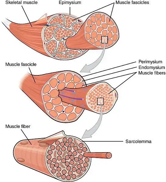
Voluntary muscles are skeletal muscles whose movement can be consciously controlled. Most attach to bones through tendons and produce actions such as walking, lifting, speaking, maintaining posture, and changing facial expression.
- Their long, cylindrical muscle fibers have many nuclei and a striated appearance under a microscope.
- Actin and myosin are arranged into repeating contractile units called sarcomeres.
- Conscious movement is initiated in the central nervous system and carried to skeletal muscle by somatic motor neurons. Sensory, or afferent, nerves return information about muscle length, tension, and position to the central nervous system.
- Skeletal muscle fibers require stimulation at the neuromuscular junction to contract; they are not normally myogenic.
- They can contract rapidly and forcefully, but sustained intense activity can cause fatigue.
What are involuntary muscles?
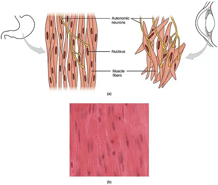
Involuntary muscles contract without conscious direction. This group includes smooth muscle in the walls of hollow organs and blood vessels, and cardiac muscle in the heart. Their activity supports continuous processes such as circulation, digestion, airway regulation, and movement of material through internal organs.
- Smooth muscle cells are spindle-shaped, usually contain one central nucleus, and do not show visible striations.
- Cardiac muscle is striated, usually has one central nucleus per cell, and contains intercalated discs that link neighboring cells.
- Smooth muscle is regulated by the autonomic nervous system, hormones, local chemical conditions, and stretching. Autonomic nerve fibers commonly release neurotransmitters from swellings called varicosities.
- Cardiac muscle is myogenic: pacemaker cells generate the heart’s rhythm. Autonomic nerves modify the rate and force of contraction rather than initiating every heartbeat.
- Many involuntary contractions are slower and more sustained than skeletal muscle contractions and can continue for long periods with relatively little fatigue.
Examples of voluntary and involuntary muscles
Biceps and other limb muscles
The biceps brachii, triceps, quadriceps, hamstrings, and most other muscles that move the limbs are voluntary skeletal muscles. The biceps brachii flexes the elbow and helps turn the forearm so the palm faces upward. These actions can be started and stopped consciously through the somatic nervous system.
Diaphragm
The diaphragm is skeletal muscle with both automatic and voluntary control. The brainstem normally drives breathing automatically through the phrenic nerves, but a person can temporarily alter breathing during speaking, singing, or breath-holding. For this reason, the diaphragm should not be described as exclusively voluntary.
Smooth muscle
Smooth muscle occurs in the walls of the stomach, intestines, urinary bladder, uterus, airways, and blood vessels. Single-unit smooth muscle cells often contract together because they are electrically connected, while multiunit smooth muscle permits finer, more independent control. Examples of multiunit smooth muscle include muscles associated with the iris of the eye.
Cardiac muscle
Cardiac muscle forms the muscular wall of the heart. It is involuntary but striated. Its branched cells are connected by intercalated discs, allowing coordinated contraction. Electrical activity generated by pacemaker cells triggers calcium-dependent interactions between actin and myosin, producing each heartbeat.
Similarities between voluntary and involuntary muscles
- Both are muscle tissues specialized to produce force and movement.
- Both use actin, myosin, calcium ions, and chemical energy from ATP during contraction.
- Both receive nerve input, although the somatic and autonomic nervous systems have different roles.
- Both help maintain normal body function by moving the body itself or materials within the body.
Voluntary vs involuntary muscles: comparison table
| Basis of comparison | Voluntary muscles | Involuntary muscles |
|---|---|---|
| Definition | Skeletal muscles that can be consciously activated | Smooth and cardiac muscles that work without conscious direction |
| Primary location | Usually attached to bones; also present in structures such as the tongue and upper esophagus | Walls of internal organs and blood vessels; cardiac muscle occurs only in the heart |
| Nervous control | Somatic motor nervous system | Autonomic nervous system regulates smooth and cardiac muscle; cardiac rhythm originates in pacemaker tissue |
| Appearance | Striated | Smooth muscle is non-striated; cardiac muscle is striated |
| Cell shape | Long, cylindrical, unbranched fibers | Smooth cells are spindle-shaped; cardiac cells are shorter and branched |
| Nuclei | Many nuclei near the edge of each fiber | Usually one central nucleus per smooth or cardiac muscle cell |
| Sarcomeres | Present | Absent in smooth muscle; present in cardiac muscle |
| Intercalated discs | Absent | Present in cardiac muscle but absent in smooth muscle |
| Contraction | Often rapid and forceful | Smooth muscle is generally slow and sustained; cardiac muscle contracts rhythmically |
| Fatigue | Can fatigue during sustained or intense activity | Generally resistant to fatigue; cardiac muscle normally works continuously |
| Examples | Biceps, triceps, quadriceps, hamstrings, and muscles used for facial expression | Muscle in the intestines, bladder, uterus, airways, blood vessels, and heart |