

Spread plate method is a microbiological plating method where a measured volume of liquid microbial suspension is spread over the surface of solidified agar medium. It is also called spread plate technique or spread plating. The agar medium is already solidified before inoculation. The microbial suspension remains on the surface, and after suitable incubation visible colonies are formed on the agar surface.
The colonies do not grow inside the medium.
The method is mainly used for isolation and enumeration of microorganisms. A diluted microbial sample is commonly used when the number of microorganisms is high. Serial dilution is often carried out before plating. It gives well-separated colonies on the solid agar surface.
Each visible colony is counted as a colony-forming unit (CFU). CFU may develop from a single viable cell or from a group of cells, not always from one cell. The number of colonies is used to estimate the culturable microorganisms present in the original sample.
Spread plate method is a quantitative method used for viable plate counting. It can also be used for qualitative examination of colony appearance and isolation of distinct microbial colonies.
Objectives of Spread Plate Method
- To spread a measured volume of liquid microbial suspension over the surface of a solidified agar medium.
- To obtain well-separated microbial colonies on the agar surface. The spreading helps in distributing the inoculum evenly, so that individual colony-forming units can develop as separate colonies after incubation.
- To estimate the number of viable and culturable microorganisms present in a sample. The colonies formed on the plate are counted and expressed as colony-forming units (CFU).
- To isolate microorganisms from a diluted mixed microbial suspension.
- To observe the colony characters of microorganisms growing on the surface of the agar medium. These characters may include colony size, shape, colour, margin and surface appearance.
- To check the microbial load in different samples such as water, food, soil, clinical material, or laboratory culture.
- To use the method in viable plate count, where only living microorganisms capable of forming colonies under the given culture condition are counted. Not all microorganisms present in the sample will grow on the selected medium.
Principle of Spread Plate Method
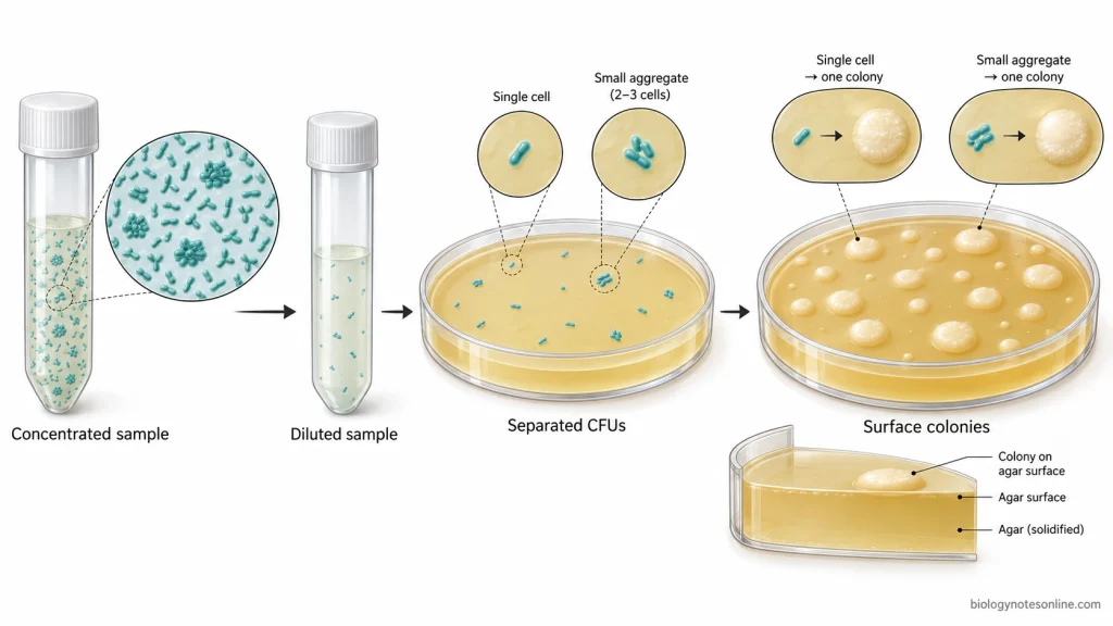
The principle of spread plate method is based on the separation of viable and culturable microorganisms on the surface of a solidified agar medium. A measured diluted suspension is spread over the agar surface. In this condition, microbial cells or cell groups remain apart from each other and after incubation they form visible surface colonies.
Dilution has a direct role in this method. If the suspension is concentrated, the microorganisms remain very close and the colonies become crowded. In a suitable dilution, the colony-forming units (CFU) are separated as individual colony producing units on the plate surface. Each CFU can multiply and gives rise to one visible colony under the selected culture condition.
A CFU is not always one microbial cell. It may be one viable cell, pair of cells, chain, clump, or any group that can produce one colony. So the result is written as CFU and not as exact cell number.
The colonies formed on a countable plate are used for quantitative estimation of viable microorganisms in the original sample. The count depends on the culture medium, incubation temperature, incubation time, oxygen condition and the ability of the microorganisms to grow in that selected condition. Only those microorganisms which grow and form colonies are counted.
Requirements for Spread Plate Method
- Suitable pre-solidified agar plates are required as the solid culture medium. The agar surface should be sterile and dry.
- Liquid microbial sample or suspension is used as the inoculum. It contains the microorganisms which are to be spread on the agar surface.
- Sterile diluent is used for preparing serial dilution of the sample. It reduces the microbial concentration.
- Dilution tubes are required to hold the sterile diluent and diluted samples. Each tube should be labelled properly.
- Micropipette and sterile tips are used to transfer a measured amount of inoculum to the agar plate.
- A sterile L-shaped glass spreader or disposable spreader is used to spread the inoculum over the agar surface. It is the spreading instrument, not the medium.
- Sterile glass beads may be used in some methods for spreading the inoculum. The beads are rolled over the agar surface.
- Incubator is required for keeping the inoculated plates at the required temperature. During incubation, visible colonies are formed.
- Labelling materials are used to mark the sample name, dilution, date and medium on the plate.
- Protective equipment such as lab coat, gloves and eye protection are required for safe handling of microbial sample. Aseptic condition is also needed.
Procedure of Spread Plate Method
workflow = Sample suspension → Serial dilution → Measured inoculum → Solidified agar plate → Spreading → Absorption → Incubation → Surface colonies → Colony examination
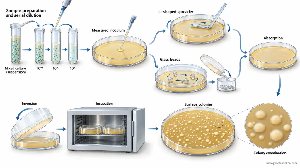
Preparation of Sample and Serial Dilution
- Prepare the microbial sample or suspension in sterile condition. The sample should be mixed properly before dilution.
- Take sterile dilution tubes containing sterile diluent. Label each tube with dilution value.
- Transfer a measured volume of sample into the first dilution tube. Mix it well.
- Transfer the measured volume from first dilution to the next dilution tube. Continue the serial dilution as required.
- Select suitable dilution for plating. Highly concentrated sample gives crowded growth and colonies cannot be counted properly.
Inoculation and Spreading of the Sample
- Take a sterile pre-solidified agar plate. The agar surface should be dry before inoculation.
- Transfer a measured volume of diluted sample on the centre of agar surface with sterile micropipette tip. Usually small volume is used for spread plating.
- For spreading with a L-shaped glass spreader or disposable spreader, hold the spreader gently on the agar surface. Spread the inoculum evenly over the surface while rotating the plate slowly.
- The spreader should cover the full agar surface without cutting the agar. The liquid sample forms a thin film over the plate.
- For spreading with sterile glass beads, add sterile beads on the inoculated agar plate. Move the plate gently in circular or back-and-forth motion.
- The beads roll over the agar surface and distribute the inoculum. After spreading, remove the beads aseptically.
- Keep the plate for short time so that the inoculum is absorbed into the agar surface.
Incubation of Spread Plates
- Invert the plates after absorption. This prevents condensed water from falling on the agar surface.
- Incubate the plates at suitable temperature and time according to the microorganism and medium used.
- During incubation, viable and culturable microorganisms multiply on the agar surface. Visible surface colonies are formed.
- After incubation, examine the plates for colony growth. Count plates with well-separated colonies and observe colony characters such as size, shape, colour, margin and surface.
Precautions and Common Procedural Errors
- Sterile plates, sterile tips and sterile spreader should be used. If the spreader or tip touches non-sterile surface, it should not be used again.
- The Petri plate lid should not be opened fully or for long time. Open it only enough to add the sample or spread the inoculum.
- The agar surface should be dry before inoculation. Wet agar surface gives uneven spreading and colonies may run together.
- Use a measured volume of inoculum. Too much inoculum remains as liquid on the agar surface and makes spreading difficult.
- The dilution should be mixed properly before taking the sample. Poor mixing gives uneven number of microorganisms in the transferred volume.
- Use a fresh sterile tip for each dilution and plate. Same tip can carry organisms from one dilution to another.
- The glass spreader should be sterilized and cooled before touching the inoculum. Hot spreader can kill microbial cells.
- The inoculum should be spread gently and evenly over the agar surface. Too much pressure can scratch the agar.
- The spreader should not push the liquid only to one side or edge of the plate. This gives uneven colony distribution.
- In glass bead spreading, only sterile beads should be used. The beads should roll over the agar surface and should be removed aseptically after spreading.
- The plate should be kept for short time after spreading. The inoculum should be absorbed before the plate is inverted.
- Plates should be incubated in inverted position. This prevents condensed water from falling on the agar surface.
- Plates should be labelled on the bottom part of the Petri plate. Sample name, dilution, medium and date should be written properly.
- Crowded plates should not be used for accurate colony count. Merged colonies and spreading colonies can give wrong count.
- Same incubation condition should be maintained for comparable plates. Change in temperature, time, medium, or oxygen condition can change colony development.
Results of Spread Plate Method
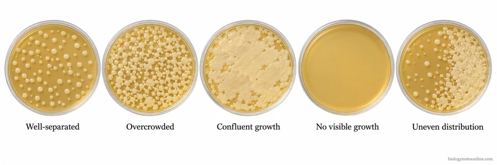
Observation of Isolated Colonies
The incubated spread plate is first observed for colonies on the agar surface. In a proper plate, the colonies are visible and separated. They are surface colonies.
The colony number is noted. Colony characters are also observed such as size, shape, colour, margin, elevation, surface and texture. These characters gives only a primary idea about the colony present on the plate.
It is not final identification.
Colony appearance may change with medium, incubation time and temperature.
Interpretation of Colony Growth Patterns
Well-separated surface colonies shows a good spread plate result. The colonies remain distinct from each other. These colonies can be counted or picked for further study.
In overcrowded growth, many colonies remain very close. Some colonies join with each other. In confluent growth, the agar surface may be covered with continuous microbial growth. Individual colonies are not clearly seen.
Such plate is not used for accurate colony count.
A plate with no visible growth shows that no colony has developed under the selected culture condition. It may occur due to absence of viable organisms, unsuitable medium, wrong incubation condition, inhibitory condition, or procedural error.
Irregular colony distribution occurs when the inoculum is not spread evenly. Colonies may collect on one side, near the edge, or in streak-like patches. The plate gives poor interpretation.
For example, a diluted sample plate with many separate, round, cream-coloured colonies on the agar surface is an interpretable spread plate result. The colonies can be described and counted. Exact microbial species cannot be confirmed from this appearance only. Microscopic, biochemical, molecular, or other identification tests are required.
CFU Calculation Using Spread Plate Method
Colony-forming unit (CFU) is used for viable and culturable microorganisms. In the spread plate method, the colonies counted from a selected plate are converted into CFU/mL of the original suspension.
Colony number on the plate is only the observed colony count. CFU/mL is the calculated value after correcting the colony count with dilution and plated volume.
Selection of Countable Plates
After incubation, a plate with clear and separated colonies is selected. The colonies should be easy to count.
Overcrowded plates are not used for accurate calculation. Very few colonies also gives poor counting result. The acceptable colony-count range depends on the analytical protocol, such as food, water, clinical, or teaching laboratory method.
It is not a universal CFU/mL threshold.
The selected plate must have known dilution and known plated volume. These two values are required.
CFU/mL Formula
The formula is-
CFU/mL = Number of colonies ÷ (Dilution × Volume plated in mL)
Here, number of colonies means the colonies counted on the selected spread plate. Dilution means the dilution plated, such as 10⁻³ or 10⁻⁴. Volume plated means the amount of diluted sample added on the agar surface.
If 0.1 mL is plated, the value is corrected to 1 mL. The dilution is also corrected back to the original suspension.
Example of CFU Calculation
Suppose 85 colonies are counted from a spread plate of 10⁻⁴ dilution. The volume plated is 0.1 mL.
CFU/mL = 85 ÷ (10⁻⁴ × 0.1)
CFU/mL = 85 ÷ 10⁻⁵
CFU/mL = 8.5 × 10⁶ CFU/mL
The original suspension contains 8.5 × 10⁶ CFU/mL under the selected culture condition. This value represents colony-forming units, not exact number of microbial cells.
Applications of Spread Plate Method
- The spread plate method is used to count viable microorganisms in food, milk, water and environmental samples. The results are expressed as colony-forming units (CFU).
- To isolate microorganisms from mixed cultures and obtain pure cultures for identification and biochemical studies.
- Nitrogen-fixing bacteria can be enriched from soil using nitrogen-deficient media.
- In genetic engineering, transformed bacterial cells having antibiotic-resistance genes are selected on agar containing suitable antibiotics.
- Microbial mutants are screened using indicator media. The colonies are distinguished by their colour or other visible reactions, such as blue-white screening in molecular cloning.
- Used to determine antimicrobial activity by comparing the surviving microbial colonies in treated and untreated samples.
Limitations of Spread Plate Method
- Usually, a small volume of sample (0.1 mL) is used in the spread plate method. Samples containing very few microorganisms may not be suitable.
- Only culturable microorganisms are counted. The viable microorganisms which cannot grow on the selected culture medium remain undetected.
- Oxygen-sensitive microorganisms may not survive under normal aerobic plating conditions and require special anaerobic conditions for their growth.
- When the microbial concentration is high, colonies may overlap and become difficult to count. The sample needs to be diluted before plating.
- Excess moisture on the agar surface may cause the colonies to spread and merge together.
- Bacterial cells present in chains or clumps may form a single colony, resulting in an underestimation of the actual cell number.
- The incubation period may take several days for colony development. Slow-growing microorganisms require even longer time.
Advantages of Spread Plate Method
- The spread plate method is simple and easy to perform using solidified agar plates.
- The microorganisms are not exposed to the high temperature of molten agar, making it suitable for heat-sensitive microorganisms.
- All the colonies develop on the agar surface. They can be easily observed, counted and picked up for further studies.
- The method favours the growth of aerobic microorganisms as the colonies develop on the surface of the agar medium.
References
- Bush, P., & Martin, K. (n.d.). Standard plate count. In WPUNJ microbiology laboratory manual. William Paterson University. https://press.wpunj.edu/microbiologylabmanual/chapter/standard-plate-count/
- Frazier, A. (n.d.). Microbial growth. In Medical microbiology for nursing and allied health. LOUIS: The Louisiana Library Network. https://louis.pressbooks.pub/microbiology/chapter/microbial-growth/
- Miller, E. (2024). Colony morphology. In Microbiology laboratory manual. Open Oregon Educational Resources. https://openoregon.pressbooks.pub/microbiologylaboratorymanual/chapter/background-theory-5/
- Pakpour, N., & Horgan, S. (2021, August 1). Lab 9: Standard plate count. In General microbiology lab manual. Biology LibreTexts. https://bio.libretexts.org/Learning_Objects/Laboratory_Experiments/Microbiology_Labs/Book:General_Microbiology_Lab_Manual(Pakpour_and_Horgan)/Lab_09:_Standard_Plate_Count
- Sanders, E. R. (2012). Aseptic laboratory techniques: Plating methods. Journal of Visualized Experiments, 63, e3064. https://doi.org/10.3791/3064
- Thomas, P., Sekhar, A. C., Upreti, R., Mujawar, M. M., & Pasha, S. S. (2015). Optimization of single plate-serial dilution spotting (SP-SDS) with sample anchoring as an assured method for bacterial and yeast CFU enumeration and single colony isolation from diverse samples. Biotechnology Reports, 8, 45–55. https://doi.org/10.1016/j.btre.2015.08.003
- Thompson, M. (2027). Lab 13 food microbiology. In Medical microbiology for nursing and allied health lab manual. LOUIS: The Louisiana Library Network. https://louis.pressbooks.pub/microbiology-lab/chapter/lab-13-food-microbiology/
- University of Wisconsin–Madison Department of Bacteriology. (n.d.). Spread plates and dilution plating. In Virtual microbiology. https://instruction.bact.wisc.edu/book/displayarticle/7
- University of Wisconsin–Madison Department of Bacteriology. (n.d.). Viable plate counts. In Virtual microbiology. https://instruction.bact.wisc.edu/book/displayarticle?aid=106
- Wingfield, D., Bess, J., & Whitlock, J. (n.d.). Exercise 16: Serial dilution & standard plate count. In Microbiology lab manual (Hillsborough College Dale Mabry). Pressbooks. https://pressbooks.hcfl.edu/microlabmanual/chapter/exercise-16-serial-dilution-standard-plate-count/
Wonderful
Thank You