

Red blood cells (RBCs) and white blood cells (WBCs) are two major cellular elements of blood. They are also referred to as erythrocytes and leukocytes, respectively.
Both RBCs and WBCs are produced from hematopoietic stem cells, mainly in the bone marrow. However, their structure and functions are very different.
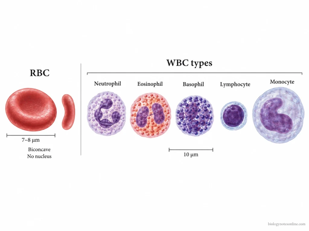
Red blood cells (RBCs)– RBCs are far more numerous in the blood. Mature human RBCs are biconcave cells without a nucleus and most of the cell organelles. They contain hemoglobin which is mainly used for carrying oxygen from the lungs to different body tissues. Hemoglobin also takes part in carbon dioxide transport.
White blood cells (WBCs)– WBCs are nucleated cells and occur in different forms. These include granulocytes, monocytes, and lymphocytes. They perform the immune and defensive functions of the body against pathogens and other harmful agents.
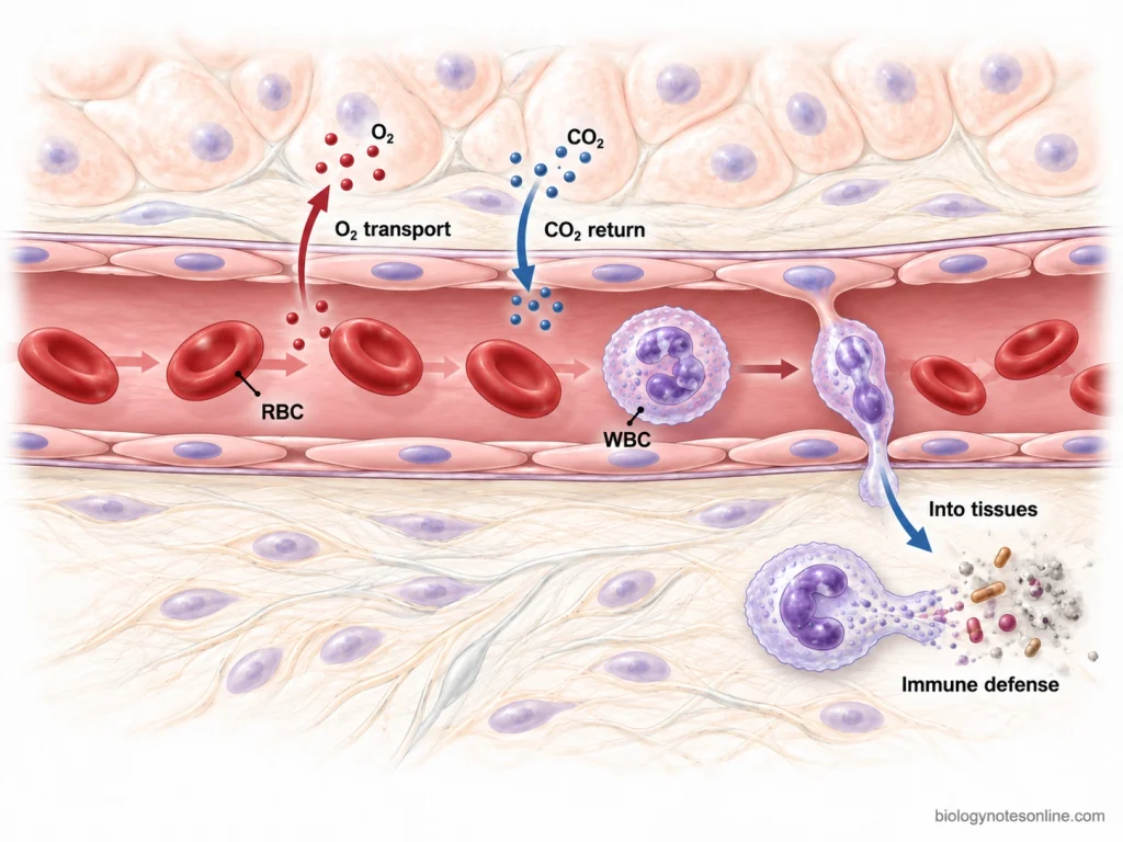
WBCs are much fewer in the circulating blood compared to RBCs. Many WBCs can move out from the blood vessels into tissues, whereas RBCs normally remain within the vascular circulation.
RBC vs WBC Comparison Table
| Basis of Difference | RBC (Red Blood Cell) | WBC (White Blood Cell) |
|---|---|---|
| Other name | RBCs are also called erythrocytes. | WBCs are also called leukocytes. |
| Colour | They appear red because of the presence of hemoglobin. | They are colourless or pale cells and do not contain hemoglobin. |
| Shape | Mature RBC is a biconcave disc, thinner at the centre. The cells are highly flexible. | Shape is not the same for all WBCs. It varies according to the different leukocyte types. |
| Nucleus | Nucleus is absent in mature human RBCs. | A nucleus is present. Its shape differs among the different WBCs. |
| Cell organelles | Mature RBC loses the nucleus and most of its cell organelles during development. | Cell organelles are present and the cells carry out different cellular and immune activities. |
| Size | Usually about 7–8 µm in diameter. | Generally larger than RBCs. Depending on type, WBCs are roughly 8–20 µm in diameter. |
| Number in blood | Present in millions. Normal adult values are roughly 4.2–6.2 million cells/µL, with differences between males and females and laboratory reference ranges. | Much fewer. Normal adult WBC count is usually about 4,500–11,000 cells/µL. |
| Relative abundance | RBCs are the most abundant formed elements in blood. | WBCs occur in much smaller numbers compared to RBCs. |
| Hemoglobin | Hemoglobin is present in large amount. It gives the characteristic red colour to the cell. | Hemoglobin is absent. |
| Major function | Mainly used for the transport of oxygen from lungs to tissues. They also take part in transport of carbon dioxide back toward the lungs. | Mainly concerned with immunity and body defense, including responses against pathogens, damaged cells and other foreign materials. |
| Types | Mature circulating RBC represents one principal erythrocyte cell form. An immature circulating form is called a reticulocyte. | Five major types are present, i.e. neutrophils, lymphocytes, monocytes, eosinophils, and basophils. |
| Granules | Cytoplasmic granules characteristic of leukocytes are absent. | Some WBCs contain easily visible cytoplasmic granules and are called granulocytes. Others are grouped as agranulocytes. |
| Movement | RBCs circulate with blood and normally remain inside the vascular system. | WBCs circulate in blood, but many can move out from blood vessels into tissues during an immune or inflammatory response. |
| Formation | They are formed from hematopoietic cells in the red bone marrow through erythropoiesis. | WBCs also originate from hematopoietic stem cells. Different leukocyte lineages develop and mature through different pathways. |
| Life span | The average life span of a normal human RBC is about 120 days. | Life span is highly variable. Some WBCs survive for a short period, whereas certain lymphocyte populations, particularly memory cells, may survive for years. |
| Removal from circulation | Old or damaged RBCs are mainly removed by macrophages, particularly in the spleen and liver. | Removal and fate varies with the type of WBC. Many migrate into tissues where they perform their particular immune functions. |
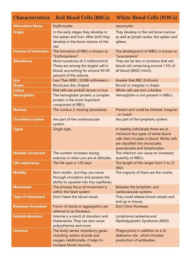
Differences between red blood cells (RBCs) and white blood cells (WBCs)
The major differences between red blood cells (RBCs) and white blood cells (WBCs) are as follows-
- Other name- Red blood cells are referred to as erythrocytes, while white blood cells are also called leukocytes.
- Function- The main function of RBC is the transport of oxygen from lungs to the different tissues. They also take part in carrying some carbon dioxide back towards lungs. WBCs, on the other hand, are mainly involved in body defense and immune reactions against microorganisms and other foreign materials.
- Shape- Mature RBCs are biconcave disc-shaped cells, thinner at their center. There is no single characteristic shape for all the WBCs. Different types have different cellular and nuclear shapes.
- Nucleus- Nucleus is absent in mature human RBC. WBCs are nucleated cells. The nucleus may also differ considerably according to the leukocyte type.
- Hemoglobin- RBCs contain a large amount of hemoglobin. It gives the cells their red colour and is used for oxygen transport. Hemoglobin is absent in WBCs.
- Number- RBCs are present in very large number, usually millions in a microliter of blood. The reference value is about 4.3-5.9 million/µL in adult males and 3.5-5.5 million/µL in adult females. WBCs are much fewer, around 4,500-11,000 cells/µL in adults (reference ranges can differ between laboratories and physiological conditions).
- Size- RBC is a small blood cell of approximately 7-8 µm diameter. WBCs are generally larger. Their size is not fixed and varies with different leukocyte types.
- Cell organelles- During maturation, RBC loses the nucleus and practically all of the usual cell organelles. In WBCs, cellular structures are retained which are required for their different immune functions.
- Types- Mature circulating RBC represents one principal erythrocyte cell form. White blood cells are of five major types, namely neutrophils, eosinophils, basophils, lymphocytes, and monocytes.
- Movement- RBCs normally remain within the blood vessels, moving along with circulating blood. Many WBCs can get out from the blood vessels and enter the tissues. Their defensive activity is then performed at these tissue sites.
- Formation- Both RBCs and WBCs are derived ultimately from hematopoietic stem cells in the bone marrow. RBC formation is called “erythropoiesis” and is strongly regulated by erythropoietin. Formation of different WBC lineages involves different growth factors and cytokines.
- Life span- Normal RBCs have an average life span of about 120 days. The life span is highly variable in WBCs. Some remain for hours to days, whereas certain lymphocytes can survive for years.
Similarities between RBCs and WBCs
The following are some of the similarities between red blood cells (RBCs) and white blood cells (WBCs)–
- Both RBCs and WBCs are cellular formed elements of blood. They are present in the circulating blood along with platelets and plasma.
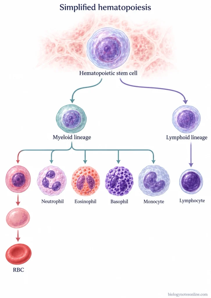
- Both are developed from a common hematopoietic stem cell. In adults, these blood-forming stem cells occur mainly in the red bone marrow.
- The formation of both cells takes place through hematopoiesis. During this process, stem cells give rise to progenitor cells which further differentiate into their particular blood cell types. RBCs and most WBC lineages follow different pathways after this common origin.
- RBCs as well as WBCs are released into blood after their development and circulate through the vascular system. WBCs may later leave the blood vessels and enter tissues, whereas RBCs normally remain in circulation.
- Both populations are continuously produced. Mature blood cells do not remain permanently, so new RBCs and WBCs are formed to replace the cells that are removed or die. Some WBCs, particularly memory lymphocytes, can remain for much longer periods.
- Their production is regulated by different hematopoietic growth factors. Erythropoietin mainly stimulates erythrocyte production, while colony-stimulating factors and several cytokines are involved in development of leukocyte lineages.
- Both RBC and WBC numbers can be measured from a blood sample. They are important components of the complete blood count (CBC), where red cell count and white cell count are determined separately.
- Both can also be examined in peripheral blood for their number and cellular appearance. Blood smear examination is commonly used along with cell counts when the morphology of circulating blood cells has to be studied.
References
- Alberts, B., Johnson, A., Lewis, J., Raff, M., Roberts, K., & Walter, P. (2002). Renewal by multipotent stem cells: Blood cell formation. In Molecular biology of the cell (4th ed.). Garland Science. https://www.ncbi.nlm.nih.gov/books/NBK26919/
- Barbalato, L., & Pillarisetty, L. S. (2022, November 14). Histology, red blood cell. In StatPearls. StatPearls Publishing. https://www.ncbi.nlm.nih.gov/books/NBK539702/
- Betts, J. G., Young, K. A., Wise, J. A., Johnson, E., Poe, B., Kruse, D. H., Korol, O., Johnson, J. E., Womble, M., & DeSaix, P. (2013). Anatomy and physiology. OpenStax. https://openstax.org/books/anatomy-and-physiology/pages/1-introduction
- Betts, J. G., Young, K. A., Wise, J. A., Johnson, E., Poe, B., Kruse, D. H., Korol, O., Johnson, J. E., Womble, M., & DeSaix, P. (2022). Anatomy and physiology 2e. OpenStax. https://openstax.org/books/anatomy-and-physiology-2e/pages/1-introduction
- Dean, L. (2005). Blood and the cells it contains. In Blood groups and red cell antigens. National Center for Biotechnology Information. https://www.ncbi.nlm.nih.gov/books/NBK2263/
- El Brihi, J., & Pathak, S. (2024, June 8). Normal and abnormal complete blood count with differential. In StatPearls. StatPearls Publishing. https://www.ncbi.nlm.nih.gov/books/NBK604207/
- Institute for Quality and Efficiency in Health Care. (2023, March 16). In brief: What does blood do? In InformedHealth.org. https://www.ncbi.nlm.nih.gov/books/NBK279392/
- Open Resources for Nursing. (2024). Blood terminology. In K. Ernstmeyer & E. Christman (Eds.), Medical terminology (2nd ed.). Chippewa Valley Technical College. https://www.ncbi.nlm.nih.gov/books/NBK607436/
- Sharma, R., & Sharma, S. (2023, April 10). Physiology, blood volume. In StatPearls. StatPearls Publishing. https://www.ncbi.nlm.nih.gov/books/NBK526077/
- Thiagarajan, P., Parker, C. J., & Prchal, J. T. (2021). How do red blood cells die? Frontiers in Physiology, 12, Article 655393. https://doi.org/10.3389/fphys.2021.655393
- Tigner, A., Ibrahim, S. A., & Murray, I. V. (2026, February 14). Histology, white blood cell. In StatPearls. StatPearls Publishing. https://www.ncbi.nlm.nih.gov/books/NBK563148/