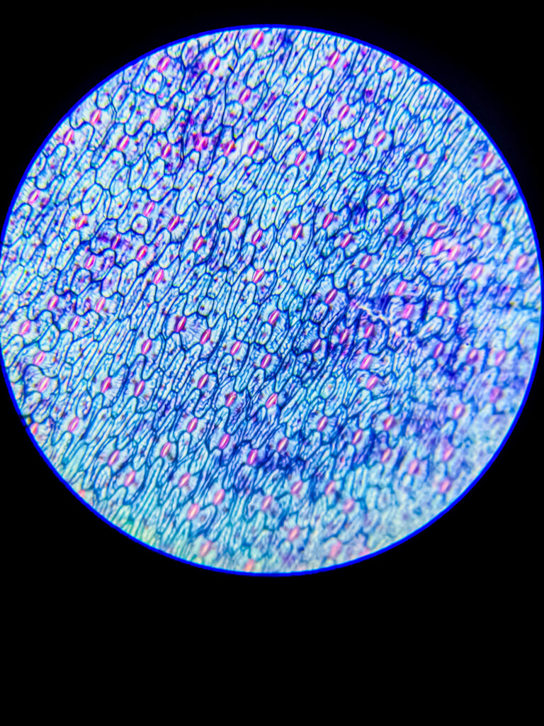
BNO-IMG-075397-01
- Magnification
- 100x
- Image dimensions
- 2560 × 1920 px
- Caption
- guard cell under 100x

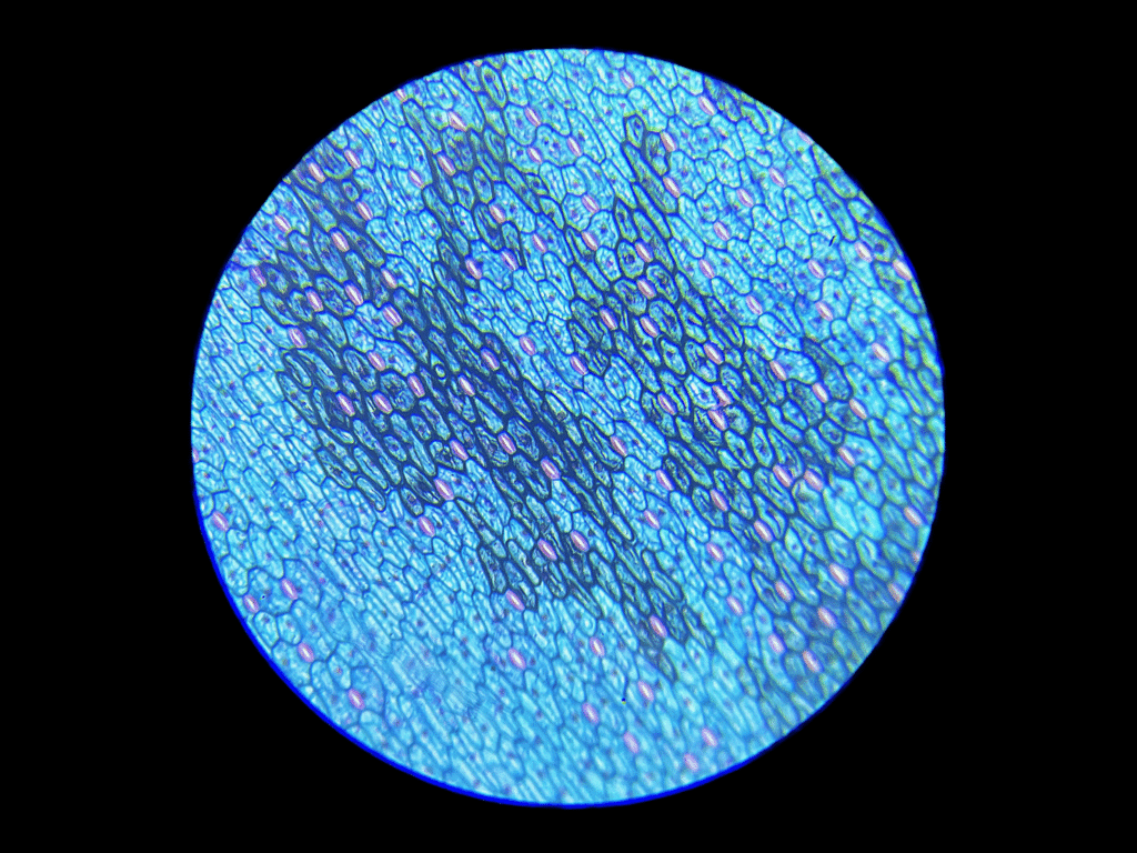
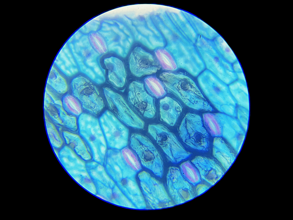
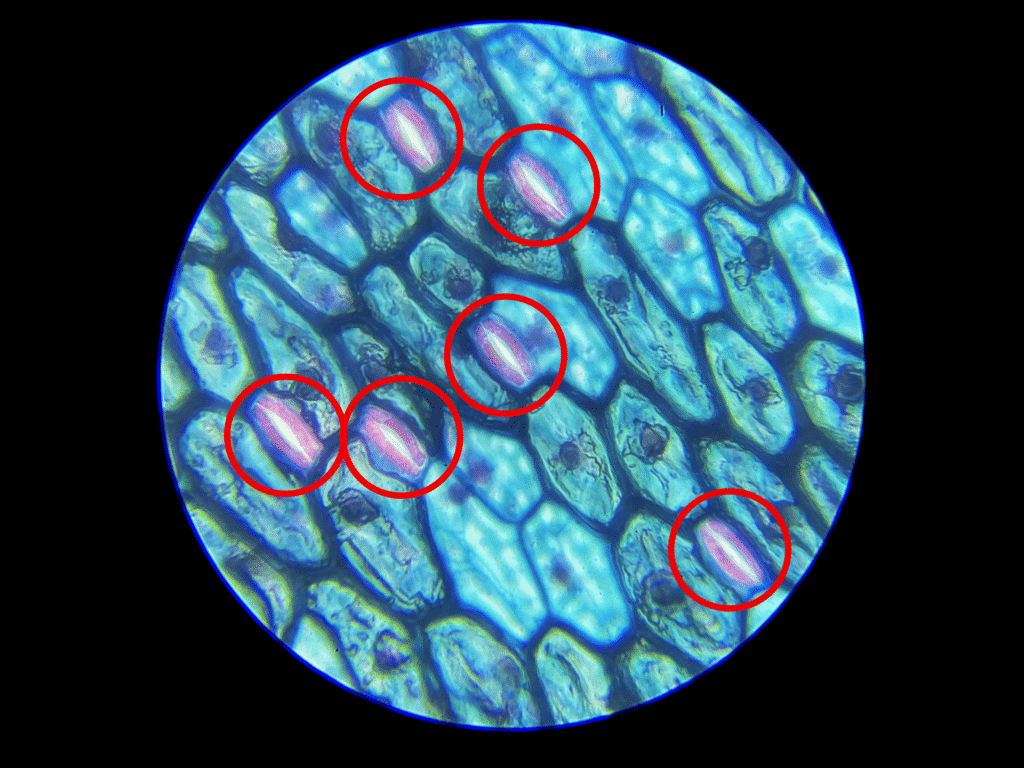
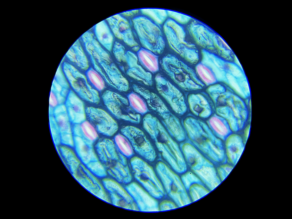
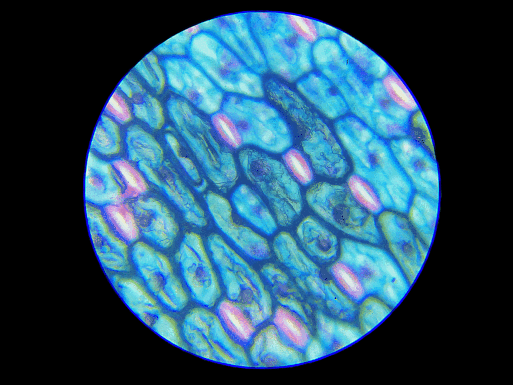
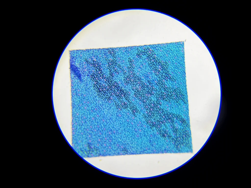

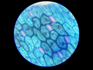
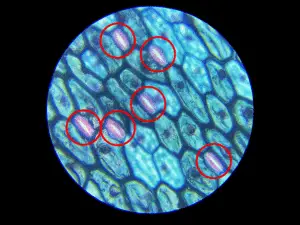
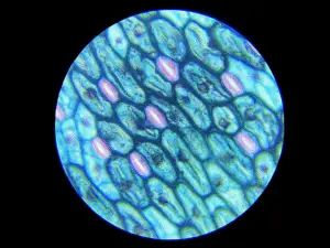
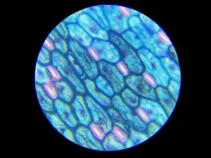
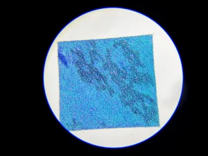
Under the microscope, guard cells appear as specialized, kidney-shaped (in dicots) or dumbbell-shaped (in monocots) pairs of cells that flank a central pore called the stoma (plural: stomata) on the surface of leaves, especially on the lower epidermis.
Microscopic Appearance of Guard Cells:
Guard cells are usually kidney-shaped in dicotyledonous plants (e.g., bean, sunflower) and dumbbell-shaped in monocotyledonous plants (e.g., grass, maize).
They are located in pairs, forming an elliptical opening (stomatal pore) between them.
The cell wall is unevenly thickened—the inner wall (towards the stoma) is thicker and less elastic, while the outer wall is thinner and more flexible, allowing the stoma to open and close with changes in turgor pressure.
Guard cells have visible nuclei, which helps distinguish them from surrounding epidermal cells.
Chloroplasts are present in guard cells, unlike most other epidermal cells, making them appear greener or more granular under higher magnification and proper lighting.
Under high magnification (typically 400x or more), cytoplasmic streaming and the green chloroplasts are often visible within the guard cells.
The surrounding epidermal cells are more elongated and irregular, and they lack chloroplasts, which helps highlight the contrast.
When stained with iodine or safranin, the cell walls and nuclei become more prominent for clearer observation.
In open stomata, the stoma appears as a clear slit or pore between the two guard cells; in closed stomata, the guard cells appear pressed together with little or no visible gap.
Observation Tips:
Best observed using a peel from the lower epidermis of a leaf (e.g., from Rhoeo, Tradescantia, or Hibiscus) mounted in water or glycerine.
Use bright-field microscopy at 100x–400x magnification.
Apply safranin or iodine stain for better contrast if unstained cells are difficult to differentiate.
Guard cells are more abundant on the lower epidermis than on the upper surface.
Key Identification Features:
Kidney or dumbbell shape.
Present in pairs, forming a pore between them.
Contain chloroplasts (green coloration under natural light).
Unequal cell wall thickness.
Surrounded by non-green epidermal cells.
Sourav Pan. (2025). Guard cells Under Microscope [BNO-IMG-075397]. Biology Notes Online. https://biologynotesonline.com/microscopic-image/guard-cells-under-microscope/
This microscopic image is an original work created by Sourav Pan for Biology Notes Online and published on June 14, 2025.
Scientific information associated with this image is provided for educational and student reference. Microscopy details such as specimen, stain, microscopy technique and magnification are shown where available.
The image may be cited using the citation information on this page. For copyright and reuse conditions, see Image Licensing.
If you notice an identification, labeling or scientific accuracy issue, please report it so the record can be reviewed.
 Sourav Pan