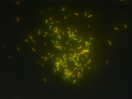
What is Acid Fast Stain? Objective of Acid Fast Stain Principle of Acid Fast Stain The principle of the Acid Fast Stain revolves around the unique characteristics of certain bacterial cells, particularly their resistance to conventional staining methods due to the presence of mycolic acid in their outer membrane. This expository explanation aims to elucidate … Read more
What is Chlamydia? Distribution of Chlamydia species Chlamydia species have a wide distribution across the world, and their prevalence varies in different regions. The family Chlamydiaceae, which includes the genus Chlamydia, is believed to have originated from the Order Chlamydiales approximately seven million years ago. However, evidence of Chlamydia trachomatis infections has been found dating … Read more
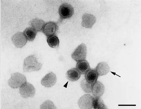
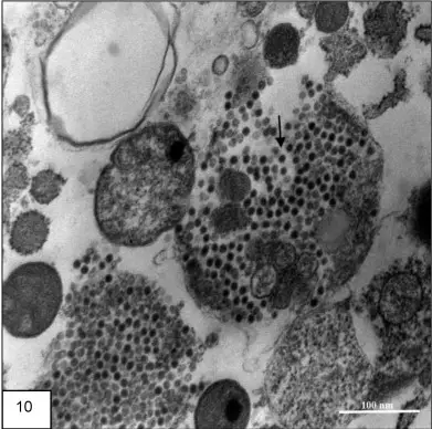
Negative staining is a method used to visualize viruses and other small particles under a microscope. In this method, a drop of the sample is placed on a flat surface and a small amount of a negatively charged stain is added to the sample. The stain surrounds the virus particle, but does not adhere to … Read more
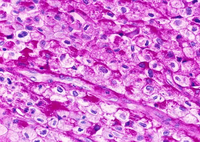
What is Periodic Acid-Schiff (PAS) Staining? Periodic Acid-Schiff (PAS) staining is a laboratory staining technique used to detect the presence of specific sugars and carbohydrates in tissue samples. The PAS stain highlights the presence of sugars, glycoproteins, and mucopolysaccharides in biological tissues and cells. The staining process is often used in the diagnosis of diseases … Read more
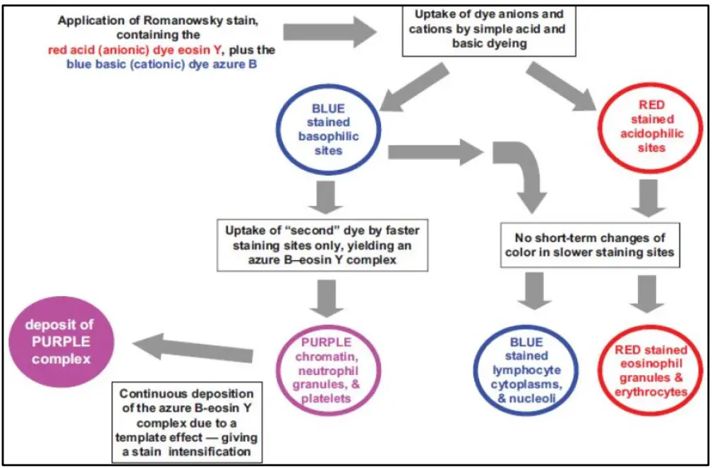
Romanowsky staining, also known as Romanowsky–Giemsa staining, is a classic staining technique that paved the way for several different but similar stains widely used in haematology (the study of blood) and cytopathology (the study of cells) (the study of diseased cells). Romanowsky stains are used to distinguish cells for microscopic study in pathological materials, particularly … Read more
What is Wheatley Trichrome Staining? In parasitology, intestinal protozoans are typically detected and identified from faeces samples using a specialised permanent technique called Wheatley Trichrome staining. Wheatley Trichrome Stain is suggested for use in qualitative methods for the detection, identification, and distinction of intestinal protozoa from background material. Typically, PVA-fixed or Schaudinn’s solution-preserved faecal specimens … Read more
Since the late 1950s and early 1960s, the positive staining approach has been employed to increase the contrast of biological materials (tissues and cell structures, viruses, etc.). The samples are treated in heavy metal salt solutions that react with cellular structures using this technique and negative staining. Uranyl acetate and lead citrate are the most … Read more
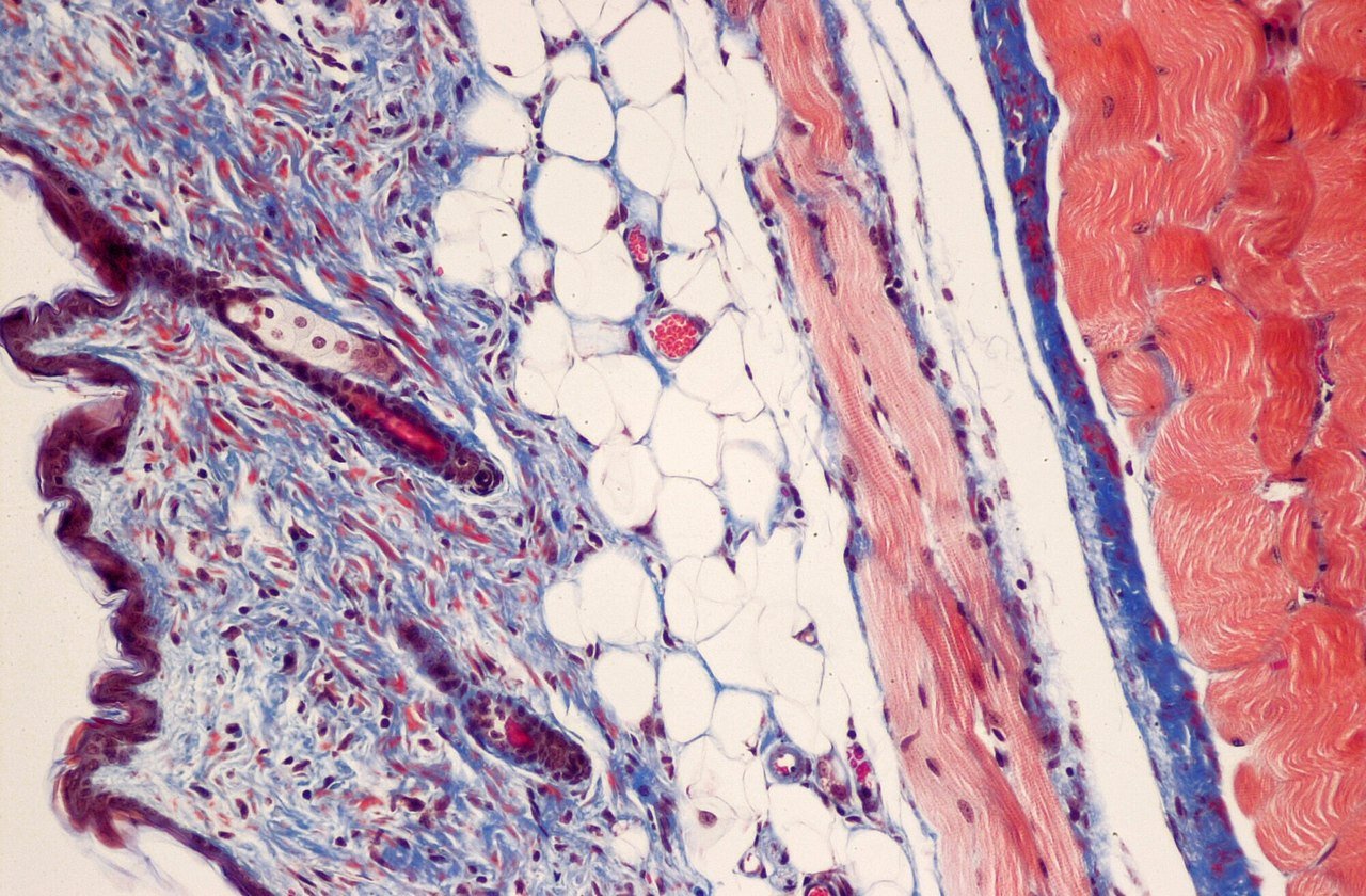
Trichrome stains, such as Masson’s Trichrome, derive its name from the three dyes utilised in the staining process. These three dyes are used to selectively stain muscle, collagen fibres, fibrin, and erythrocytes using acid-base chemistry. Initial placement of tissue slices in Bouin’s solution. As a mordant, Bouin’s solution links the dye to the specific tissue … Read more
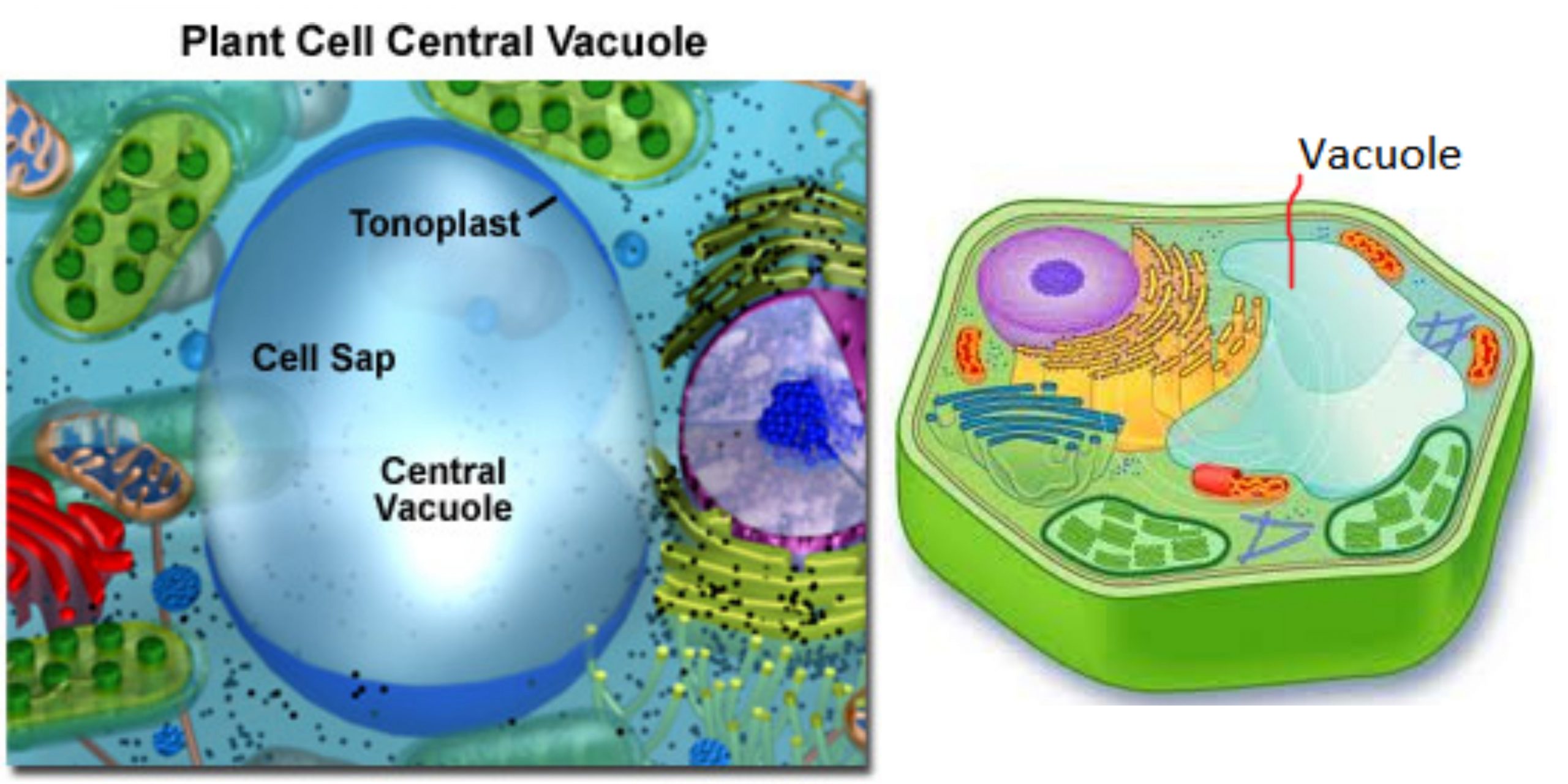
A light microscope may be used to see and examine the vacuole’s structure. While the vacuole doesn’t stain as the other organelles of the cell (because it does not contain many constituents that stain) tests have demonstrated that staining is possible for this organelle because the vacuole’s sap absorbs and stores dyes that are colored.
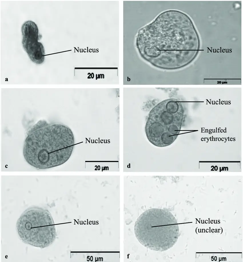
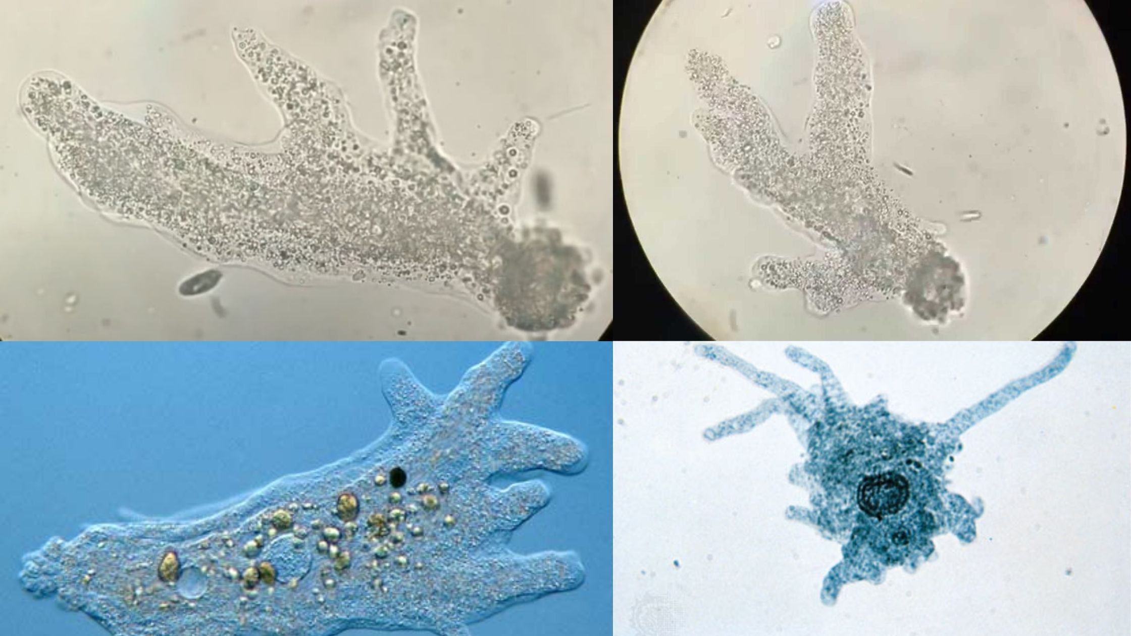
Amoeba (less often spelled ameba, or amoeba, plural am(o)ebas or am(o)ebae) is often referred to as an amoeboid, is kind of unicellular organism that has the capability of changing its form, mostly by retracting and expanding pseudopods. Amoebae do not belong to a single taxonomic class, They are found in all major lineages of the … Read more
⚠️
Click on your ad blocker icon in your browser's toolbar
Select "Pause" or "Disable" for this website
Refresh the page if it doesn't automatically reload