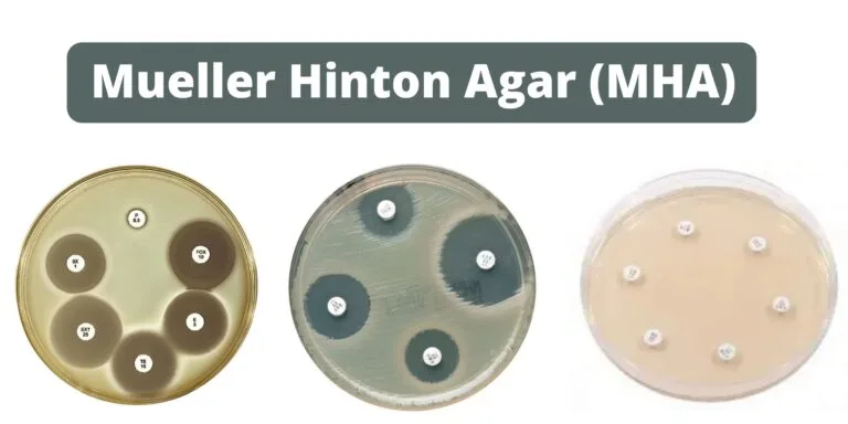
What is Mueller Hinton Agar (MHA)? Why MHA is used for antibiotic susceptibility testing (AST)? Mueller Hinton Agar (MHA) is commonly used for antibiotic susceptibility testing due to several key reasons: Mueller Hinton Agar (MHA) Principle The principle of Mueller Hinton Agar (MHA) revolves around its composition and the controlled levels of various components to
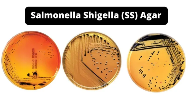
Salmonella Shigella (SS) Agar can be moderately specific and a differing medium used for the cultivation, isolation as well as differentiation Salmonella spp. and a few varieties that belong to Shigella spp.
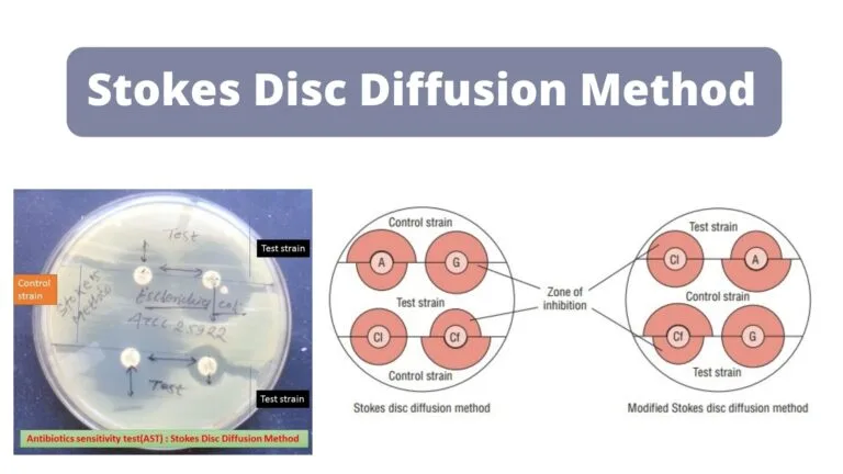
Stokes disc diffusion method isn’t as well-standardized as Kirby-Bauer’s method and is utilized in labs especially where the exact amount of antimicrobial present in discs isn’t known because of the difficulty in getting discs and correctly storing them or when other requirements needed for the Kirby-Bauer method cannot be fulfilled.
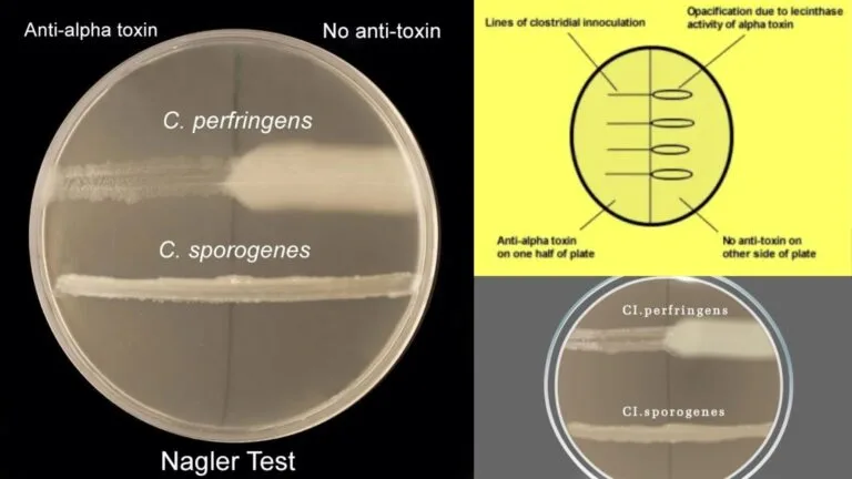
Nagler’s Reaction or Lecithinase test is a test in biochemistry used to detect organisms that produce the phospholipases (lecithinases) e.g. Clostridium perfringens. Its alpha (a) toxin from C. perfringens exhibits the phospholipase enzyme activity, which aids in the distinction between C. perfringens and the other Clostridium species that generate the enzyme lecithinase (C.baratti, C.absonum, C.bifermantans, C.sordelli, and C.novyi) through neutralization of lecithin C activity with an antitoxin.
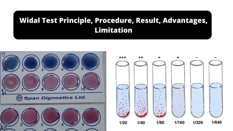
The Widal test is an agglutination test that determines any antibodies present in the patient’s serum that are generated against the causal agents of the enteric febrile illness (Salmonella Typhi, and Salmonella paratyphi B, A as well as C).
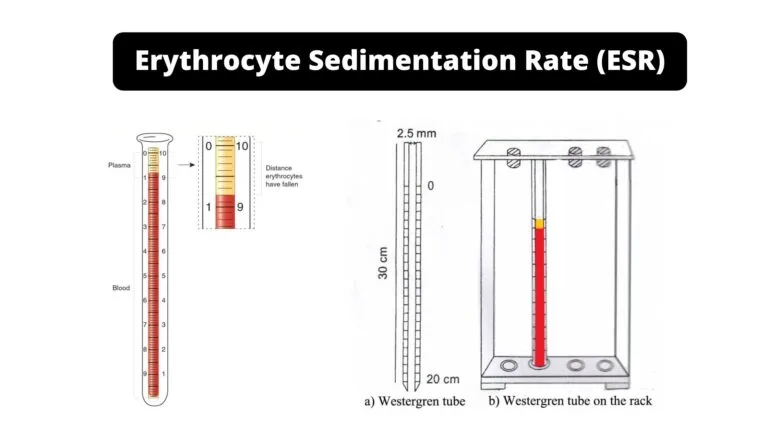
Erythrocyte Sedimentation Rate (ESR) can be described as a commonly used test used to detect non-specific signs of inflammation result of infection cancers, or certain autoimmune disorders. It is defined as the rate that Red Blood Cells (RBCs) accumulate over an hour.
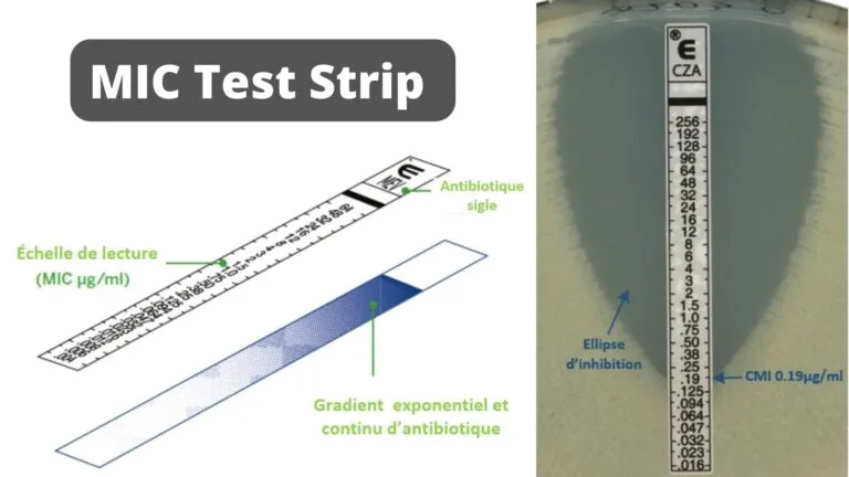
The Minimum Inhibitory Concentration (M.I.C.) of antimicrobial drugs against bacteria and the detection of resistance mechanisms can be found with the help of a MIC Test Strip, a quantitative assay.
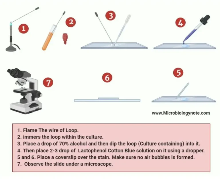
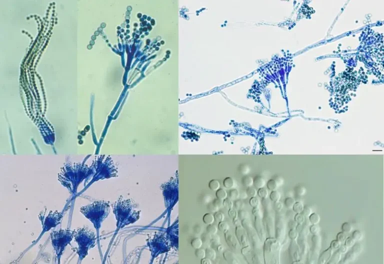
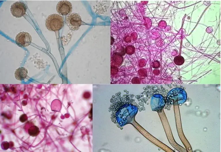
The Lactophenol Cotton Blue solution stains the chitinous cell wall of the fungal cell, which makes them different from the surrounding environments and can easily distinguishable. LCB is made of three main components such as Phenol, Lactic acid, and Cotton blue.