We've just released a new feature: Text Annotation & Highlight - Add notes and highlights to articles! Login required.

Microscope Slides Preparation Step By Step Guide

Microscope Slides Preparation Step By Step Guide

Objects are magnified under compound microscopes are mounted onto microscope slides. Made from plastic or glass Slides measure approximately 1×3 inches in size and range from 1mm-1.2 millimeters thick.

History of the Microscope

History of the Microscope

Microscopes have been around for the ages. Roman philosophers had mentioned “burning glass” within their works. However, the first microscope of this type was not invented until the 1300’s. Two lenses were set on opposite sides of the tube. This tube of magnifying power was the basis for our modern-day microscope.

Digital Microscope – Principle, Parts, Uses, Advantages

USB Microscope - Principle, Definition, Parts, Examples, Uses

Digital microscopes are modern microscope which does not have an eyepiece. This is a huge contrast from an optical microscope. Digital microscopes have an electronic camera that acts as the detector as well as the imaging output gadget. It displays the images takes place via a computer’s screen or monitor, which defines the scope of the microscope’s digital.

USB Microscope – Principle, Parts,Uses, Advantages

USB Microscope Principle, Definition, Parts, Examples, Uses

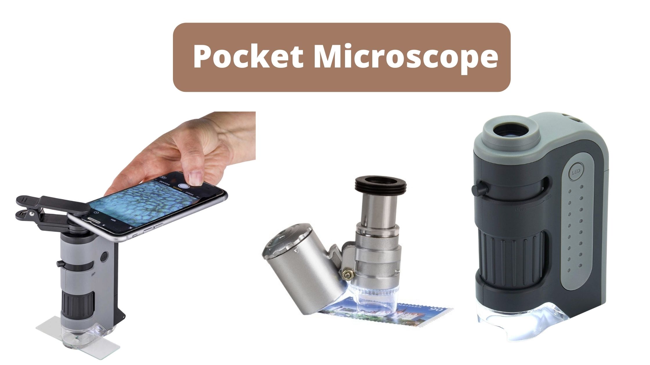
This is a form of digital Microscope which is connected to a computer using an USB port. It operates on low power. The camera is fitted with a powerful macro lens. Additionally, they have an integrated LED light that is built into the lens.

Atomic Force Microscope (AFM) – Principle, Parts, Procedure, Uses

Atomic Force Microscope (AFM) - Definition, Principle, Application

The AFM, also known as the atomic force microscope (AFM) is a sort scanner probe. Its principal functions include measuring characteristics like height, magnetism and friction.

Oil Immersion Technique – Objectives, Procedure, Types

Oil Immersion Technique

The oil immersion method enhances the resolving power of a microscope, enabling light microscopy to distinguish finer details. Immersion oil—clear and colorless, having a refractive index of about 1.515—is placed in between the lens and the specimen. The refractive index is higher; therefore, less light is refracted. If less light is refracted, then less is … Read more

Transmission Electron Microscope (TEM) – Principle, Parts, Uses

Transmission Electron Microscope (TEM)

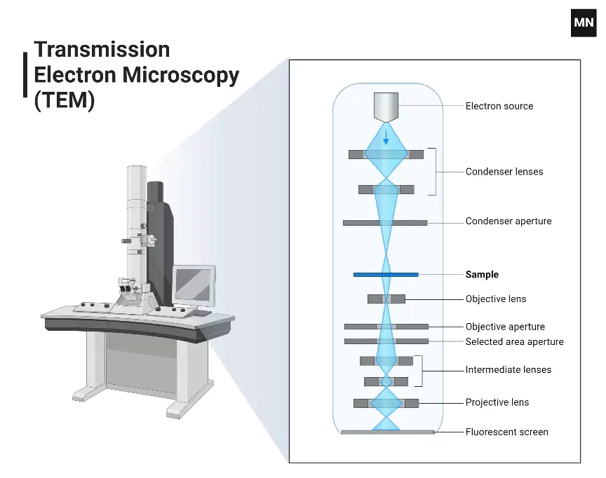
What is transmission electron microscope? A transmission electron microscope (TEM) is a high-resolution imaging tool that uses a beam of electrons instead of light to see very small details in thin samples. It can make structures up to several million times bigger, which lets scientists see things like viruses, cell organelles, or even macromolecular complexes. … Read more

Scanning Electron Microscope – Principle, Parts, Uses

Scanning Electron Microscope

A scanning electron microscope (SEM) produces high-resolution images from an electron beam that scans a focused beam over the surface of a specimen. The benefit of using electrons as opposed to an optical light microscope is that, due to electronegativity, the resultant interactions with the orbitals surrounding the atom create signals that indicate surface morphology … Read more

Phase Contrast Microscopy- Principle, Parts, Uses

Phase Contrast Microscopy

What is phase contrast microscopy? Okay, so picture this: back in the 1930s, scientists were struggling to study things like living cells or bacteria under a microscope. Why? Because those tiny critters are practically see-through! You couldn’t get a clear look without dunking them in harsh dyes—which, you know, kinda killed them. Not exactly helpful if you … Read more

Confocal Microscope – Principle, Parts, Uses

Confocal Microscope Principle, Uses, Parts, Advantages, and Disadvantages.

Confocal microscopy offers some pretty cool perks compared to regular optical microscopes. For starters, it gives you a razor-thin focus depth, cuts out that annoying blurry background glow, and lets you snap crisp, detailed “slices” of thick samples—one after another. This makes it a go-to tool in biomedicine, especially for studying cells and tissues, whether … Read more

Ad Blocker Detected
We've detected that you're using an ad blocker. Some content may not display properly.
Why are you seeing this? Ad blockers can prevent certain content and features from loading correctly on our website.

To continue with the best experience: