

Blood Collection Tubes are evacuated, sterile and single-use medical devices that are used to collect human blood samples for laboratory testing.
These tubes are used by trained healthcare professionals during blood collection. It helps to collect, contain, preserve and transport the blood sample safely. The tubes are generally made up of glass or shatter-resistant plastic such as polyethylene terephthalate (PET).
Blood collection tubes work by a pre-calibrated vacuum system. This vacuum automatically draws a fixed volume of blood during venipuncture. So, the required amount of blood is collected inside the tube.
These tubes may contain different chemical additives. Anticoagulants are used to prevent clotting. Clot activators are used to promote clotting. Some preservatives are also present to maintain the sample during storage and transport.
The caps of blood collection tubes are colour-coded. This colour coding helps to identify the type of additive present inside the tube and to select the correct tube for a particular diagnostic test.
Components of Blood Collection Tube
The following are the components of Blood Collection Tubes–
- Glass or Plastic Tube
Glass or plastic tube is a clear evacuated tube which is used to hold the blood sample. It is made up of glass or shatter-resistant latex-free plastic such as polyethylene terephthalate (PET). - Closure Assembly (Stopper)
Closure assembly is the stopper of the tube. It seals the tube and maintains the vacuum present inside the tube. - Safety Closure
Safety closure is a special type of stopper. In Hemogard™ closure, the rubber stopper is present inside a protective plastic shield. It protects the user from blood exposure. - Additives
Additives are chemical reagents present inside the tube. These are used to preserve the blood sample or prepare the sample for laboratory test. - Anticoagulants
Anticoagulants are additives which prevent clotting of blood. Examples include EDTA, sodium citrate and heparin. - Clot Activators
Clot activators are substances which promote clotting of blood. Silica and thrombin are used as clot activators. - Preservatives
Preservatives are used to maintain some analytes in blood sample. Sodium fluoride prevents the breakdown of glucose. - Separation Gel
Separation gel is a polymer gel present in some blood collection tubes. During centrifugation, it forms a barrier between blood cells and serum or plasma.

Order of Draw in Blood Collection Tube
The following are the order of draw in Blood Collection Tubes–
- Blood Culture Tubes or Bottles
Blood culture tubes or bottles are drawn first. It is used first to maintain maximum sterility. Example is yellow top tube with SPS. - Sodium Citrate Tubes
Sodium citrate tubes are drawn after blood culture tubes. It is used for coagulation tests. Example is light blue top tube. - Serum Tubes
Serum tubes are drawn after sodium citrate tubes. These tubes may contain clot activators and gel separators. Examples include red, gold or speckled top tubes. - Heparin Tubes
Heparin tubes are drawn after serum tubes. These may contain gel or without gel. Examples include dark green, light green or speckled green top tubes. - EDTA Tubes
EDTA tubes are drawn after heparin tubes. These may be with or without gel separators. Examples include lavender, pink, pearl or tan top tubes. - Glycolytic Inhibitor Tubes
Glycolytic inhibitor tubes are drawn last. These tubes contain sodium fluoride and potassium oxalate. Example is gray top tube.
Color of the Cap and Its Purpose
The following are the color of cap and its purpose-
- Red Cap
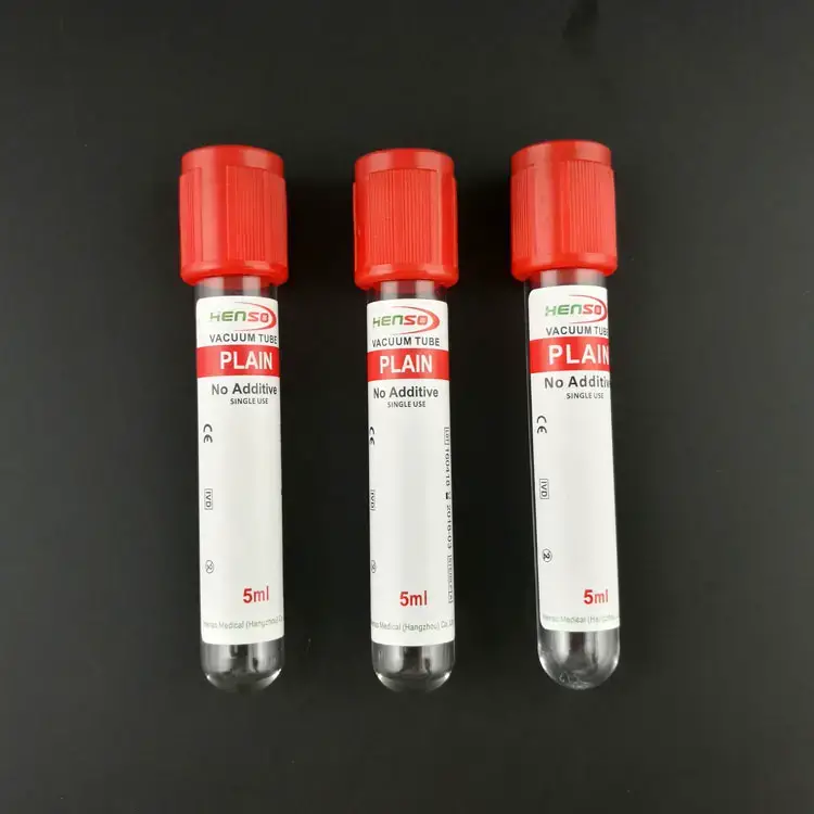
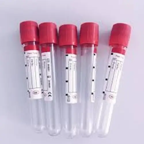
Red cap tube contains no additive or clot activator. It is used to collect serum for chemistry, serology and blood banking tests. - Gold or Mottled Red/Gray Cap
Gold or tiger top tube contains clot activator and gel separator. It is also called Serum Separator Tube (SST). It is used for chemistry panel, thyroid test and liver function test. - Light Blue Cap
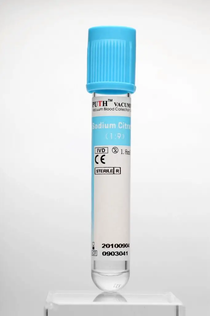
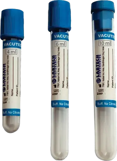
Light blue cap tube contains sodium citrate. It is an anticoagulant that binds calcium. It is used for coagulation tests such as PT, aPTT, INR and D-dimer. - Green Cap
Green cap tube contains sodium heparin or lithium heparin. It prevents clotting of blood. It is used for routine chemistry test, electrolytes, ammonia and blood gas analysis. - Light Green Cap
Light green cap tube contains lithium heparin and gel separator. It is also called Plasma Separator Tube (PST). It is used for rapid plasma chemistry tests. - Lavender or Purple Cap
Lavender cap tube contains EDTA. It prevents clotting and preserves cell shape. It is used for whole blood hematology tests like Complete Blood Count (CBC), HbA1c and peripheral blood smear. - Pink Cap
Pink cap tube contains EDTA. It is mainly used in blood bank for blood typing, cross matching and antibody screening. - Gray Cap
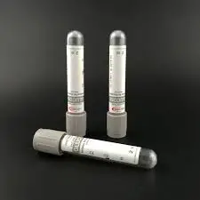
Gray cap tube contains sodium fluoride and potassium oxalate. Sodium fluoride prevents glucose breakdown and potassium oxalate prevents clotting. It is used for glucose, lactate and blood alcohol test. - Yellow Cap
Yellow cap tube contains SPS (sodium polyanethol sulfonate) or ACD (acid citrate dextrose). SPS is used for blood culture to detect bacteria. ACD is used for DNA, tissue typing and paternity testing. - Royal Blue Cap
Royal blue cap tube is a trace-element-free tube. It may contain EDTA, heparin or no additive. It is used for toxicology and trace metal analysis like zinc, copper and mercury. - Tan Cap
Tan cap tube contains certified lead-free EDTA. It is used for blood lead level testing. - Orange Cap
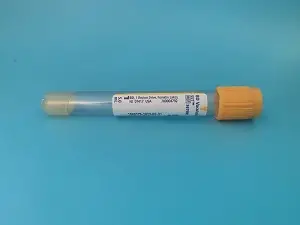
Orange cap tube contains thrombin-based clot activator. It is used for STAT serum chemistry tests where rapid clotting is required. - Black Cap
Black cap tube contains sodium citrate. It is used for Westergren Erythrocyte Sedimentation Rate (ESR) testing. - White or Pearl Cap
White or pearl cap tube contains EDTA and gel separator. It is also called Plasma Preparation Tube (PPT). It is used for molecular diagnostic tests and PCR, such as viral load testing.


Uses of Blood Collection Tubes
The following are the uses of Blood Collection Tubes–
- Red, gold or tiger top tubes are used for chemistry panels, serology, immunology and therapeutic drug monitoring. These tubes allow the blood to clot and serum is separated from the clotted blood.
- Lavender, purple or pink top tubes are used for hematology tests. It is used for Complete Blood Count (CBC), HbA1c, blood typing and cross-matching in blood banks.
- Light blue top tubes are used for coagulation studies. It is used for Prothrombin Time (PT), aPTT, INR and D-dimer tests to evaluate bleeding disorders.
- Green or light green top tubes are used to obtain plasma for routine chemistry tests. It is also used for blood gas analysis, electrolyte test and ammonia level checking.
- Gray top tubes are used for blood glucose testing. It is also used for lactate screening and blood alcohol testing because it prevents the glucose of the sample from breaking down.
- Yellow top tubes or special bottles are used in microbiology. It is used to detect bacterial or fungal infection in blood such as sepsis and also used for genetic or DNA testing.
- Royal blue or tan top tubes are used for toxicology and trace metal testing. It is used for testing metals like zinc, copper or mercury. Tan tubes are specially used for lead testing.
- Black top tubes are used to measure Erythrocyte Sedimentation Rate (ESR). This test helps in diagnosis and monitoring of inflammation.
- White or pearl top tubes are used for molecular diagnostic tests and PCR testing. It is used for viral load testing and genetic testing.
Advantages of Blood Collection Tubes
The following are the advantages of Blood Collection Tubes–
- Blood collection tubes provide a closed collection system. It reduces manual handling of blood sample and minimizes the risk of contamination.
- These tubes help to preserve the quality of the specimen. So, the sample remains suitable for laboratory testing.
- The tubes contain a pre-calibrated vacuum. This vacuum draws the required amount of blood automatically.
- It helps to maintain accurate blood-to-additive ratio. This gives proper and consistent test result.
- Blood collection tubes reduce the risk of occupational exposure to bloodborne pathogens.
- It also reduce the chance of needlestick injuries during blood collection.
- Safety closures such as Hemogard™ closure prevent blood splatter and direct contact with residual blood.
- These tubes allow quick filling of multiple tubes during a single blood draw. It makes blood collection faster in high-volume laboratory settings.
- It eliminates the need of manual aspiration. So, the collection process becomes more smooth and easy.
- The caps of the tubes are colour-coded. This helps in quick identification of additives and the use of the tube.
- Colour coding helps to reduce selection error during blood collection.
- It also helps to improve laboratory workflow and prevent diagnostic error.
Limitations of Blood Collection Tubes
The following are the limitations of Blood Collection Tubes–
- The vacuum pressure of evacuated tube cannot be changed. It may give more pressure on small, fragile or difficult veins.
- In pediatric and geriatric patients, the vein may collapse due to the fixed vacuum pressure. It may also cause hemolysis, where red blood cells are ruptured.
- Blood collection tubes need exact amount of blood. If the tube is underfilled, it is called short draw.
- In short draw, the blood-to-additive ratio becomes improper. Excess additive may change the test result.
- In sodium citrate tube, short draw may cause falsely prolonged clotting time.
- Additive carryover may occur when the needle passes from one tube to another tube. Small amount of additive may enter into the next tube.
- If the correct order of draw is not followed, cross-contamination may occur. It may cause false result like high potassium or low calcium.
- Some additives do not act immediately. Sodium fluoride may take time to stop glycolysis completely.
- Delay in processing of glucose sample may cause falsely low blood sugar reading.
- Standard tubes cannot preserve blood cells for long time. White blood cells may start to rupture within 6 to 24 hours.
- This may damage the sample used for molecular diagnostic tests such as cell-free DNA testing.
- Plastic tubes are shatter-resistant but they may lose vacuum slowly over time. This is because plastic tubes are more gas-permeable than glass tubes.
- Normal blood collection tubes may contain trace amount of metals from tube material or rubber stopper.
- These tubes are not suitable for trace element testing like lead, zinc or copper. Special metal-free tubes are required for these tests.
References
- American Certification Agency for Healthcare Professionals. (2017). New CLSI venipuncture guidelines. ACAreer, 22(2), 1-4.
- Bayot, M. L., & Tadi, P. (2023). Laboratory tube collection. In StatPearls. StatPearls Publishing.
- Becton, Dickinson and Company. (n.d.). Blood collection points to practice – BD.
- Brooks, N. (2025). The importance of proper color coding for blood collection tubes: A guide for healthcare professionals. Needle.Tube.
- Carter, E. (2025). Effects of underfilling a sodium citrate tube: Implications on coagulation test results. Needle.Tube.
- Center for Phlebotomy Education. (n.d.). The order of draw: Do I have to follow it?.
- Centers for Disease Control and Prevention. (n.d.). Blood lead testing.
- Chauhan, A. (2024). Vacutainer colour coding guide for blood collection. Flabs.
- Clinical and Laboratory Standards Institute (CLSI). (2019). Order of blood draw tubes and additives.
- Dr.Oracle. (2025). What effect can under-filled blue top (blood collection) tubes have on coagulation test results?.
- Fisher Healthcare. (n.d.). Evacuated blood collection tubes.
- Gamage, S. (2025). The order of draw and rationale of blood collection tubes: A review of guidelines and pre-analytical considerations. Centre for Research – National Hospital Kandy.
- Gambino, R., Piscitelli, J., Ackattupathil, T. A., Theriault, J. L., Andrin, R. D., Sanfilippo, M. L., … (2009). Acidification of blood is superior to sodium fluoride alone as an inhibitor of glycolysis. Clinical Chemistry, 55, 1019-1021.
- Greiner Bio-One. (n.d.). Tube chart: Venous blood collection tubes.
- Gupta, S., Gupta, A. K., Verma, M., Singh, K., Kaur, A., Chopra, B., & Kaur, V. (2016). A study to compare the plasma glucose levels obtained in sodium fluoride and citrate buffer tubes at a tertiary care hospital in Punjab. International Journal of Applied and Basic Medical Research, 6(1), 50–53.
- Harris, A. (2025). What is the significance of a tan top tube in lead testing? Needle.Tube.
- Henry Schein. (n.d.). The BD Vacutainer® system.
- Kang, Q., Henry, N. L., Paoletti, C., Jiang, H., Vats, P., Chinnaiyan, A. M., Hayes, D. F., Merajver, S. D., Rae, J. M., & Tewari, M. (2016). Comparative analysis of circulating tumor DNA stability In K3EDTA, Streck, and CellSave blood collection tubes. Clinical Biochemistry, 49(18), 1354-1360.
- Labcorp. (n.d.). Blood specimens: Chemistry and hematology.
- Labpedia.net. (2025). Blood samples, color coding of the test tubes.
- MBP Inc. (2024). Understanding the different types of blood collection tubes: A comprehensive guide.
- McLendon Clinical Laboratories. (2025). Evacuated blood collection tube guide. UNC Medical Center.
- Michigan Medicine Laboratories (MLabs). (n.d.). Order of draw for multiple tube collections.
- MYCO Medical. (2026). Battle of the blood draw: Syringe or evacuated tube system (ETS)?.
- Odabasi, M. S., & Kara, Z. M. Y. (2024). Are tube fill volumes below 90% a rejection criterion for all coagulation tests? Laboratory Medicine, 55(4), 442-446.
- Phlebotomy Geeks. (n.d.). Collection tubes for phlebotomy.
- PreAnalytiX. (n.d.). Validate cell-free DNA (cfDNA) workflow sensitivity.
- Sarstedt. (n.d.). Colour coding.
- Simundic, A. M., Cornes, M. P., Grankvist, K., Lippi, G., Nybo, M., Ceriotti, F., Theodorsson, E., & Panteghini, M. (2014). Colour coding for blood collection tube closures – a call for harmonisation. Clinical Chemistry and Laboratory Medicine (CCLM).
- Siny Medical. (2023). Blood collection tube additives: What they are and how they affect your samples.
- Siny Medical. (2024). Why fluoride tube is used for glucose test.
- Siny Medical. (2025). Sodium citrate tube short draw: Impact on test results.
- The National Blood Test Clinic. (2025). Blood test bottle colours explained | Complete phlebotomy guide 2025.
- Turner, J. (2025). The role of sodium fluoride in measuring glucose: Inhibition of glycolysis and preservation of blood samples. Needle.Tube.
- Ullius, A., Provencher, E., & Voss, T. (2020). Abstract 1965: Multimodal analysis of circulating cell-free RNA (ccfRNA), cell-free DNA (ccfDNA) and genomic DNA (gDNA) from blood samples collected in PAXgene Blood ccfDNA Tubes. Cancer Research, 80(16_Supplement), 1965.
- U.S. Food & Drug Administration. (2023). 510(k) Premarket notification: BD Vacutainer K2EDTA and K3EDTA blood collection tubes.
- Unknown Author. (n.d.). Comprehensive analytical report on clinical blood collection systems: Standardization, biochemical mechanisms, and pre-analytical variable management.
- Unknown Author. (n.d.). Inhibition of glycolysis for glucose estimation in plasma: Recent guidelines and their implications.
- University of Washington. (n.d.). Blood collection tubes | Laboratory test guide. UW Medicine Pathology.
- Warton, K., Yuwono, N. L., Cowley, M. J., McCabe, M. J., So, A., & Ford, C. E. (2017). Evaluation of Streck BCT and PAXgene stabilised blood collection tubes for cell-free circulating DNA studies in plasma. Molecular Diagnosis & Therapy, 21(5), 563-570.
- Warton, K., Yuwono, N. L., Cowley, M. J., McCabe, M. J., So, A., & Ford, C. E. (n.d.). Evaluation of Streck BCT and PAXgene Stabilised Blood Collection Tubes for Cell-Free Circulating DNA Studies in Plasma. ResearchGate.
- WEGO Medical. (2024). What do color-coded blood collection tubes mean.