The Onion Peel Cell Experiment is a popular and educational activity used to observe and understand the structure of plant cells. This experiment focuses on the onion, a eukaryotic plant known for its multicellular composition. As we delve into this experiment, we explore the essential components that make up a cell, the building blocks of life.
In the realm of biology, a cell is considered the fundamental unit of life, both in terms of structure and function. It is responsible for constructing and sustaining living organisms, serving as the basic building material for all life forms.
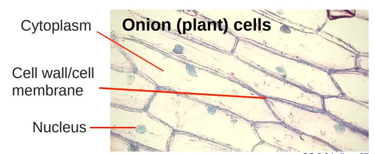
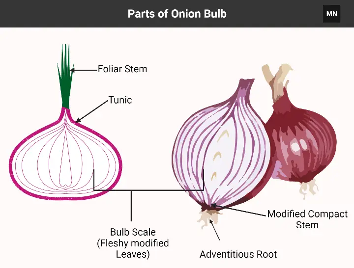
An onion bulb, though commonly used as a culinary ingredient, presents an excellent opportunity for observing plant cell structures due to its unique composition. The bulb is formed from modified leaves, making it an ideal candidate for this experiment. Just like other plant cells, onion cells feature a rigid cell wall that encapsulates the cell membrane, housing the essential components of the cell, such as the cytoplasm and the nucleus.
Specifically, the experiment focuses on the onion’s epidermal cells, which constitute a single layer that acts as a protective skin. This layer effectively separates the thick, juicy scale leaves of the onion, playing a vital role in the bulb’s formation.
When observing the epidermal cell of an onion bulb under a microscope, it appears simple and transparent. This characteristic provides an introduction to the general anatomy of plant cells and their arrangement.
To visualize the onion’s epidermal cells, firm and medium-sized onions are typically chosen. These onions offer a clear view of the cellular structure, making it easier for students to observe and comprehend the intricacies of plant anatomy.
Now, let’s delve into the theory, requirements, and procedure of the Onion Peel Cell Experiment. The theory behind the experiment revolves around the idea of examining the cellular structure of plants and understanding the role of different components within the cell. By using onions, students get a practical understanding of how plant cells are organized and how they differ from animal cells.
The requirements for the experiment are relatively simple and readily available. All that is needed are firm and medium-sized onions, a sharp blade or scalpel, a microscope, a glass slide, a cover slip, and a dropper of water.
To conduct the experiment, the outermost thin layer of the onion is gently peeled using the blade or scalpel. This thin layer, which consists of epidermal cells, is carefully removed and placed on the glass slide. A drop of water is added to the onion peel to ensure it remains hydrated and prevents it from drying out under the microscope. Once the onion peel is properly positioned, the cover slip is gently placed over it.
With the microscope set to the appropriate magnification, students can now observe the onion peel cells in detail. They can identify and study the cell wall, cell membrane, cytoplasm, and nucleus, gaining insights into the structural organization of a plant cell.
The experiment’s observation reveals the simple yet vital characteristics of onion epidermal cells, offering students a firsthand look at plant cell anatomy. This hands-on experience fosters a deeper understanding of plant biology, sparking curiosity and interest in further scientific exploration.
After conducting the experiment, the students discuss their observations and draw conclusions about the onion peel cell structure. The result of the experiment solidifies their knowledge of plant cell components and their functions.
However, as with any scientific experiment, precautions should be taken to ensure accurate and safe results. Students should handle the scalpel or blade with care to avoid injuries. Additionally, proper microscopy techniques should be followed to achieve clear and accurate observations.
In conclusion, the Onion Peel Cell Experiment is an engaging and informative way to introduce students to plant cell structures and plant anatomy. By exploring the onion’s epidermal cells, students gain a deeper understanding of the cell’s components and their functions. This experiment paves the way for further scientific exploration and sparks curiosity about the wonders of the natural world.
Objective of this Experiment
The primary objective of conducting the onion peel cell experiment is to observe and study the arrangement and structural components of the onion epidermis. This experiment holds educational value as it provides students with essential insights into the structure of plant cells.
One significant advantage of using the epidermis of the onion bulb for this experiment is that it consists of a single layer of tissue that is easily separable. This simplicity in isolation makes onion peel an ideal choice for educational and experimental purposes, enabling students to focus on the specific characteristics of the plant cell structure.
Another benefit of using onions for this experiment is the relatively large size of their cells. The larger size of onion cells allows them to be examined under low magnification settings on a microscope. This feature is advantageous for students as they can clearly visualize and identify the different components of the cell without requiring high magnification, making the observation process more straightforward.
Furthermore, the onion peel cell experiment is considered a simple and manageable activity for students to perform. Its simplicity allows students to efficiently carry out the experiment without the need for complex procedures or specialized equipment. In addition to observing plant cell structures, this experiment provides an excellent opportunity for students to practice using a microscope, an essential skill in the field of biology and scientific research.
Overall, the main objective of the onion peel cell experiment is to provide students with a hands-on experience in studying plant cell structures, foster an understanding of their arrangement, and familiarize them with using a microscope. This practical approach to learning enhances students’ knowledge of plant biology and encourages them to explore and appreciate the wonders of the microscopic world.
Requirements
To prepare a temporary slide of an onion peel for the observation of its cellular structure, the following glassware and reagents are required:
Materials required to separate onion skin:
- Medium-sized onion: A firm and medium-sized onion is needed to obtain the onion peel for the experiment. The onion should be fresh to ensure the integrity of the cells.
- Knife: A sharp knife or scalpel is necessary to carefully peel off the outermost thin layer of the onion, which constitutes the epidermal cells.
- Forceps: Forceps are used to handle the onion and hold it in place while peeling off the epidermal layer.
Materials needed to stain and mount the onion peel:
- Petri Plate: A Petri dish is required to hold the onion peel during the staining and mounting process.
- Distilled Water: Distilled water is used to hydrate the onion peel, ensuring that the cells remain in a suitable environment for observation.
- Safranin: Safranin is a red dye or stain used to enhance the visibility of the cell structures under the microscope. It helps to highlight the various components of the plant cell.
- Clean glass slide: A clean glass slide acts as the base on which the stained onion peel will be mounted for microscopic observation.
- Glycerine: Glycerine is used as a mounting medium to place the stained onion peel on the glass slide and preserve it for a longer duration. Glycerine also helps in improving the clarity of the microscopic image.
- Coverslip: A coverslip is placed over the stained and mounted onion peel to protect it and provide a flat surface for easy observation under the microscope.
- Blotting paper: Blotting paper is used to remove excess water from the onion peel before mounting it on the glass slide, preventing excess moisture from affecting the observation.
- Compound microscope: A compound microscope is essential for observing the onion peel at various magnifications. This instrument allows students to visualize the cellular structures in detail and study the different components of the plant cells.
By gathering these necessary materials and reagents, students can effectively prepare and observe the onion peel cell under the microscope, enhancing their understanding of plant cell structures and their organization. The use of staining and mounting techniques in combination with a compound microscope provides an opportunity for students to explore the microscopic world of plant biology in an educational and engaging manner.
Principle
The principle of the onion peel cell experiment is to observe and understand the characteristic features of plant cells, which are evident in onion cells due to their multicellular nature. As a plant, onions share common features with other plant cells, such as the presence of a rigid cell wall and a large vacuole, which contribute to their structural organization.
The first key element to consider is the cell wall. The cell wall is a rigid and protective coat that surrounds the cell membrane, encompassing all the internal components of the onion cell. This rigid structure plays a crucial role in maintaining the shape of onion cells and contributes to the compact arrangement of the epidermal cells in the onion. The presence of the cell wall is a characteristic feature of plant cells and distinguishes them from animal cells.
Moving inward from the cell wall, we encounter the cell membrane, which lies interior to the cell wall. The cell membrane forms a boundary around the cytoplasm and its internal structures, acting as a selective barrier that controls the movement of substances in and out of the cell.
The cytoplasm is the inner space of the cell that appears jelly-like and fills the area between the cell membrane and the nucleus. Within the cytoplasm, there are various organelles and cytosolic materials. A fascinating phenomenon called “cytoplasmic streaming” can also be observed in plant cells, including onion cells. This process involves the movement of cytosolic material around the cell, facilitating the distribution of nutrients, organelles, and other essential substances.
Located near the periphery of the cytoplasm, the nucleus serves as the control center of the cell. It is the largest organelle in the cell and houses the cell’s genetic material in the form of DNA. The nucleus plays a vital role in regulating cellular activities and determining the cell’s characteristics and functions.
One prominent feature that stands out within the onion cell is the large vacuole, which is prominently seen at the center of the cell. Vacuoles are unique to plant cells and are responsible for storing solid and liquid contents. The size and shape of the vacuole can vary depending on the specific needs of the cell. This organelle contributes to the regulation of water content, nutrient storage, and waste management within the cell.
In summary, the principle of the onion peel cell experiment revolves around studying the characteristic features of plant cells, which are exemplified in onion cells. The presence of a rigid cell wall, cell membrane, cytoplasm, nucleus, and large vacuole in onion cells provides valuable insights into the structural organization and functions of plant cells. By observing these features under a microscope, students can gain a better understanding of plant biology and appreciate the complexity of cellular structures in the natural world.
Onion Peel Cell Experiment Procedure
Steps to separate an onion peel
To separate an onion peel for the experiment, follow these steps:
- Begin by taking a medium-sized onion and carefully remove its outermost peel. To do this, use a knife to cut the onion into two equal halves.
- After chopping the onion into halves, select one fleshy scale leaf from the chopped onion bulb. To facilitate the separation, gently split the chosen scale leaf into two parts.
- Now, with the help of forceps, carefully pull a thin and transparent epidermal peel from the convex surface of the scale leaf. The epidermal peel is the outermost layer of the onion cell, which is suitable for the experiment.
- Once you have the thin epidermal peel, place it in a Petri plate containing water. This step ensures that the onion peel remains hydrated during the experiment, allowing for better observation under the microscope.
- If necessary, you can further cut the onion peel into small rectangular pieces using a blade. This process helps in preparing smaller and manageable sections of the peel for mounting on the glass slide.
By following these steps, you can successfully separate an onion peel and prepare it for the onion peel cell experiment. The obtained onion peel will serve as the basis for observing and studying the structure of plant cells under the microscope, providing valuable insights into the arrangement and components of onion epidermal cells.
Steps to stain and mount an onion peel
To stain and mount an onion peel for microscopic observation, follow these step-by-step instructions:
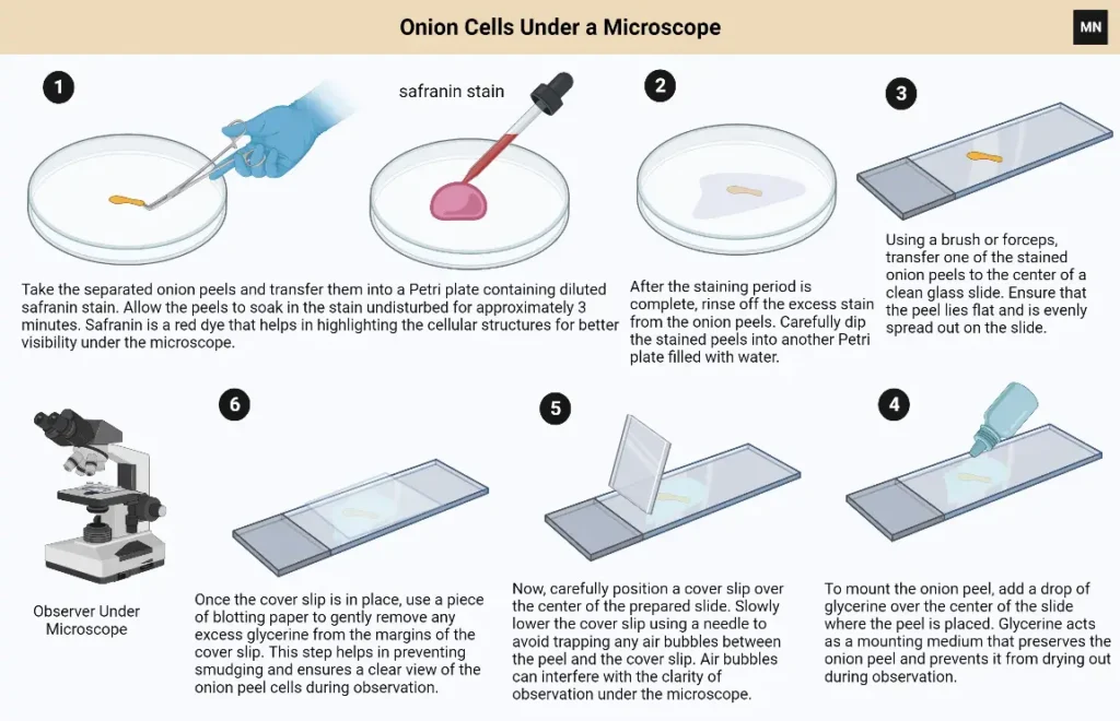
- Take the separated onion peels and transfer them into a Petri plate containing diluted safranin stain. Allow the peels to soak in the stain undisturbed for approximately 3 minutes. Safranin is a red dye that helps in highlighting the cellular structures for better visibility under the microscope.
- After the staining period is complete, rinse off the excess stain from the onion peels. Carefully dip the stained peels into another Petri plate filled with water. This step ensures that only the necessary amount of stain remains on the peels, preventing over-saturation.
- Using a brush or forceps, transfer one of the stained onion peels to the center of a clean glass slide. Ensure that the peel lies flat and is evenly spread out on the slide.
- To mount the onion peel, add a drop of glycerine over the center of the slide where the peel is placed. Glycerine acts as a mounting medium that preserves the onion peel and prevents it from drying out during observation.
- Now, carefully position a cover slip over the center of the prepared slide. Slowly lower the cover slip using a needle to avoid trapping any air bubbles between the peel and the cover slip. Air bubbles can interfere with the clarity of observation under the microscope.
- Once the cover slip is in place, use a piece of blotting paper to gently remove any excess glycerine from the margins of the cover slip. This step helps in preventing smudging and ensures a clear view of the onion peel cells during observation.
By following these steps, you will successfully stain and mount an onion peel for microscopic examination. The staining process enhances the contrast of the cellular structures, allowing for a detailed study of the onion epidermal cells. The mounted peel will be ready for observation under a compound microscope, providing valuable insights into the arrangement and components of plant cells in the onion epidermis.
Observe the temporary slide under the compound microscope
To observe the temporary slide of the stained and mounted onion peel under a compound microscope, follow these steps:
- Turn on the microscope’s light source and ensure that the low objective lens is in line with the optical tube. Adjust the light intensity as needed for better visibility.
- Carefully place the prepared slide containing the stained onion peel on the stage of the microscope. Secure the slide in place using the stage clips or holders to prevent movement during observation.
- Looking from the side (not through an eyepiece), use the coarse focus knob to lower the tube until the end of the objective lens is just above the cover glass. Be cautious not to apply excessive force, as it may crack the cover glass or damage the objective lens.
- Now, look through the eyepiece and adjust the smaller, fine focusing knob to move the optical tube upwards. Gradually turn the knob until an image of the onion peel cells comes into focus. This process allows you to achieve a clear and sharp image of the cellular structures.
- Once the cells are in focus with the low objective lens, you can proceed to swap the objective lens to a higher magnification, such as the high objective lens. Switching to a higher magnification will allow you to observe the cells in greater detail and explore specific features of the onion epidermal cells.
- While observing the onion peel cells under the microscope, prepare an observation table to record your findings. Note down the various features you observe in the cells, such as the cell wall, cell membrane, cytoplasm, nucleus, and the large vacuole. Additionally, you can document any specific characteristics, shapes, or sizes of the cells and their organelles.
By following these steps and using the compound microscope effectively, you will be able to examine the stained onion peel cells in detail. The microscope’s magnification capabilities enable you to explore the intricate structures of the onion epidermal cells and gain valuable insights into the organization and components of plant cells. The observation table serves as a useful reference to document and organize your observations, making it easier to analyze and compare the different features seen under the microscope.
Method 2 Onion Peel Cell Experiment
The procedure for the onion peel cell experiment is as follows
Materials needed:
- A thin onion membrane (epidermal peel)
- Microscopic glass slides
- Microscopic cover slips
- Needle
- Blotting paper
- Dropper
- Iodine solution (or methylene blue)
- Water
- Microscope
Procedure
- Add a drop of water at the center of a clean microscopic glass slide. This drop of water will act as a medium to suspend the onion membrane for examination.
- Take a thin membrane (epidermal peel) from the onion layer and carefully lay it at the center of the microscopic slide. The drop of water will help to flatten the membrane and hold it in place for observation.
- Add a drop of iodine solution (or methylene blue) onto the onion membrane. The staining solution will enhance the visibility of the cellular structures under the microscope.
- Gently place a microscopic cover slip on top of the onion membrane, making sure to press it down gently to avoid trapping air bubbles. The cover slip helps to protect the onion membrane and holds it in place for observation.
- Use a needle to carefully remove any air bubbles that may have formed between the cover slip and the onion membrane.
- Touch a blotting paper on one side of the slide to drain any excess iodine solution or water. This step ensures that the slide remains clean and avoids smudging during observation.
- Place the prepared slide on the microscope stage under low power magnification. Start by looking from the side of the microscope (not through the eyepiece) and lower the optical tube to bring the objective lens as close to the slide as possible. This prevents accidents and damage to the slide or microscope lenses.
- Once the objective lens is in a safe position, look through the eyepiece and adjust the focus using the fine focusing knob to achieve clarity and a clear image of the onion peel cells.
- Observe the onion peel cells under low power magnification to study their general structure and arrangement.
- Students can make another slide without adding the stain to observe the difference between a stained slide and a non-stained slide. This comparison helps in understanding the importance of staining in microscopic observation.
By following these steps, students can successfully conduct the onion peel cell experiment and explore the cellular structure of onion epidermal cells under the microscope. The procedure allows for a hands-on learning experience, enhancing students’ understanding of plant cell anatomy and the role of staining techniques in microscopy.
Observation
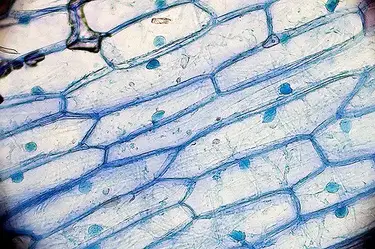
During the observation of the stained and mounted onion peel under the compound microscope, several key features of the onion epidermal cells become apparent. These observations provide valuable insights into the structural organization and components of the plant cells.
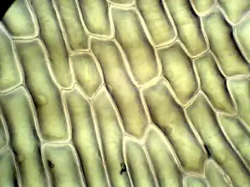
- Shape of cells: The onion epidermal cells appear rectangular in shape. This regularity in cell shape is a common characteristic of plant cells, and it contributes to the compact arrangement of cells in the onion epidermis.
- Arrangement of cells: The cells in the onion peel exhibit a compact arrangement, with little to no inter-cellular spaces between them. This close packing of cells is typical in plant tissues and helps provide structural support to the onion bulb.
- Inter-cellular spaces: Upon observation, it is noted that there are no inter-cellular spaces present between the onion epidermal cells. The absence of significant gaps between cells further reinforces their compact arrangement.
- Nucleus: Each onion epidermal cell contains a prominent nucleus located at the cell’s periphery. The nucleus serves as the control center of the cell and houses the genetic material (DNA). Its position near the cell’s edge allows for efficient regulation of cellular activities.
- Stained portions: Under the safranin stain, the cell wall and nucleus of the onion epidermal cells appear darkly stained. The stain highlights these structures, making them more visible under the microscope. The dark staining of the cell wall reinforces its role as a rigid and protective layer surrounding the cell. Meanwhile, the nucleus stands out due to its significant role in controlling cellular functions.
- Unstained portions: In contrast to the stained cell wall and nucleus, the cell membrane and vacuole appear less stained. The cell membrane, being selectively permeable, allows for the passage of certain substances in and out of the cell. The vacuole, being a large organelle responsible for storing various materials, shows a less intense staining pattern compared to other cellular structures.
Overall, the observation of the stained onion peel cells under the microscope provides a detailed view of the arrangement and structural components of the onion epidermal cells. The rectangular shape and compact arrangement of the cells contribute to the overall structure of the onion bulb, providing a protective skin and structural support. The presence of the nucleus at the cell’s periphery highlights its crucial role in controlling cellular activities. Additionally, the selective staining of different cellular structures allows for better identification and understanding of their functions within the plant cell.
Result
The result of observing the stained and mounted onion peel under the compound microscope reveals several distinctive features of the onion epidermal cells:
- Shape and arrangement of cells: The onion epidermal cells exhibit a rectangular shape and are compactly arranged. There are no intercellular spaces between the cells, indicating a close packing arrangement typical of plant tissues.
- Cell wall: Each onion epidermal cell is encased by a distinct and rigid cell wall. The cell wall provides structural support and protection to the cell, maintaining its shape and integrity.
- Nucleus: Within each onion epidermal cell, a prominent nucleus is present. The nucleus serves as the control center of the cell, containing the genetic material (DNA) that regulates various cellular functions.
- Vacuole: The onion epidermal cells also contain a large vacuole. The vacuole is an essential organelle responsible for storing various substances, including water, nutrients, and waste products.
The observation and identification of these features provide a comprehensive understanding of the structural composition of onion epidermal cells. The rectangular shape and compact arrangement of the cells support the onion’s bulb structure and contribute to its overall integrity. The presence of a distinct cell wall ensures the cell’s rigidity and protection, while the nucleus serves as a vital center for cellular control and genetic regulation. Furthermore, the large vacuole plays a significant role in storing essential materials needed for the cell’s functions.
In conclusion, the result of the observation confirms that the onion epidermal cells possess key characteristics common to plant cells. Understanding these features is crucial for gaining insights into the organization and functions of plant tissues, as well as appreciating the intricate cellular structures that make up the onion bulb.
Precautions
When conducting the onion peel cell experiment and preparing the stained and mounted slide, it is essential to take certain precautions to ensure accurate and reliable results:
- Do not overstain the onion skin: Overstaining the onion peel with safranin can lead to excessive coloration, making it challenging to distinguish specific cellular structures. It is essential to follow the recommended staining time to achieve an appropriate level of contrast without overpowering the cell features.
- Avoid folding of the peel: Care should be taken to prevent any folding or crumpling of the onion peel during the staining and mounting process. Folding can distort the cellular structures and hinder proper observation under the microscope.
- Use dry and clean glass slide and cover slip: Before mounting the onion peel, ensure that both the glass slide and cover slip are free from any moisture or contaminants. Any impurities on the slide or cover slip may interfere with the observation and affect the clarity of the images.
- Put a coverslip carefully to avoid air bubbles: When placing the coverslip over the mounted onion peel, take care to avoid trapping air bubbles between the peel and the cover slip. Air bubbles can obstruct the view and make it difficult to observe the cellular structures clearly.
- Use blotting paper to remove extra glycerine: After mounting the coverslip, gently remove any excess glycerine from the margins using blotting paper. This step prevents smudging and ensures a clear view of the onion peel cells during microscopic observation.
By adhering to these precautions, you can enhance the accuracy and quality of the onion peel cell experiment. Maintaining proper staining, handling, and mounting techniques will allow for a clear and detailed examination of the onion epidermal cells, providing valuable insights into the structure and components of plant cells.
Onion Cells Under a Microscope Video
FAQ
What is the onion peel cell experiment?
The onion peel cell experiment is a common educational activity where students observe and study the cellular structure of onion epidermal cells under a microscope.
Why use onion peel for this experiment?
Onion peel is used because it consists of a single layer of cells that are easy to separate and observe. Additionally, the large size of onion cells allows for examination under low magnification.
What materials are needed for the experiment?
The materials needed include an onion, knife, forceps, Petri plate, distilled water, safranin stain, clean glass slide, glycerine, coverslip, blotting paper, and a compound microscope.
How is the onion peel prepared for observation?
To prepare the onion peel, a thin, transparent epidermal layer is carefully pulled from the onion bulb’s scale leaf using forceps and washed in water.
What is the purpose of staining the onion peel?
Staining the onion peel with safranin enhances the visibility of cellular structures, making it easier to observe and study the different components of the cell.
How do you mount the stained onion peel on a glass slide?
To mount the stained onion peel, a drop of glycerine is added to the center of the slide, and the peel is carefully placed on it. A cover slip is then placed over the peel to protect it and avoid air bubbles.
What precautions should be taken during the experiment?
Precautions include not overstaining the onion skin, avoiding folding of the peel, using dry and clean glass slides and coverslips, and removing excess glycerine using blotting paper.
How do you observe the temporary slide under the compound microscope?
To observe the temporary slide, turn on the microscope’s light, ensure the low objective lens is in line with the optical tube, and carefully place the slide on the stage. Adjust the focus for clarity and switch to a higher objective lens for greater magnification if needed.
What features can be observed in the onion peel cells?
Features that can be observed include the rectangular shape of cells, the compact arrangement with no intercellular spaces, the presence of a distinct cell wall, a prominent nucleus, and a large vacuole.
Why is the onion peel cell experiment valuable for students?
The experiment provides students with a hands-on opportunity to study plant cell structures and gain a deeper understanding of plant anatomy. It also helps students practice microscope usage and observation skills, fostering an appreciation for the microscopic world.